References:
Main textbook: Medical Microbiology, Jawetz, Melnick, 26th ed.,2013
1
st
lecture of Medical Virology for 3
rd
year students- college of Medicine
Presented by Dr. Mohammed J. M. Shallal


* Viruses can infect all forms of life (bacteria, plants, protozoa, fungi,
insects, fish, reptiles, birds, and mammals.
* Viral diseases are considered as one important reason of mortality and
permanent disability, especially among infants and children. Antibiotics
are effectively control most bacterial infections, while the viral infections
pose a relatively greater and less controlled threat to human health.
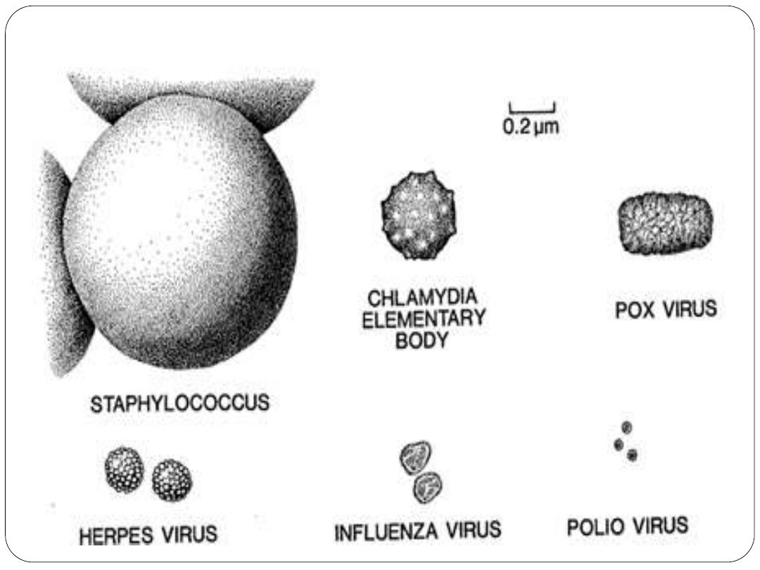
Viruses: are the smallest infectious agents (20-300nm) in size,
composed of nucleic acid surrounding by protein shell which is in
some type of viruses surrounding by lipid envelope.
Viruses are small, sub cellular that are unable to multiply outside a
living host cell (Viruses replicate only in living cells (obligatory).
Containing only one kind of nucleic acid (DNA or RNA) as their
genome
Introduction
Medical Virology
:
is the science that deals with pathogenic and infectious
viruses infect human
Nucleic acid is enclosed in a protective protein shell which may be
surrounded by lipid containing membrane (Enveloped viruses) or not
(Naked or non-enveloped viruses).
By 2000, the International Committee on Taxonomy of Viruses had
organized more than 4000 animal and plant viruses into 56 families, 9
subfamilies, and 233 genera.
However, there are only 21 families of viruses are capable of causing
human infections .
Enveloped
Naked

Viruses differ from other microorganisms by structure, biology and replication
Property
Virus
Bacteria
Size
20-300nm
1000nm
Genome(nucleic acid(
DNA or RNA, but not both
DNA and RNA
Cell wall
Envelope present in some
viruses
Have cell wall
Ribosome
No ribosome
Have ribosome
Multiplication by binary fission
(
-
)
)+(
Sensitivity to antibiotics
(
-
)
)+(
Growth in culture media
Grow only in living host cells
Grow in culture media
Virus particle = virion

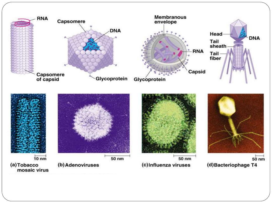
Structure of viruses:
some terms related to the structure of viruses
Capsid: The protein shell, or coat that encloses the nucleic acid
genome:
specific number of identical subunits called Capsomeres
Capsomeres: Morphologic units seen in the electron microscope on
the surface of icosahedral virus particles. Capsomeres represent
clusters of polypeptides, but the morphologic units do not necessarily
correspond to the chemically defined structural units.
Defective virus: A virus particle that is functionally deficient in some
aspect of replication.
Envelope: A lipid-containing membrane that surrounds some virus
particles. It is acquired during viral maturation by a budding process
through a cellular membrane. Virus-encoded glycoproteins are exposed
on the surface of the envelope. These projections are called peplomers.
Nucleocapsid: The protein-nucleic acid complex representing the
packaged form of the viral genome.

Virion: The complete virus particle. In some instances (eg, papilloma
viruses, picorna viruses), the virion is identical with the nucleocapsid.
In more complex virions (herpesviruses, orthomyxoviruses), includes
the nucleocapsid plus a surrounding envelope. Virion, serves to
transfer the viral nucleic acid from one cell to another.
Chemical composition of viruses:
A) Viral protein:
The protein coat have several important functions:
1- To facilitate transfer of the viral nucleic acid from one host cell to
another.
2- To protect the viral genome against inactivation by nucleases.
3- Participate in the attachment of the virus particle to a susceptible
cell.
4- Provide the structural symmetry of the virus particle.
5- Determine the antigenic characteristics of the virus.

6- Some surface proteins may also exhibit specific activities, eg, influenza
virus hemagglutinin agglutinates red blood cells.
7- Some viruses carry enzymes (proteins) are essential for the initiation
of the viral replicative cycle.
B) Viral Nucleic Acid:
Viruses contain a single kind of nucleic acid either
DNA or RNA single or double-stranded, circular or linear that
encodes the genetic information necessary for replication of the virus.
The size of the viral DNA genome ranges from 3.2 kbp
(hepadnaviruses) to 375 kbp (poxviruses). Size of RNA genome ranges
from about 7 kb (some picornaviruses and astroviruses) to 30 kb
(coronaviruses). Viral nucleic acid may be characterized by its G + C
content.
DNA viral genomes can be analyzed and compared using restriction
endonucleases (enzymes that cleave DNA at specific nucleotide
sequences.

C) Viral Lipid Envelopes
Some viruses contain lipid envelopes as part of their structure (eg,
Sindbis virus. The lipid is acquired when the viral nucleocapsid buds
through a cellular membrane in the course of maturation. Budding
occurs only at sites where virus-specific proteins have been inserted
into the host cell membrane. The specific phospholipid
composition of a virion envelope is determined by the specific type
of cell membrane involved in the budding process. For example,
herpesviruses bud through the nuclear membrane of the host cell,
and the phospholipid composition of the purified virus reflects the
lipids of the nuclear membrane.
Lipid-containing viruses are sensitive to effect of ether and other
organic solvents indicating that disruption or loss of lipid results in
loss of infectivity. On the other hand, non-lipid-containing viruses
are generally resistant to Ether.

Viral envelopes also contain glycoproteins. The envelope
glycoproteins are virus-encode. However, the sugars added to viral
glycoproteins often reflect the host cell in which the virus is grown
and have several functions, such as:.
It is the surface glycoproteins of an enveloped virus that attach the
virus particle to a target cell by interacting with a cellular receptor.
They are also often involved in the membrane fusion step of
infection.
The glycoproteins are also important viral antigens.
As a result of their position at the outer surface of the virion, they are
frequently involved in the interaction of the virus particle with
neutralizing antibody.
Extensive glycosylation of viral surface proteins may prevent effective
neutralization of a virus particle by specific antibody.

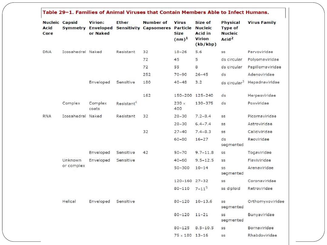
Basis of viral Classification:
Viruses are classified on the basis of morphology, chemical
composition, and mode of replication. The viruses that infect humans
are currently grouped into 21 families, reflecting only a small part of
the spectrum of the multitude of different viruses whose host ranges
extend from vertebrates to protozoa and from plants and fungi to
bacteria.
The following properties have been used as a basis for the
classification of viruses:
A- Based on chemical and physical criteria :
1- Morphology:Viruses are grouped on the basis of size and shape,
chemical composition and structure of the genome (symmetry), and
mode of replication.
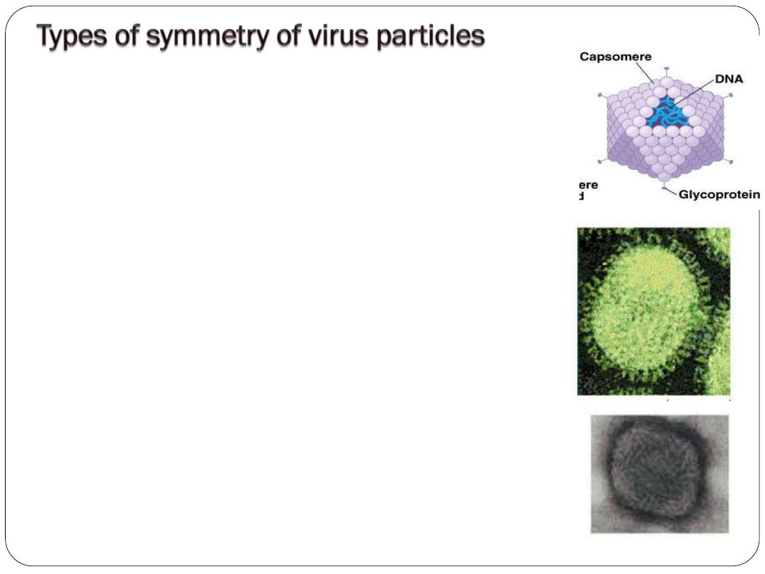
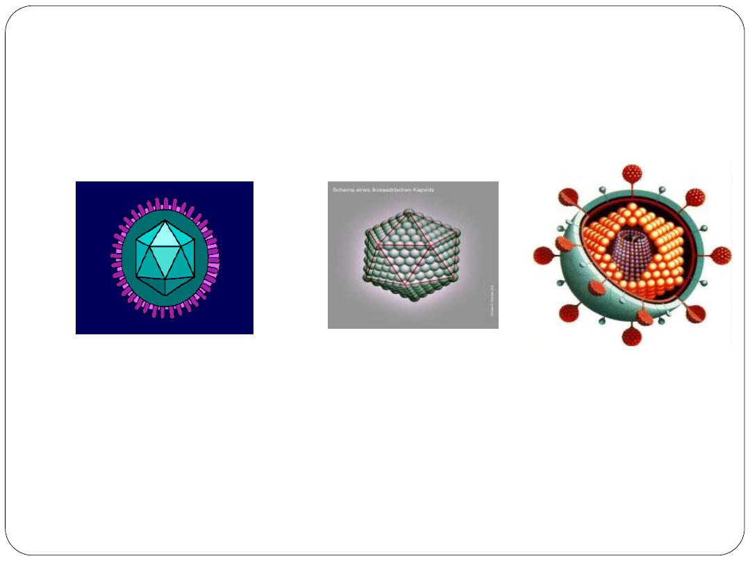
Icosahedral symmetry (cubic(:
composed
of 12 vertices and 20 equal triangular sides, with
approximate outline of sphere, e.g Herpes virus
and Adenovirus
.
Helical symmetry:
the capsomeres are
arranged like steps in a spiral strain case or hollow,
rod shaped, the helix rigid or flexible, e.g influenza
and parainfluenza viruses.
Complex viruses:
e.g Poxvirus, in which there
are many layers around the capsid.

2-Virus genome properties, including
Type of nucleic acid (DNA or RNA).
Size of genome in kilobases (kb) or base pairs (bp).
Strandedness (single or double).
Whether linear or circular, sense (positive, negative, ambisense).
Segments (number, size).
Nucleotide sequence.
G + C content.
3- Physicochemical properties of the virion, including:
Molecular mass,
pH stability,
Thermal stability,
Susceptibility to physical and chemical agents, especially Ether and
other detergents.

(4) Virus protein properties, including: number, size, and functional
activities of structural and nonstructural proteins, amino acid
sequence, modifications (glycosylation, phosphorylation), and special
functional activities (transcriptase, reverse transcriptase ,
neuraminidase and fusion acticity).
(5) Genome organization and replication, including:
gene order,
Strategy of replication (patterns of transcription, translation).
Cellular sites (accumulation of proteins, virion assembly, virion
release).
(6) Antigenic properties.
(7) Biologic properties, including: Natural host range, Mode of
transmission, Vector relationships, Pathogenicity , Tissue tropisms
and pathology.

B- Classification according to diseases they produce:
Generalized diseases: in which virus is spread throughout the body
via blood stream and in which multiple organs are affected Skin
rashes may occur, these include Measles, rubella, chicken pox,
yellow fever and enteroviruses.
Diseases primarily affected specific organs:
a- Diseases of CNS, such as poliomyelitis, rabies, aseptic meningitis
and herpes simplex.
b- Diseases of liver, such as hepatitis type A,B,C,D,E ,yellow fever
and rubella virus.
c- Diseases of skin or mucous membranes, such as herpes simplex,
molluscum contagiosum, warts and herpes zoster.
d- Diseases of Eye, such as adenovirus, herpes keratoconjunctivitis
and epidemic haemorragic conjunctivitis.

e- Diseases of the gastrointestinal tract, such as rotavirus and enteric
adenviruses.
f- Sexually transmitted diseases, such as herpes, hepatitis B virus,
papilloma viruses, reteroviruses (HIV) and cytomegalovirus.
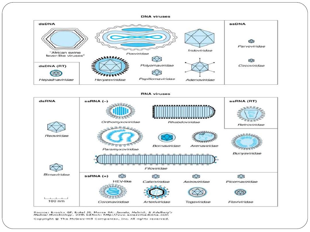
C- Classification of viruses based on nucleic acid genome
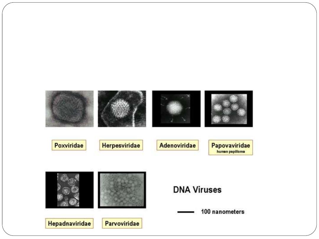
DNA viruses: divided into:
A- Enveloped DNA viruses like: Hepadnaviruses (Hepadnaviridae)
and Herpesviruses (Herpesvridae).
B-Non-enveloped DNA viruses like: Parvoviridae, Papillomaviridae
,Polyomaviridae (Papovaviridae) and Adenoviridae .
Poxviridae
:
C- Complex DNA viruses
RNA viruses divided into :
A- Non-enveloped RNA viruses such as: Picornaviruses, Astroviruses
Caliciviruses and Reoviruses.
B- Enveloped RNA viruses such as: Togaviruses, Arenaviruses
,Flaviviruses,Reteroviruses , Orthomyxoviruses, Bunyaviruses
,Rhabdoviruses, Paramyxoviruses and Filoviruses
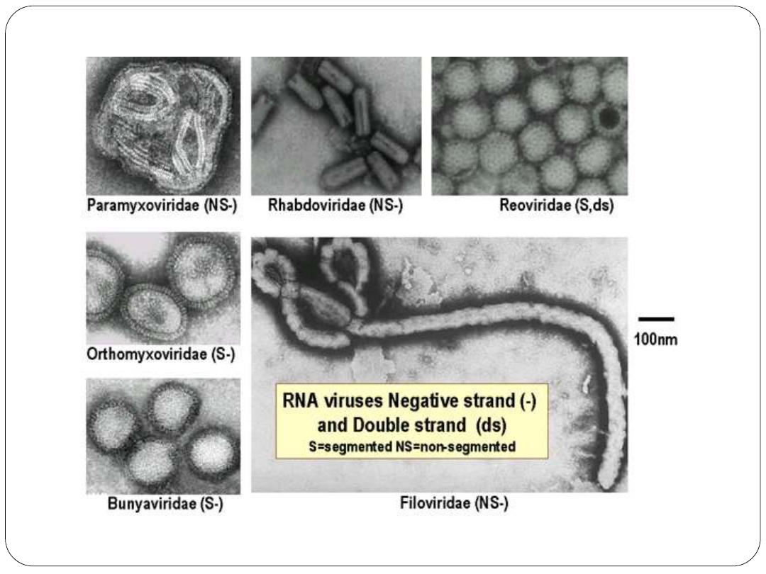
RNA viruses
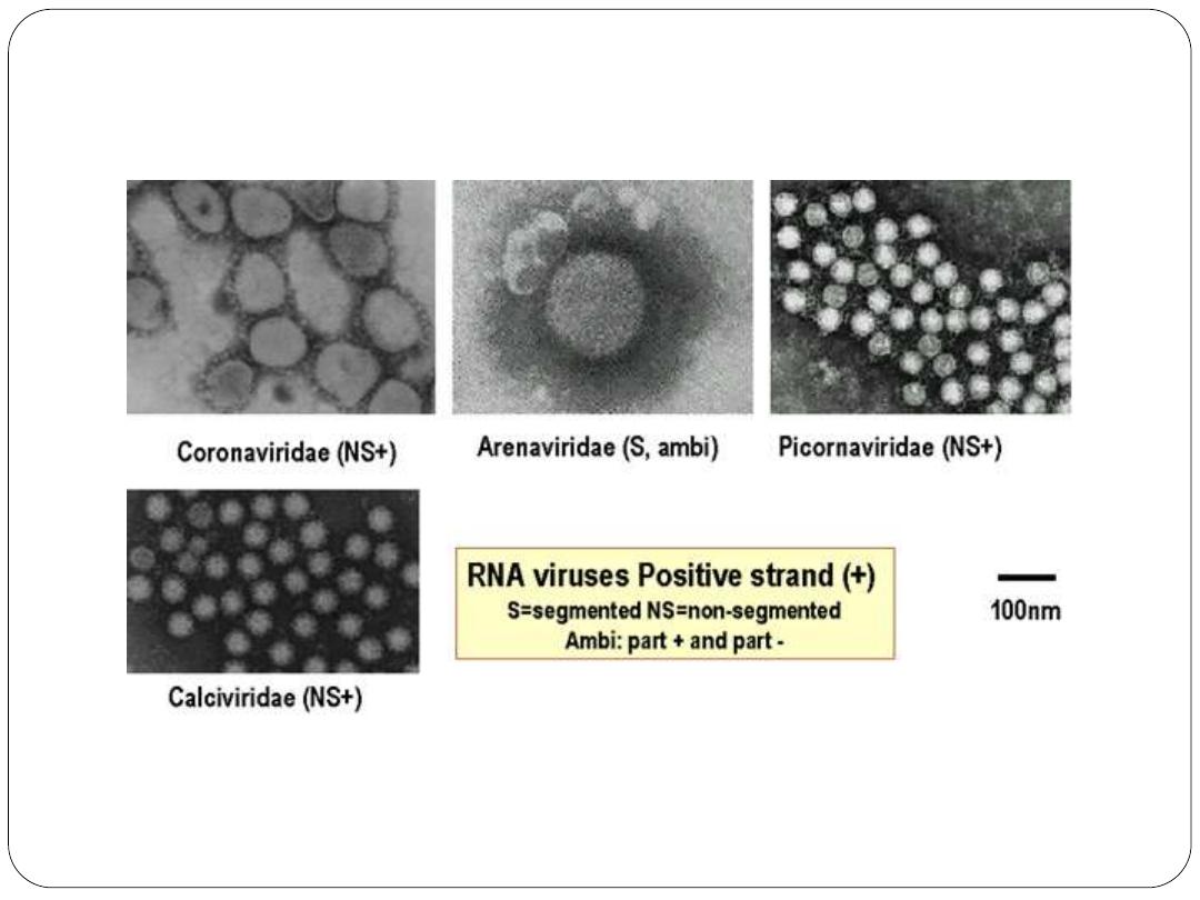
RNA viruses

Evolutionary Origin of Viruses
Three hypotheses of viral origin can be summarized as follows:
(1) Pre-cellular origin hypothesis: viruses originated before cells
(2) Escape host gene hypothesis: Viruses may be derived from DNA
or RNA nucleic acid components of host cells that became able to
replicate autonomously and evolve independently. Fragments of
cellular genomes became infectious
(3) Regressive evolution hypothesis: cells or proto-cells evolved into
virions . Viruses may be degenerate forms of intracellular parasites.
There is no evidence that viruses evolved from bacteria, though other
obligately intracellular organisms, eg, rickettsiae and chlamydiae,
presumably did so.
Non of the above hypotheses explain the origin of virus..but the Pre-
Cellular theory is most popular.

Universal System of Virus Taxonomy
A system has been established in which viruses are separated into
major groupings called families on the basis of virion morphology,
genome structure, and strategies of replication. Virus family names
have the suffix
-viridae
.
And each family is divided to subfamily
–virinae
within each
subfamily, subdivisions called genera are usually based on
physicochemical or serologic differences.
Criteria used to define genera vary from family to family. Genus
names carry the suffix
–virus.

Nomenclature of viruses
In the early days of virology, viruses were named according to common
pathogenic properties:
organ tropism
and/or
modes of transmission
, and
often also after
their discoverers
. From the early 1950s until the mid of
1960s, it was popular to compose virus names by using (abbreviations
derived from a few or initial letters. The name “
Picornaviridae
” is derived
from
pico (small) and RNA
; the name “
Reoviridae
” is derived from
respiratory, enteric, and orphan viruses
because the agents were found in
both
respiratory and enteric specimens
and were
not related to other
classified viruses; “
Papovaviridae
” is from
papilloma, polyoma, and
vacuolating agent
(simian virus 40 [SV40]); “
retrovirus
” is from
reverse
transcriptase
; “
Hepadnaviridae
” is from the
replication of the virus in
hepatocytes
and their
DNA genomes
, as seen in hepatitis B virus.

Adenoviridae
(adeno, “
gland
”; refers to the
adenoid tissue
from which
the viruses were first isolated);
Astroviridae
(
astron means star
);
Arenaviridae
(
arena “sand
”) describes the
sandy appearance
of the
virion.
Bunyaviridae
(from
Bunyamwera, the place in Africa (
where the type strain was isolated);
Calicivirus
(
calix, “cup” or “goblet
”
from the cup-shaped depressions on the viral surfaces);
Coronaviridae
(
corona, “crown
”) describes the
appearance of the peplomers
protruding
from the viral surface;
Filoviridae
(from the Latin
filum,
“thread” or “filament
”) describes the morphology of these viruses.
Herpesviridae
(
herpes, “creeping
”) describes the
nature of the lesions
;
Orthomyxoviridae
(
ortho, “true,” plus myxo
“mucus,” a substance for
which the viruses have an affinity;
Paramyxoviridae
derived from
para,
“closely resembling” and myxo
;
Parvoviridae
(parvus means, “
small
”);
Poxviridae
(
pock means, “pustule
”);
Rhabdoviridae
(
rhabdo, “rod
”
describes the shape of the viruses and
Togaviridae
(
toga, “cloak
”) refers
to the tight viral envelope.

Several viruses of medical importance still
remain unclassified
. Some
are difficult or impossible to propagate in standard laboratory host
systems and thus cannot be obtained in sufficient quantity to permit
more precise characterization.
Hepatitis E virus
, the
Norwalk virus
and similar agents that cause nonbacterial gastroenteritis in humans
are now assigned to the
calicivirus family
.
lecture 1
lecture 1


Sindbis virus (SINV) is a member of the Togaviridae family, in the Alphavirus genus.
The virus was first isolated in 1952 in Cairo, Egypt. The virus is transmitted by
mosquitoes (Culex spp.) SINV causes sindbis fever in humans and the symptoms
include arthralgia, rash and malaise.