
The SKULL (2)
Dr. Firas Al-Hameed
M.B.CH.B C.A.B.S MRCS(ENT)(England)
Thi-Qar Medical School
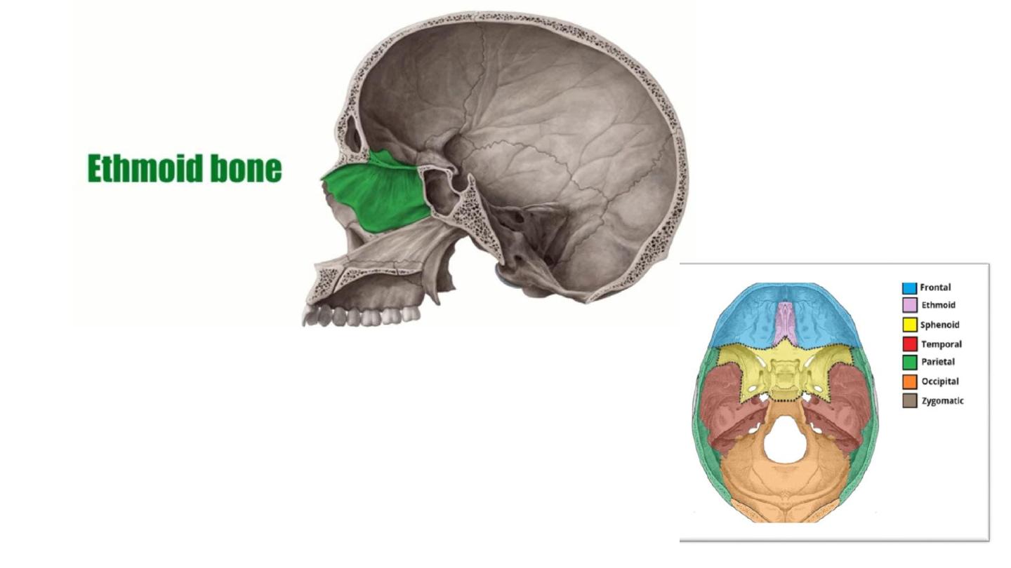
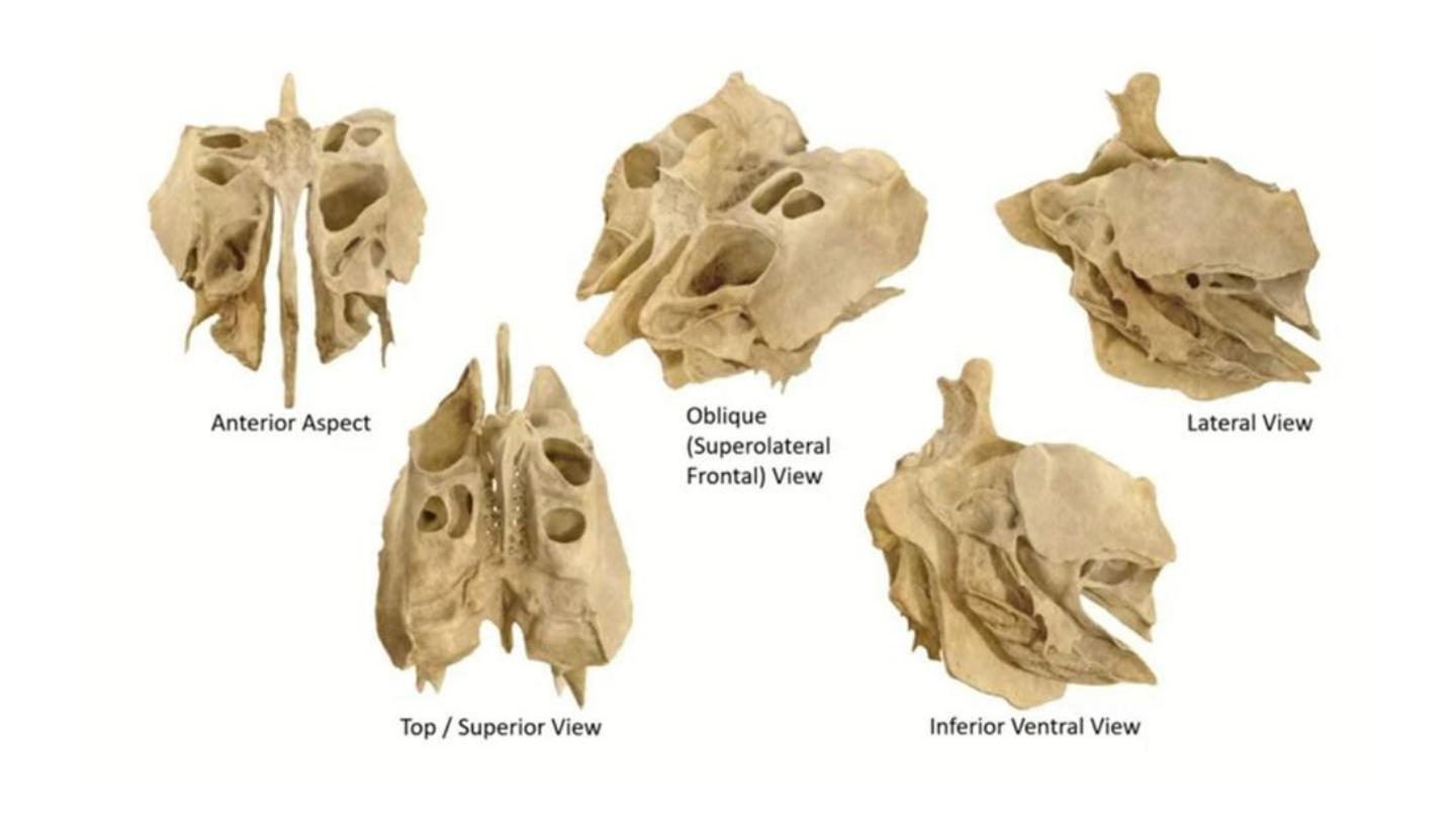
• Cribriform plate
• Perpendicular plate
• Ethmoidal labyrinth.
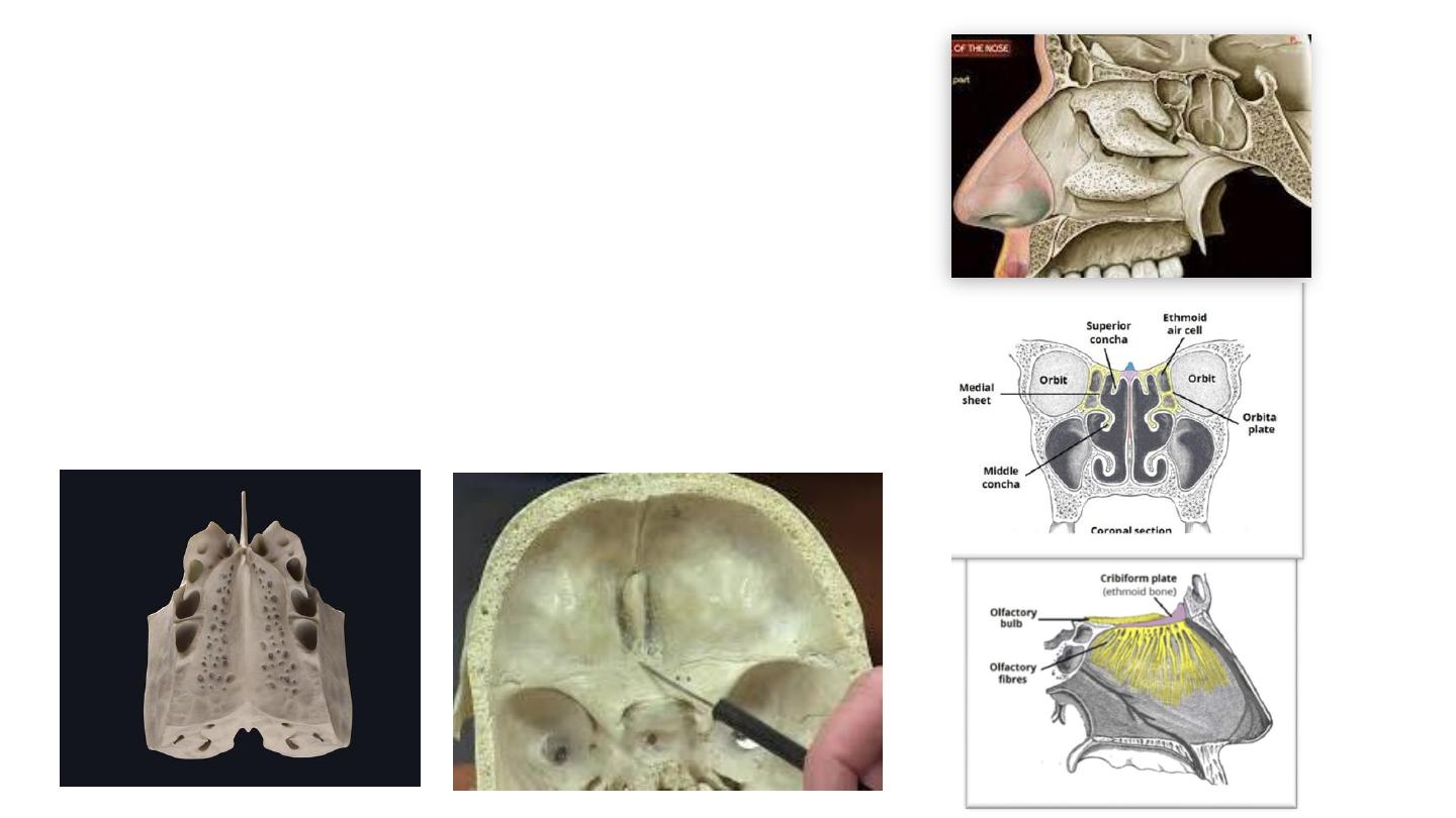
• The cribriform plate
• Forms the roof of the nasal cavity.
• Pierced by numerous olfactory nerve fibres, which
innervate the nasal cavity with the sense of smell.
• Crista galli: Projects superiorly from the cribriform
plate which provides an attachment point for the falx
cerebri (sheet of dura mater that separates the two
cerebral hemispheres).
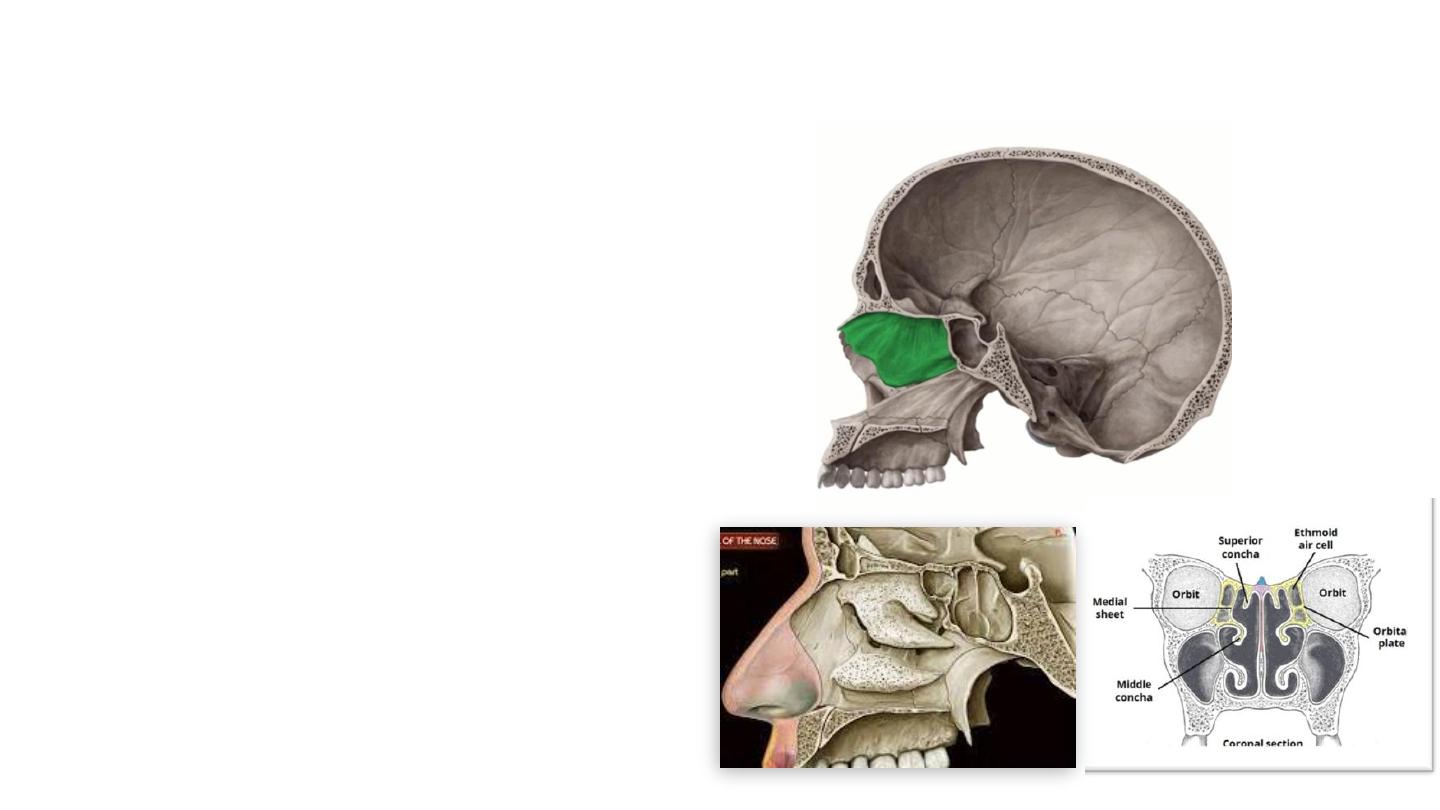
• Perpendicular plate: descends
from the cribriform plate. It
forms the superior two-thirds of
the nasal septum.
• The ethmoid bone contains two
ethmoidal labyrinths which
contain the ethmoidal air cells
(sinuses).
• Superior and middle concha
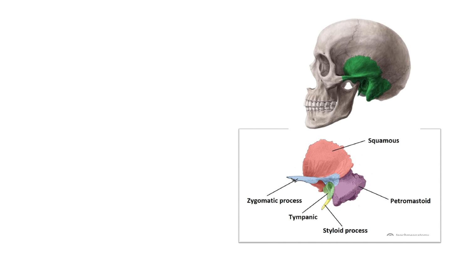
Temporal bone
• Contributes to the lower lateral walls
of the skull.
• It contains the middle and inner
portions of the ear and surround
external auditory canal.
• Crossed by the majority of the cranial
nerves.
• The temporomandibular joint of the
jaw.
• Five constituent parts: The squamous,
tympanic and petromastoid parts, with
the zygomatic and styloid processes
projecting outwards
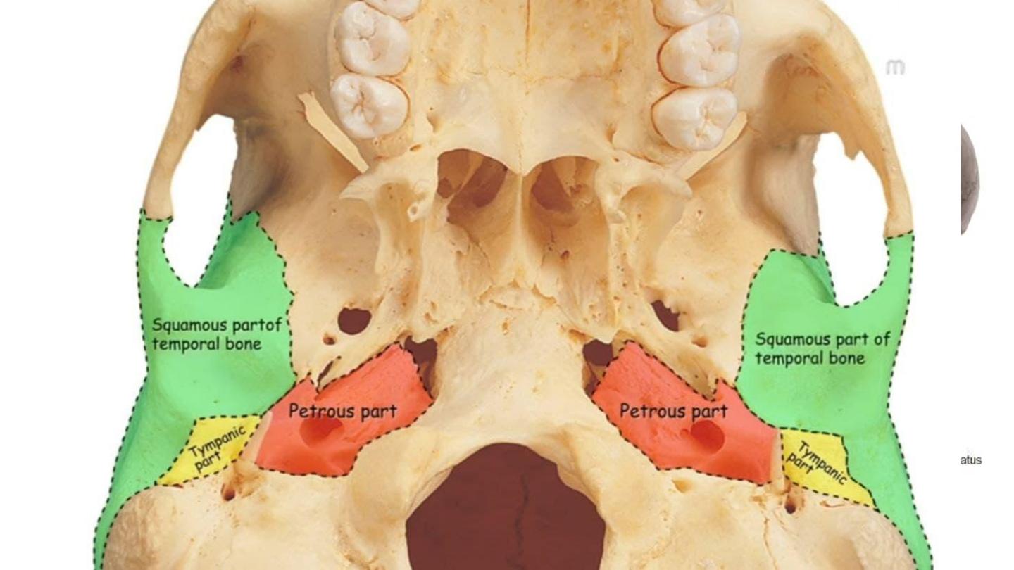
Petromastoid part
• Can be split into a mastoid and petrous
parts.
• Mastoid part:
• mastoid process, an inferior projection of bone,
palpable just behind the ear
• mastoid air cells. These are hollowed out areas
within the temporal bone. They act as a reservoir
of air, equalising the pressure within the middle
ear
• The petrous part is pyramidal shaped, and
lies at the base of temporal bone. It contains
middle and inner ear. Separate the middle
from posterior fossa
Mastoid process

Attached muscles
Muscle
Site of Attachment
Description
Temporalis
Originates from the lower part of squamous
Muscle of mastication
Masseter
Lateral zygomatic surface
Muscle of mastication
Sternocleidomastoid
Mastoid process
Superficial muscle of the neck. Involved in rotation of head and flexion of
neck. Important landmark for the anterior and posterior cervical triangles.
Posterior belly of digastric
Mastoid process
A suprahyoid muscle. Involved in processes such as swallowing.
Splenius capitis
Mastoid process
Strap-like muscle in the back of the neck. Involved in movements such as
shaking the head.
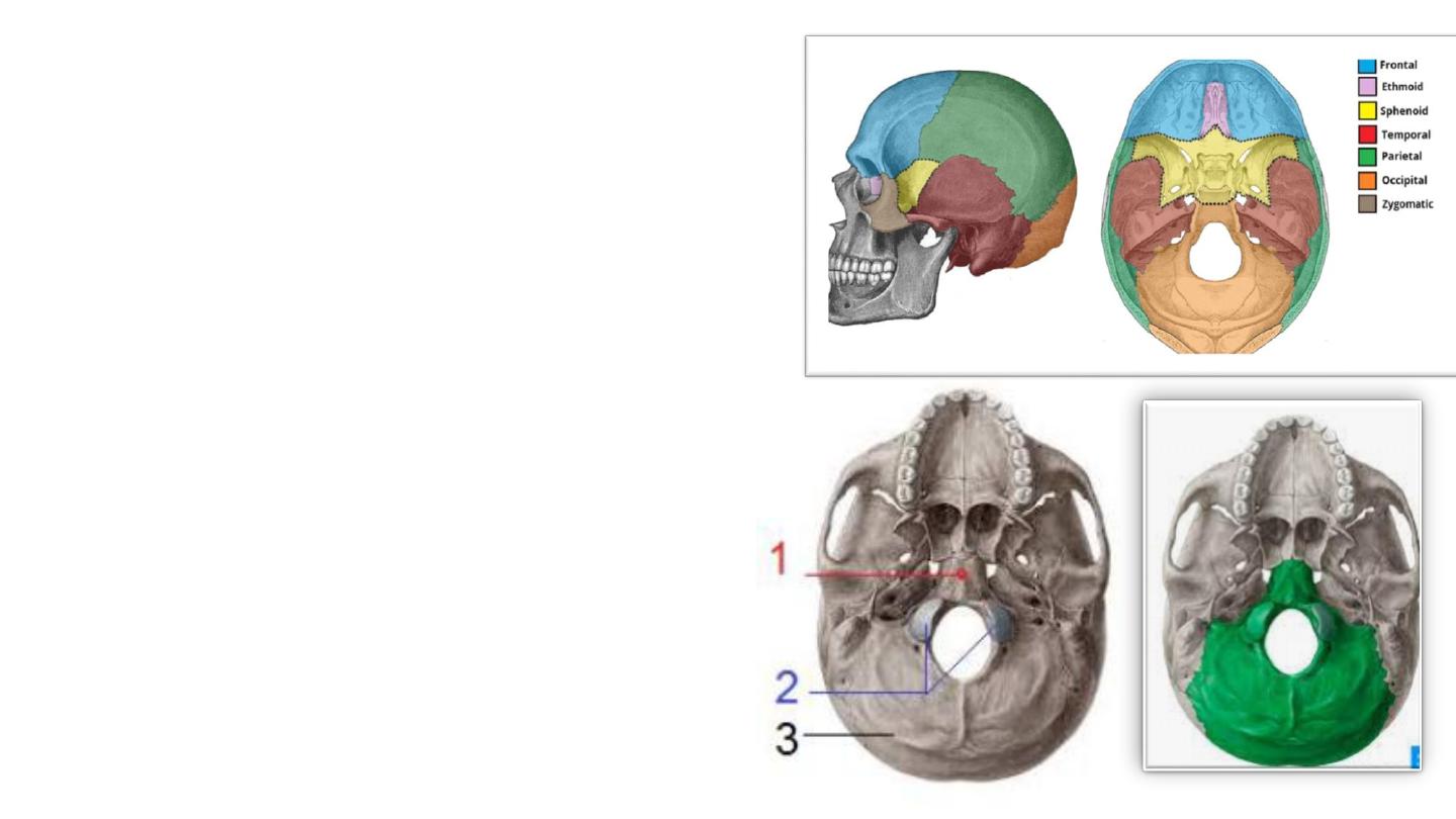
Occipital Bone
• Unpaired trapezoidal bone
• Main bone of back of the skull (occiput).
• It entirely houses the cerebellum.
• Articulates:
• Parietal bones.
• The cervical spine (only cranial bone )
• Sphenoid
• Petrous
• Parts:
• Basilar part (1)
• Condylar part (2)
• Squamous part (3)

The squamous part
• The largest; it lies posterior to foramen magnum
• The external occipital protuberance.. …attachment for the
trapezius muscle
• Three curved lines (nuchal lines)
• Highest, superior and inferior
• Medial nuchal line
• The internal surface : marked by grooves due to venous
cranial sinuses:
• The superior sagittal sinus
• The transverse sinuses
• The sigmoid sinus
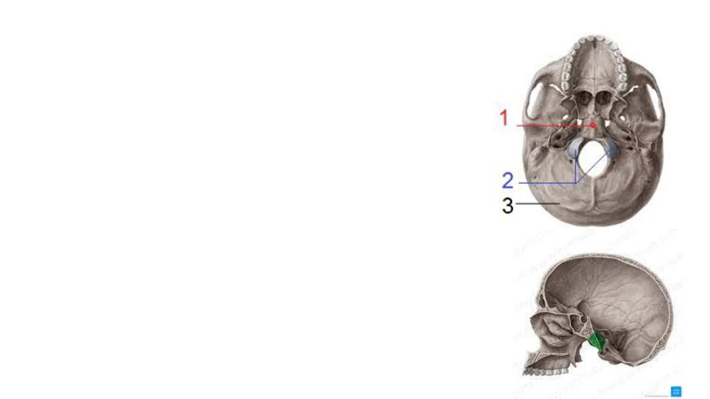
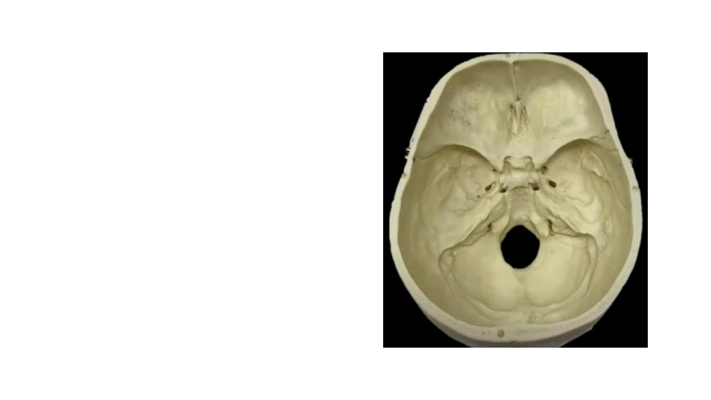
• The basilar part
• Anterior to the foramen magnum and adjacent to the
petrous part of the temporal bone.
• Clivus: anterior most part of basilar part that fuses with
sphenoid bone.
• The condylar parts (occipital condyle, Condylus occipitalis)
• Two kidney-shaped prominences.
• Lateral to the foramen magnum.
• Articulate with the first cervical vertebra (atlanto-
occipital joint).
• Hypoglossal canal pierces through the condylar part of the
occipital bone.
• The jugular foramen lies between the occipital bone and
petrous part of the temporal bone
Inner surface
• Median internal occipital crest
• Internal occipital protuberance
• Groove for the transverse sinus
runs from each side of the
protuberance.
• Groove for sigmoid sinus
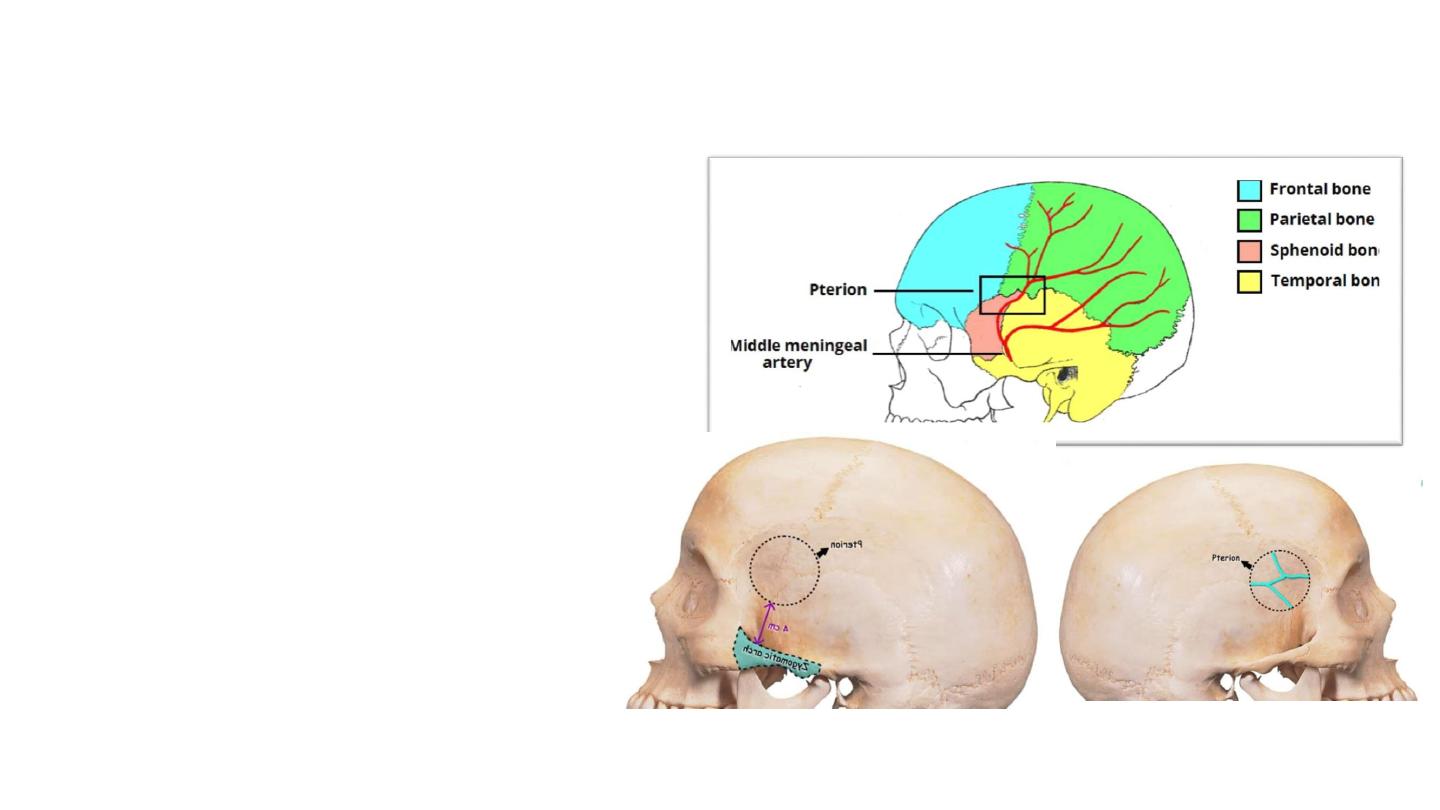
The pterion
• ‘H-shaped’ junction between
temporal, parietal, frontal and
sphenoid bones.
• The thinnest part of the skull.
• A fracture here can lacerate the
middle meningeal artery
(anterior branch), resulting in
an extradural haematoma.
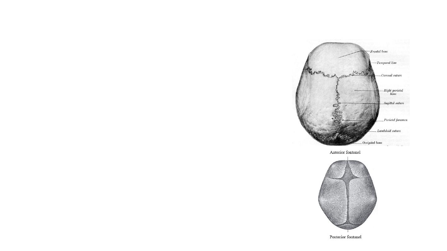
Sutures of the Skull
• Unique to the skull. Immovable junctions between
bones, and fuse completely around the age of 20.
• Clinical importance: they can be points of potential
weakness in both childhood and adulthood.
• The main sutures in adulthood are:
• Coronal
suture which fuses the frontal bone with the
two parietal bones.
• Sagittal
suture which fuses both parietal bones to each
other.
• Lambdoid
suture which fuses the occipital bone to the
two parietal bones.
• In neonates, the incompletely fused suture joints
give rise to membranous gaps between the bones,
known as
fontanelles
.
• Frontal fontanelle (located at the junction of the coronal
and sagittal sutures)
• Occipital fontanelle (located at the junction of the
sagittal and lambdoid sutures).