The Tongue, Muscles of Facial
Expression and Mastication
Dr Firas Al-Hameed
M.B.Ch.B C.A.B.S MRCS (ENT) (England)
Thi-Qar Medical School
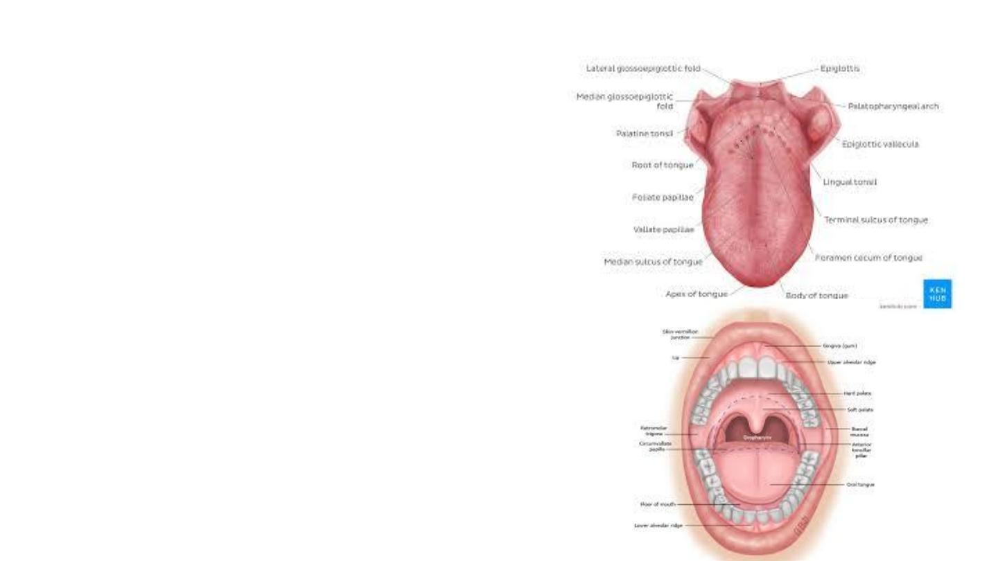
Tongue
• Muscular organ
• Attached via muscles to the hyoid bone,
mandible, styloid process, palate, and pharynx.
• Function
• Mastication
• Swallowing
• Enable speech
• Primary organ of taste
• Parts: divided into two parts by the V-shaped
sulcus terminalis
• Oral: anterior two-thirds
• Pharyngeal
• The foramen cecum at the apex of the sulcus
terminalis indicates the site of embryonic
origin of the thyroglossal duct.
• Median sagittal septum: separate the tongue
into left and right parts
• Superior surface: dorsal
• Inferior: ventral
• Base: posterior third
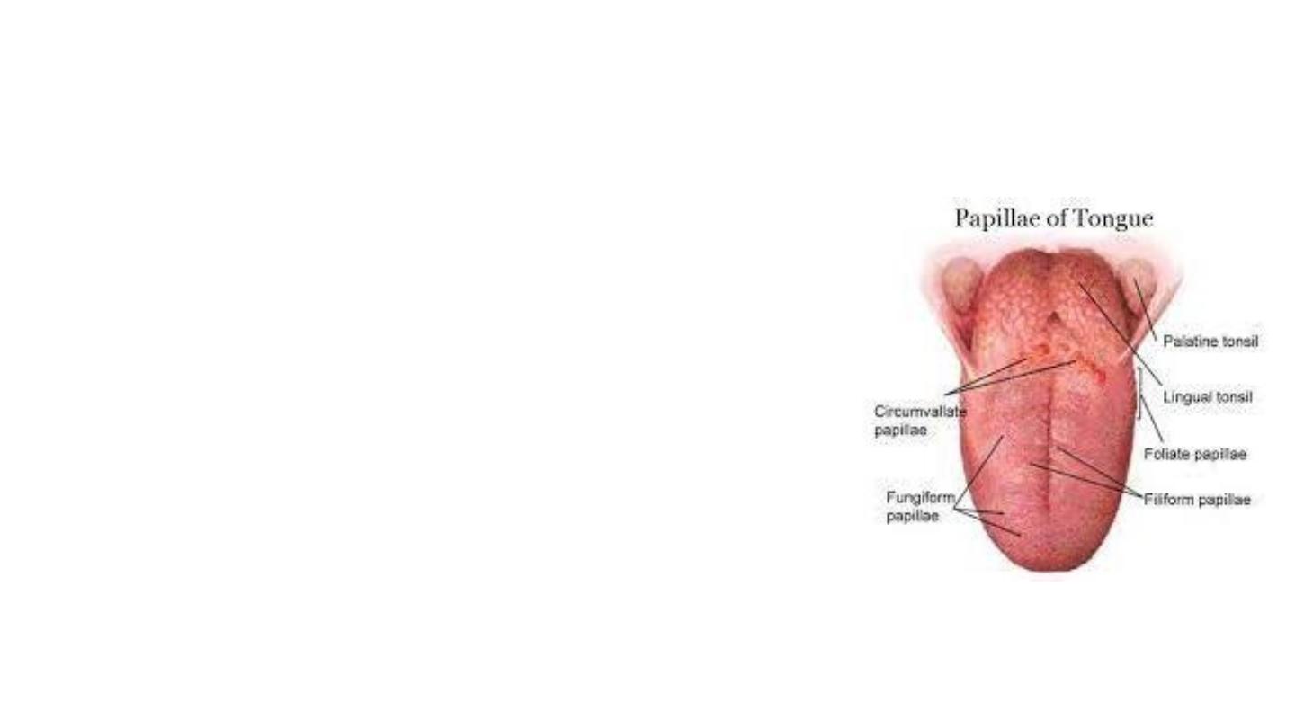
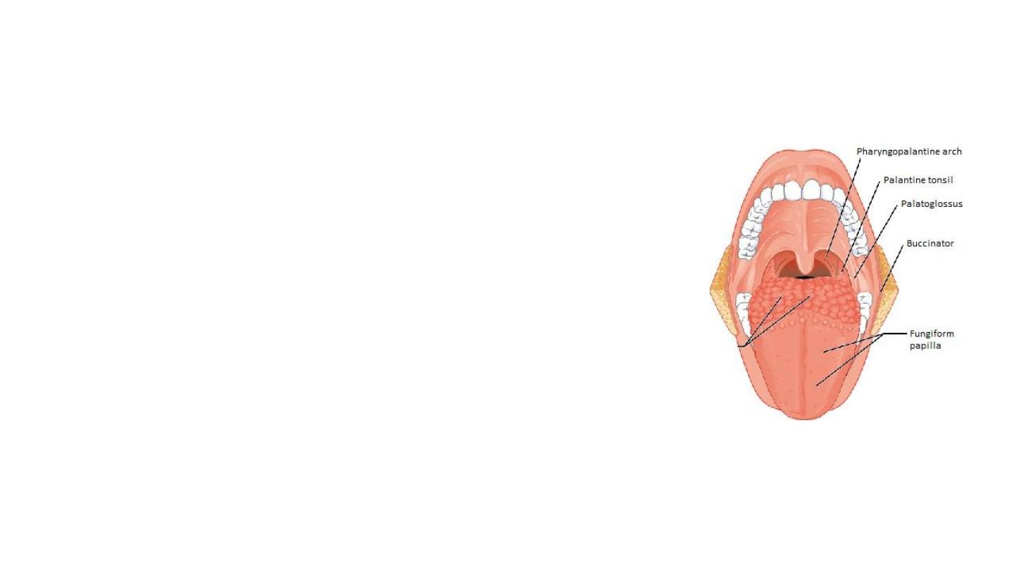
Lingual papillae
• Small, nipple-like structures on the upper surface of the tongue that give
it its characteristic rough texture
• Vallate (circumvallate) papillae
• Arranged in a V-shape arrow anterior to the sulcus terminalis.
• More than half of the taste buds are located
• Filiform papillae
• Organized in rows parallel to the sulcus terminalis.
• Most numerous
• Do not contain taste buds
• Fungiform papillae
• Mostly present on the tip and sides of the tongue.
• Foliate papillae
• Rarely found in humans (vestigial).
• Sides at the back of the tongue, just in front of the palatoglossal arch
of the fauces
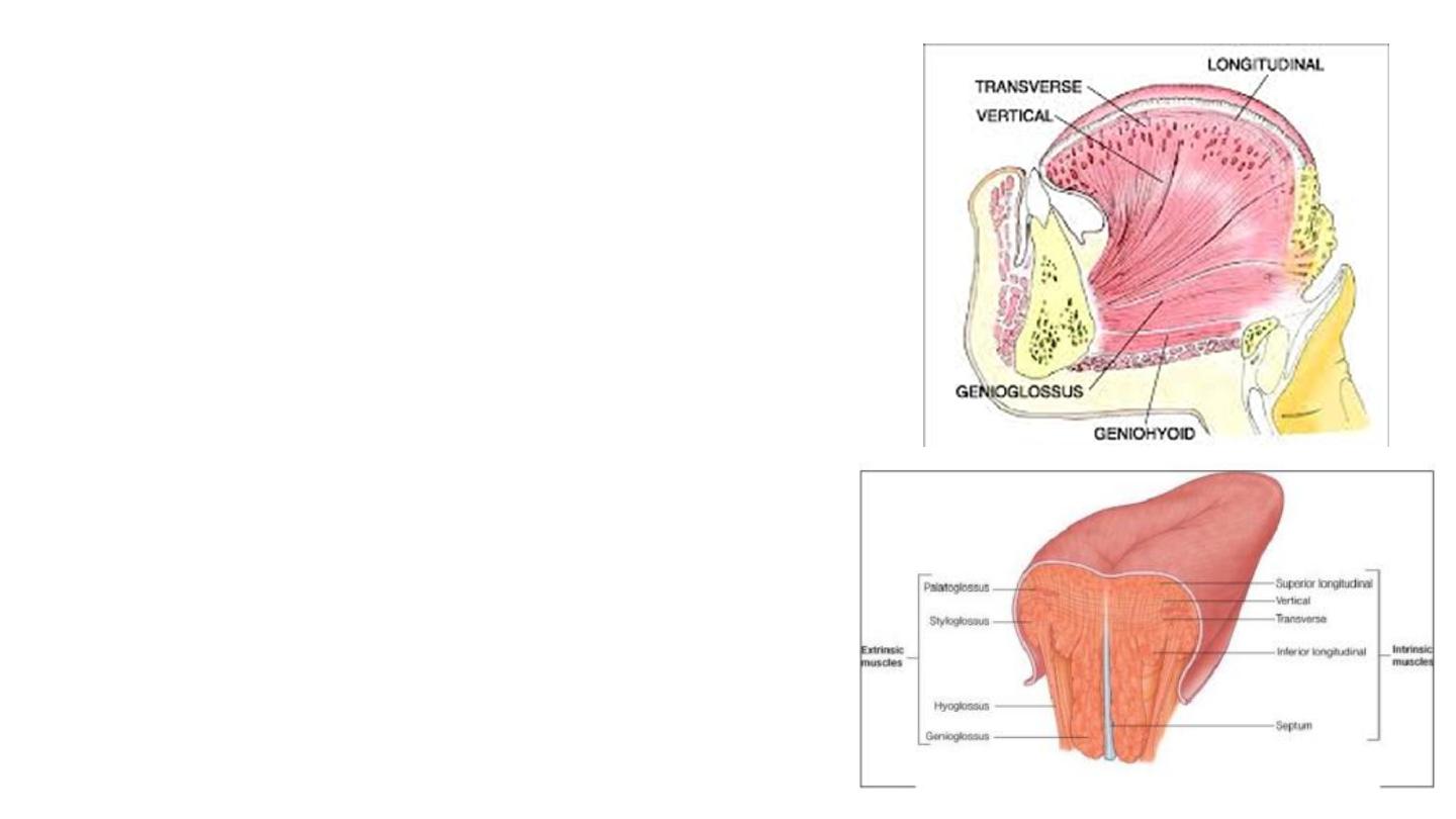
Intrinsic Muscles
• Originate and insert within the substance of the
tongue.
• There are four paired intrinsic muscles.
• They are named by the direction in which they
travel
• Longitudinal ( superior and inferior)
• Transverse
• Vertical muscles.
• Function:
• Affect the shape and size of the tongue
• Have a role in facilitating speech, eating and
swallowing.
• Motor innervation : Hypoglossal nerve (CNXII).
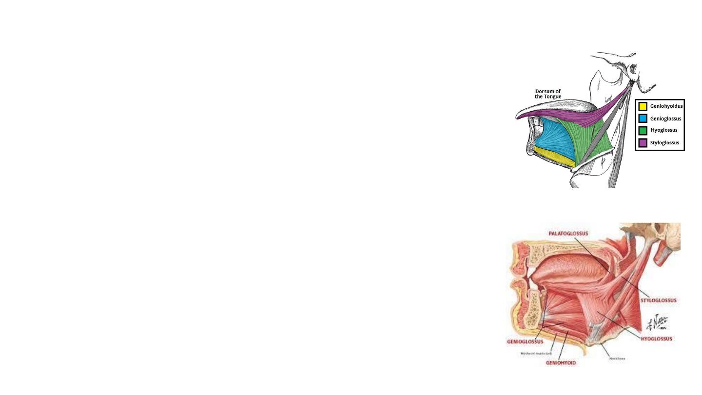
Extrinsic Muscles
Originate from structures outside the tongue and insert into it.
Genioglossus
• Attachments:
• Arises from mental spine of the mandible.
• Inserts into the body of the hyoid bone and the entire length of the tongue.
• Protrudes the tongue
Hyoglossus
• Attachments:
• Arises from the hyoid bone and inserts into the side of the tongue
• Depresses and retracts the tongue
Styloglossus
Attachments:
• Originates at the styloid process of the temporal bone and inserts into the side
of the tongue
• Draws up the sides of the tongue
Innervation
: Motor innervation via the hypoglossal nerve (CNXII).
Palatoglossus
• Attachments:
• Arises from the palatine aponeurosis and inserts broadly across
the tongue
• Elevates the posterior tongue, closes the oropharyngeal
isthmus, aids in the initiation of swallowing, and prevents
the spill of saliva from the vestibule into the oropharynx
• Innervation: Motor innervation via the vagus nerve
(CNX).
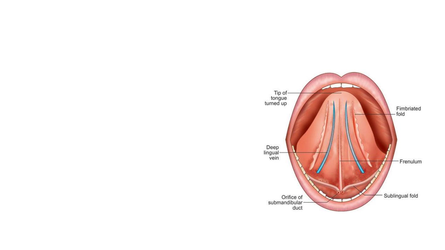
Inferior surface
• Lacks papillae
• Has a number of linear mucosal folds
• Frenulum: continuous with the
mucosa of the floor of the tongue.
• Deep lingual vein
• Fimbriated fold

Sensory innervation
• Anterior 2/3
• General sensation: lingual nerve- mandibular nerve (CN V3)- trigeminal nerve
(CNV).
• Taste: chorda tympani- the facial nerve (CNVII). The circumvallate papillae
get special afferent taste innervation from cranial nerve IX
• The posterior 1/3 :
• Both touch and taste are supplied by the glossopharyngeal nerve (CNIX).

Vasculature
• The lingual artery (main)
• A branch from the facial artery, called the tonsillar artery,
which can provide some collateral circulation.
• Drainage is by the lingual vein.
• Lymphatic Drainage
• The tip of the tongue drains into the submental lymph nodes.
• The anterior 2/3 of the tongue drains into the submandibular &
deep cervical lymph nodes.
• Lymph from the posterior 1/3 of the tongue drains into the
deep cervical lymph nodes.
• Bilateral drainage is common from central segments.

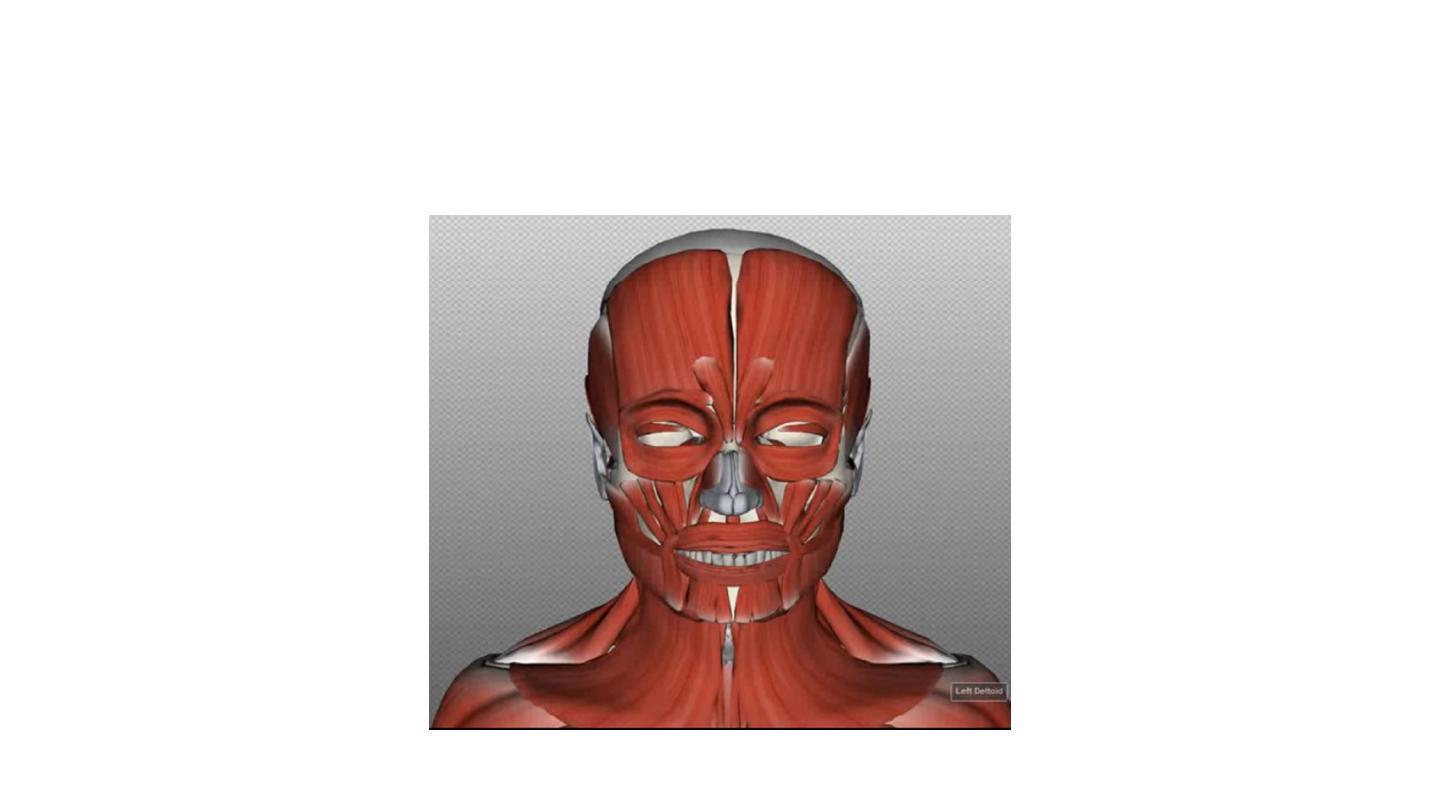
Muscles of Facial Expression
• The muscles of facial expression are located in the
subcutaneous tissue
• Originate from bone or fascia, and inserting onto the
skin.
• They are the only group of muscles that insert into skin.
• By contracting, the muscles pull on the skin and exert
their effects on the face, such as smiling, grinning and
frowning.
• These muscles have a common embryonic origin – the
2nd pharyngeal arch.
• All the muscles of facial expression are innervated by
the facial nerve.
• The facial muscles can broadly be split into: orbital,
nasal , oral and muscles of the cranium and neck.
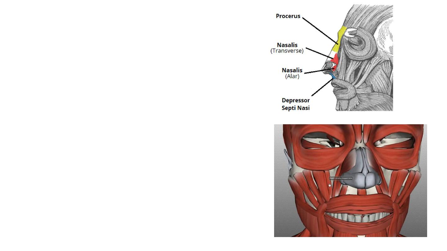
Nasal Group
• The nasal group of facial muscles are
associated with movements of the
nose, and the skin around it.
• There are three muscles in this group
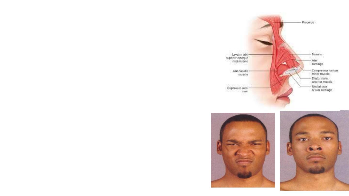
Procerus
• Origin: nasal bone and lateral nasal cartilage
• Insertion: skin between the eyebrows
• Action:
• Pulls down the medial end of the eyebrow
• Wrinkles the skin of the nose transversely in
frowning
Nasalis
• Transverse (compressor)
• Wrinkles the skin of the nose transversely in
frowning
• Alar ( dilator naris):
• Widens the nasal aperture
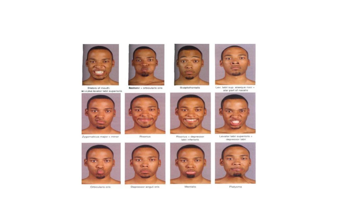
Oral group
Responsible for movements of the mouth and lips
• Orbicularis oris
• Buccinator
• Lower group of oral muscles
• Depressor anguli oris
• Depressor labii inferioris
• Mentalis
• Upper group of oral muscles
• Risorius
• Zygomaticus major and zygomaticus minor
• Levator labii superioris
• Levator labii superioris alaeque nasi
• Levator anguli oris
Oral Group
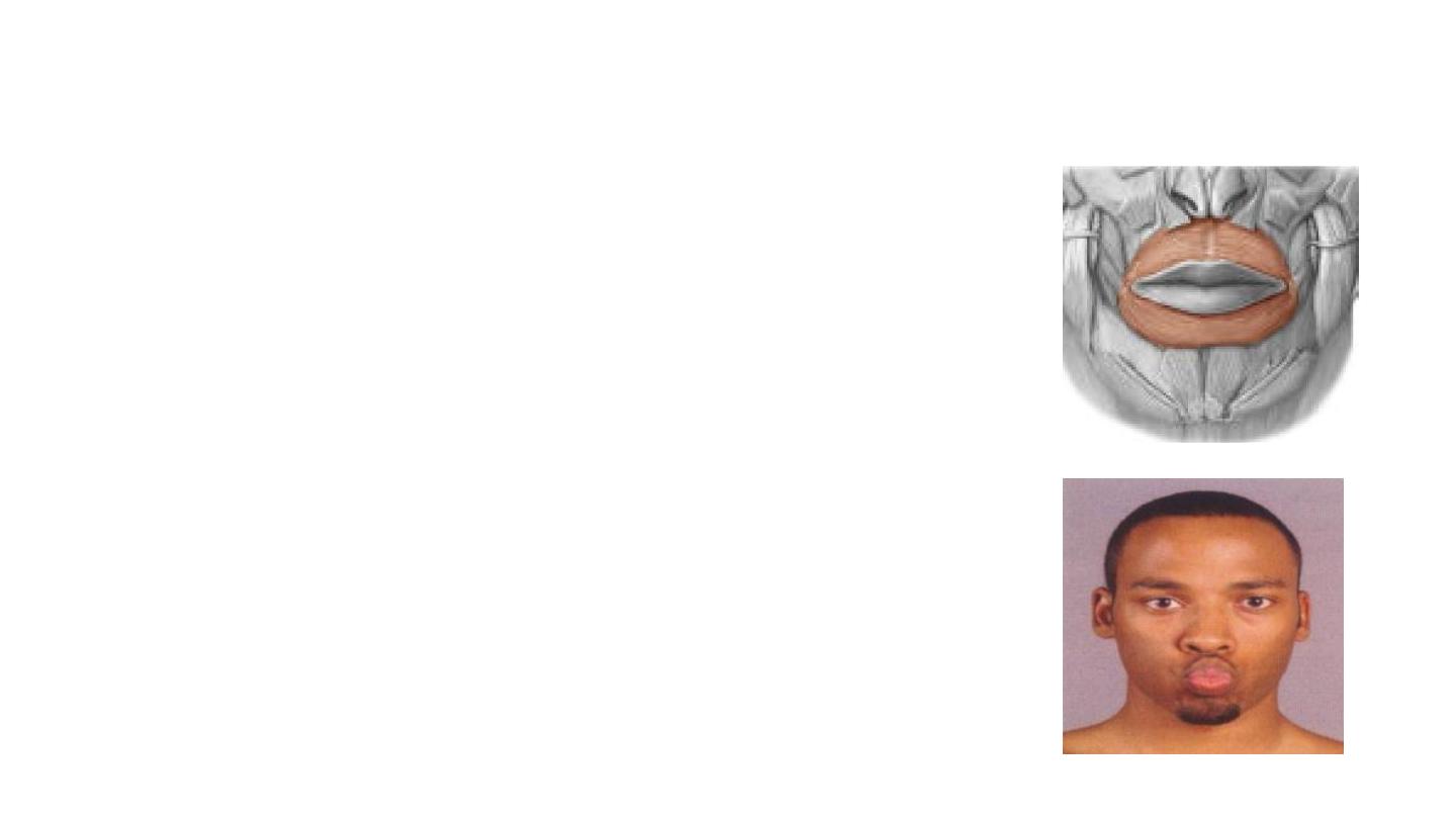
Orbicularis Oris
• Enclose the opening to the oral cavity.
• Attachments: Arises from the maxilla and from the other
muscles of the cheek. It inserts into the skin and mucous
membranes of the lips.
• Action: Purses the lips.
• Innervation: Facial nerve.
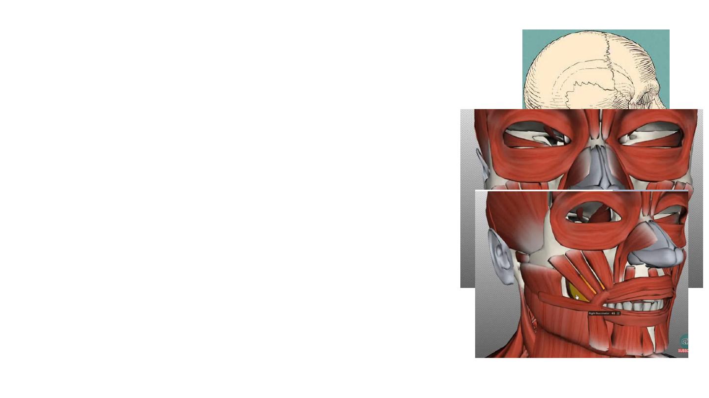
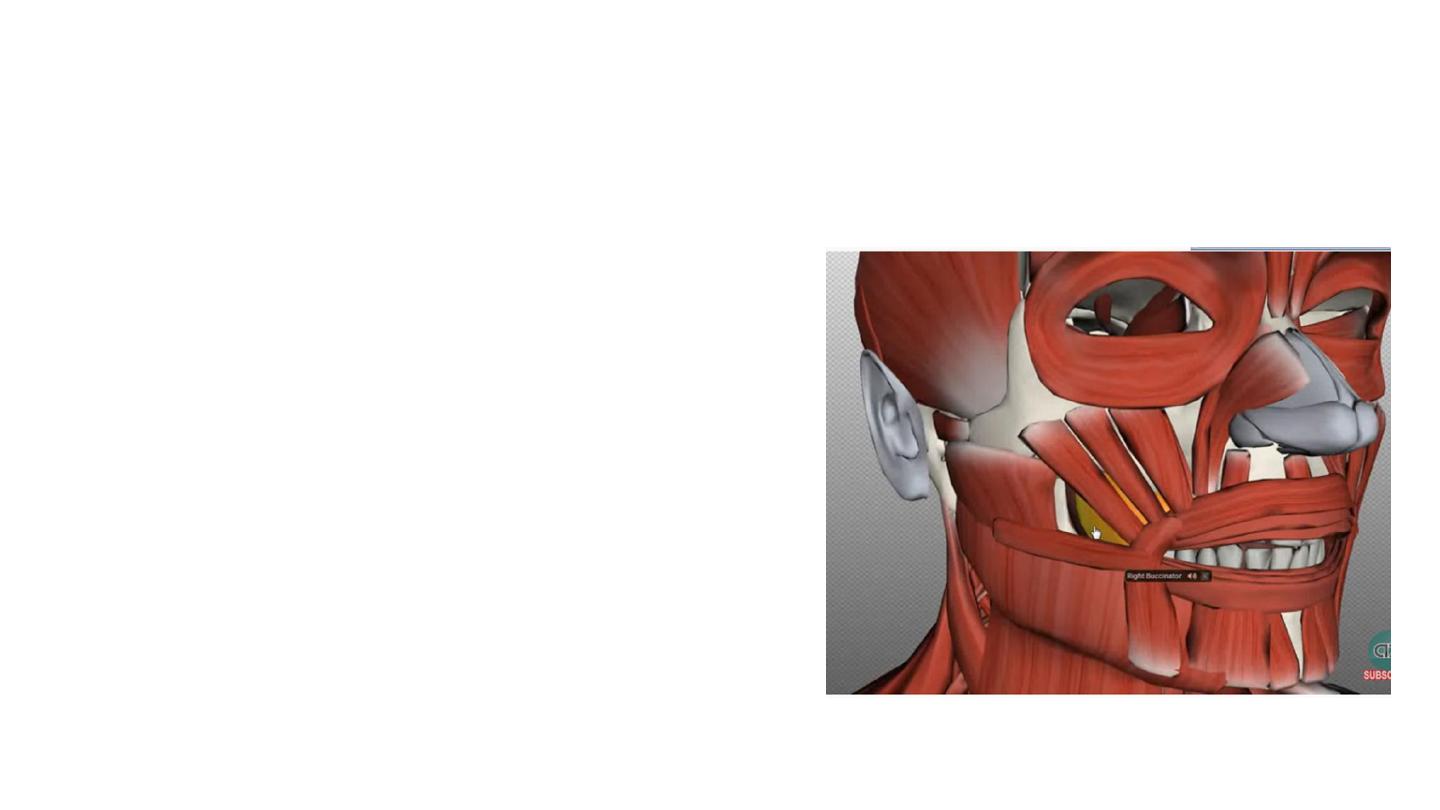
• Buccinator
• This muscle is located between the mandible and
maxilla, deep to the other muscles of the face.
• Attachments: It originates from the maxilla and
mandible. The fibres run in an inferomedial direction,
blending with the orbicularis oris and the skin of the
lips.
• Actions: The buccinator pulls the cheek inwards against
the teeth, preventing accumulation of food in that
area.
• Innervation: Facial nerve.
• Other Oral Muscles
• There are other muscles that act on the lips and
mouth.
• The lower group contains the depressor anguli oris,
depressor labii inferioris and the mentalis.
• The upper group contains the risorius, zygomaticus
major, zygomaticus minor, levator labii superioris,
levator anguli oris and levator labii superioris alaeque
nasi.
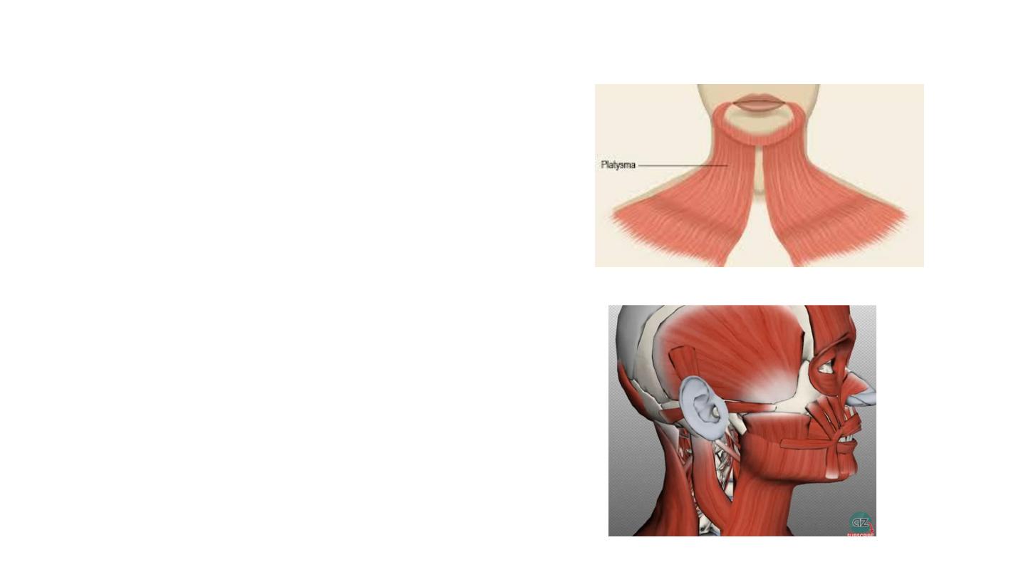
Other muscle groups
• Platysma
• Auricular (anterior, superior, and
posterior auricular muscles)
• Occipitofrontalis
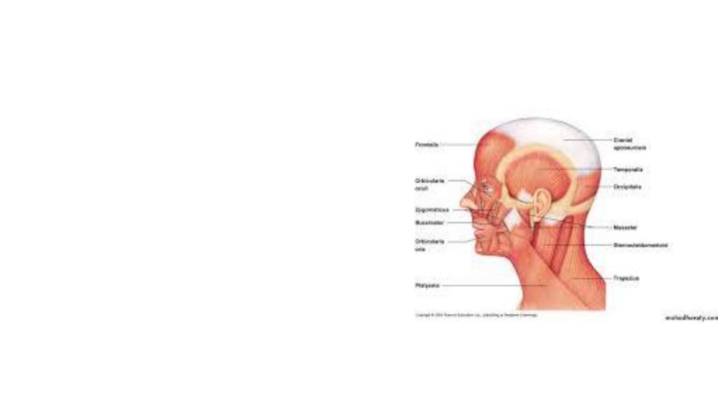
Occipitofrontalis Muscle
• It has a frontal belly anteriorly, an
occipital belly posteriorly, and an
aponeurotic
tendon
(epicranial
aponeurosis) connecting the two.
• Frontal belly
• Origin : Skin and superficial fascia
of eyebrow
• Insertion: Epicranial aponeurosis
• Occipital belly
• Origin : Highest nuchal line of
occipital bone
• Insertion: Epicranial aponeurosis
• Nerve Supply: Facial nerve
• Action: Moves scalp on skull and
raises eyebrows

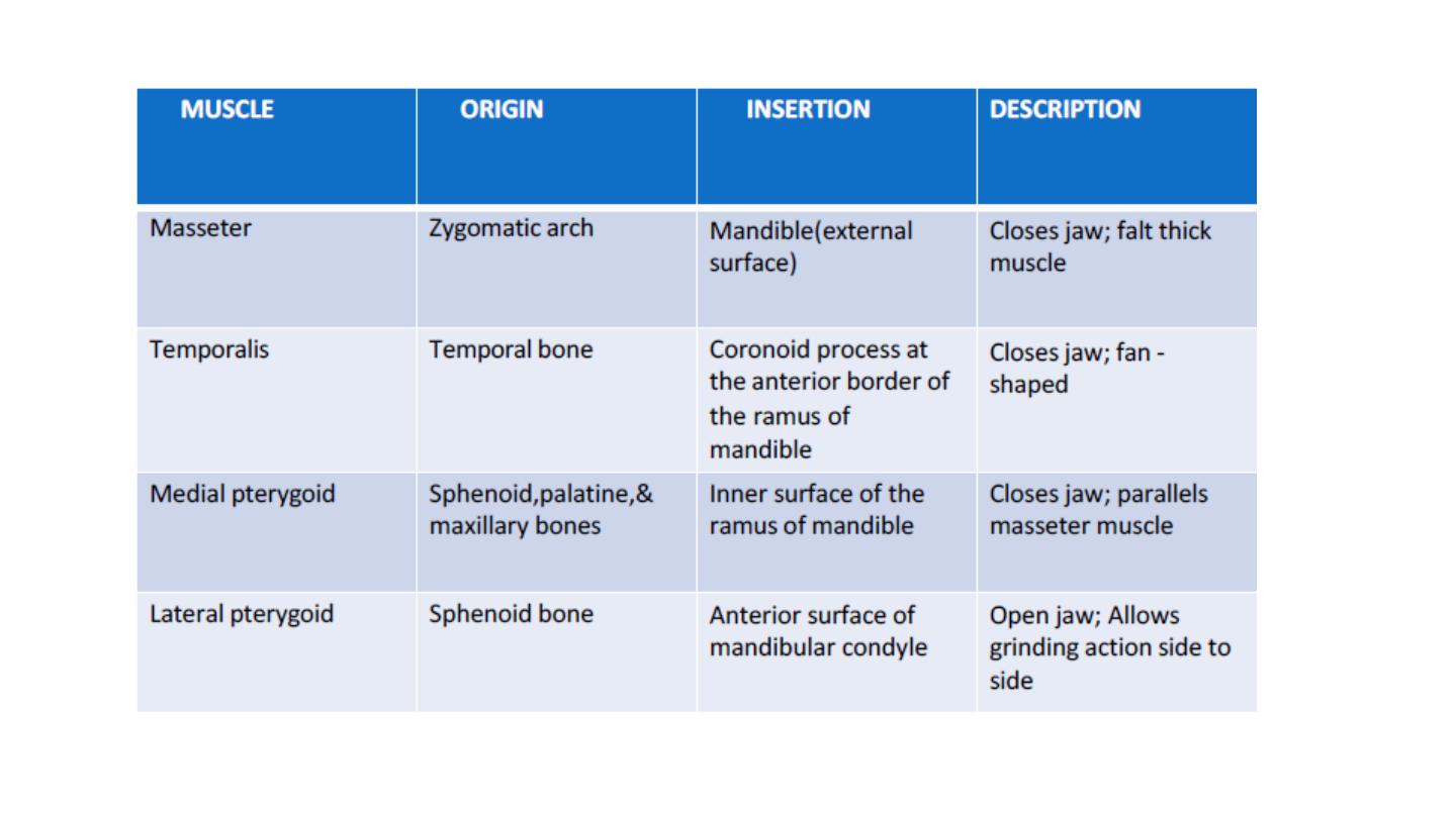
The Muscles of Mastication
• The muscles of mastication are associated with movements of the jaw
(temporomandibular joint).
• Masseter
• Temporalis
• Medial pterygoid
• Lateral pterygoid
• The muscles of mastication develop from the first pharyngeal arch.
Thus, they are innervated by a branch of the trigeminal nerve (CN V),
the mandibular nerve.
• All the muscles are bilateral structures.
Masseter
• The most powerful muscle of mastication.
• It is quadrangular in shape and has two parts:
deep and superficial.
• Attachments:
• The superficial part originates from maxillary process
of the zygomatic bone.
• The deep part originates from deep/ inferior surface
of zygomatic arch.
• Both parts attach to the ramus of the mandible.
• Actions: Elevates the mandible, closing the
mouth.
• Innervation: Mandibular nerve (V3).
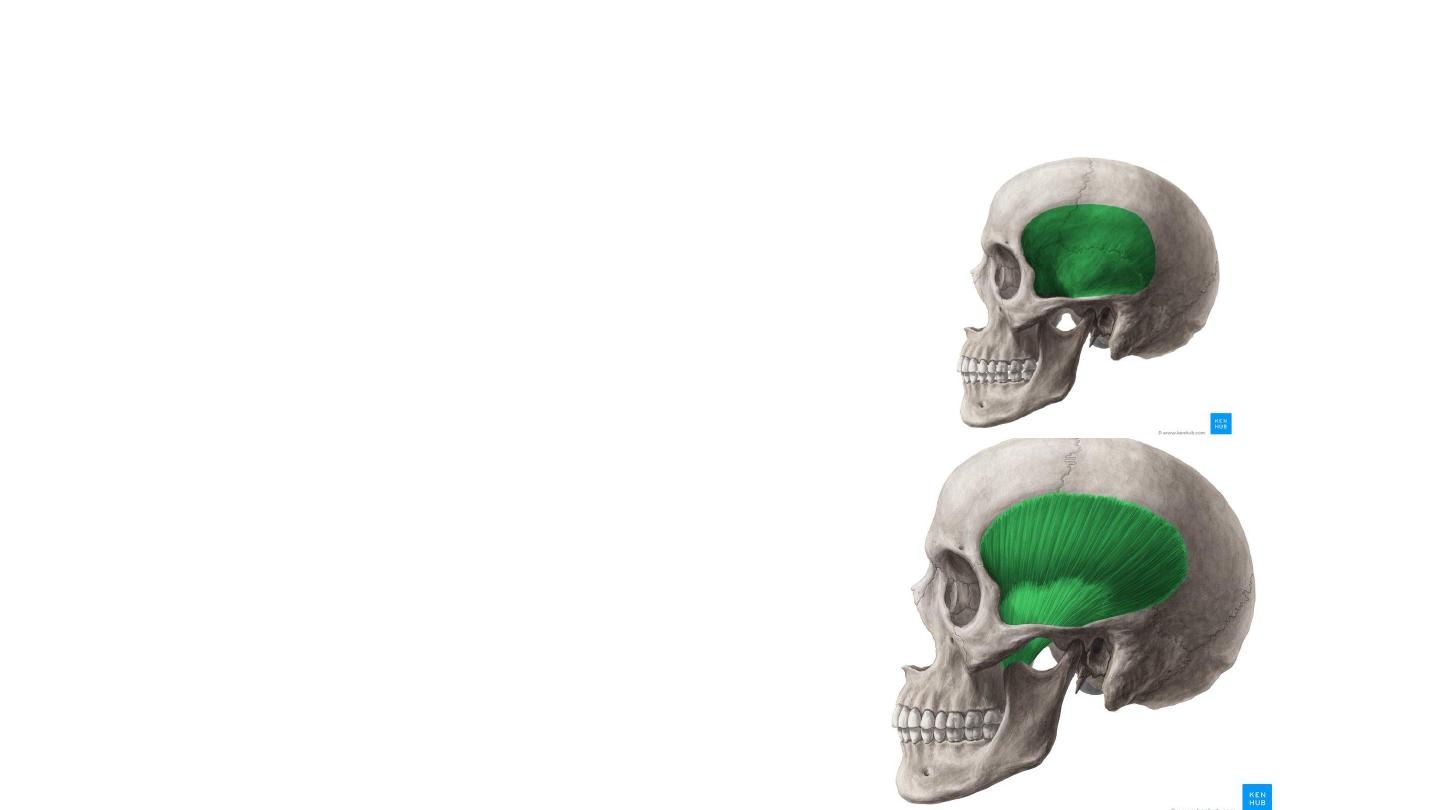
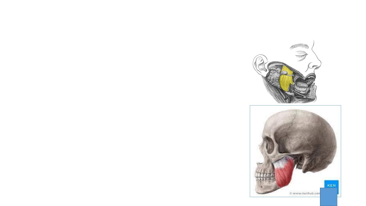
Temporalis
• Attachments: Originates from the
inferior temporal line and temporal
fossa. It condenses into a tendon, which
inserts onto the coronoid process of the
mandible.
• The muscle is covered by tough fascia
• Actions: Elevates the mandible, closing
the mouth. Also retracts the mandible,
pulling the jaw posteriorly.
• Innervation: Mandibular nerve (V3).
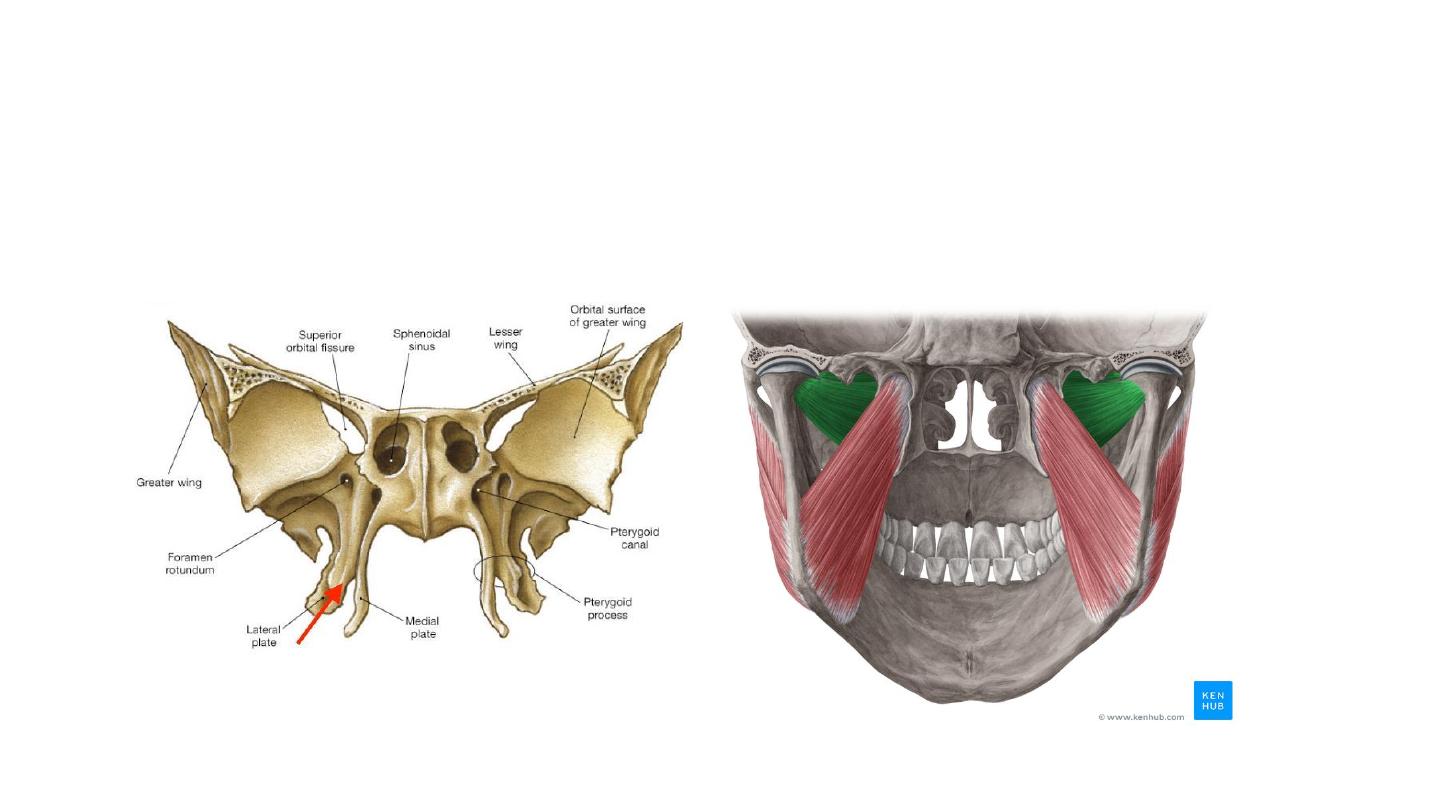
Pterygoid muscles
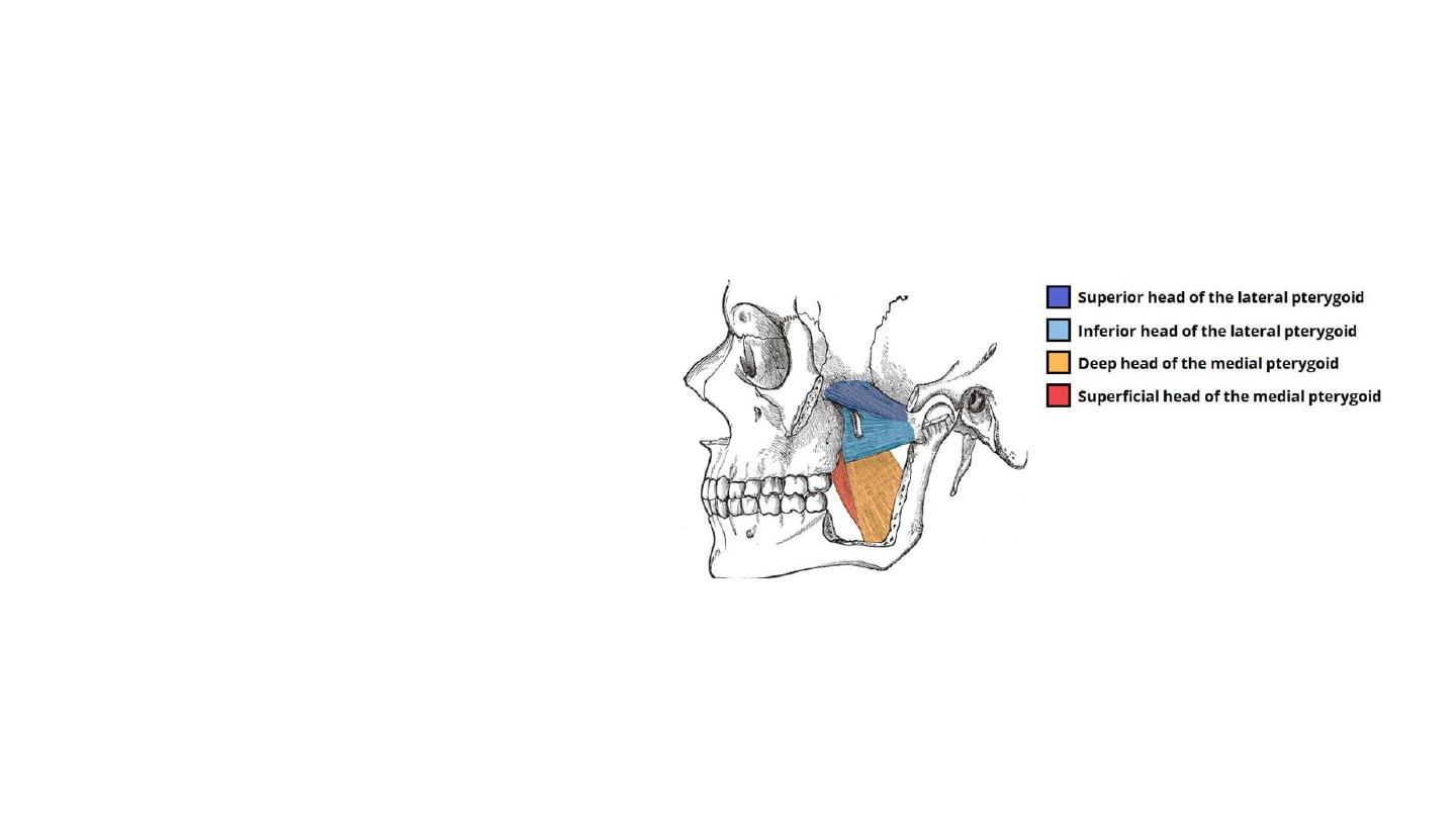
Lateral Pterygoid
• Triangular shape with two heads:
superior and inferior.
• Attachments:
• The superior head originates from
the greater wing of the sphenoid.
• The inferior head originates from
the lateral pterygoid plate of the
sphenoid.
• The two heads converge into a
tendon which attaches to the neck
of the mandible.
• Actions: Acting bilaterally, the lateral
pterygoids
protract
the
mandible,
pushing the jaw forwards. Unilateral
action produces the ‘side to side’
movement of the jaw.
• It also assists in depressing the
mandible
• Innervation: Mandibular nerve (V3).
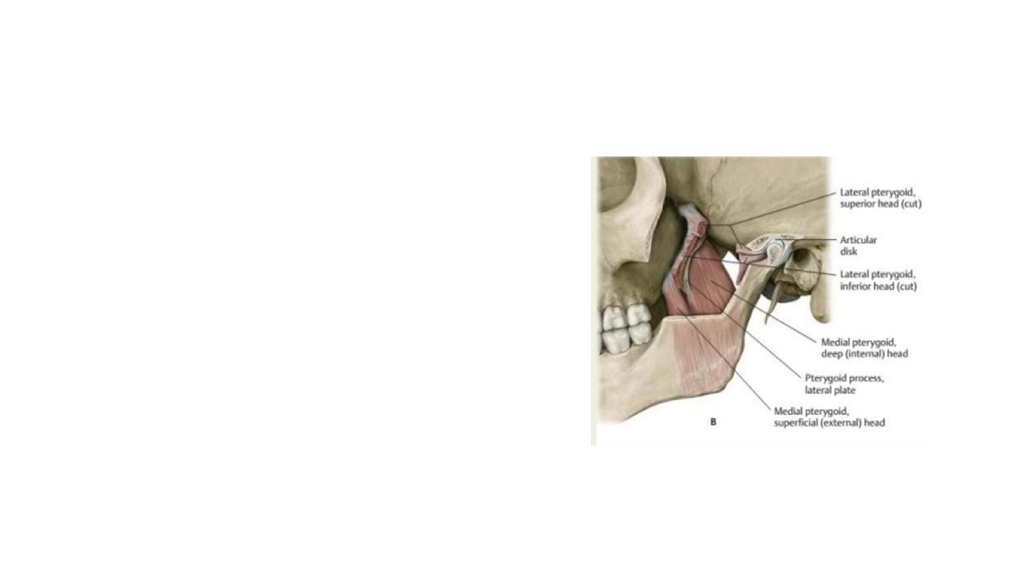
Medial Pterygoid
• The medial pterygoid muscle has a quadrangular
shape with two heads: deep and superficial.
• It is located inferiorly to the lateral pterygoid.
• Attachments:
• The superficial head originates from the maxilla and
the palatine bone.
• The deep head originates from the medial aspect of
the lateral pterygoid plate of the sphenoid bone.
• Both heads attach to the ramus of the mandible
near the angle of mandible.
• Actions: Elevates the mandible, closing the
mouth.
• Innervation: Mandibular nerve (V3).