Lecture 2 – Classification of Genetic disorders
DR RUSUL MAHDI ABID
1. Classical Genetic disease:
A) Chromosomal DisordersThe number of chromosomes or
The structure of chromosomes
And may effect autosomes or sex chromosomes
So, they could be Numerical or Structural.
Numerical chromosomal abnormalities
Are define as a gain or loss of one or more whole chromosome (s) ( whether an autosomal or sex chromosome) or a whole set of chromosomes.Any number that is not an exact multiple of haploid (n) as aneuploidy.
A gain of one or more set of chromosomes is known as polyploidy. This polyploidy may tripody when cells have (3n) or tetraploidy when cells have (4n)
Polyploidy generally result in spontaneous abortions.
A gain of one chromosomes known as trisomy.
Down syndrome is trisomy of chromosome 21
Edward syndrome is trisomy 18
Patau syndrome is trisomy 13
Kleinfelter syndrome is trisomy of sex chromosome in male
A loss of one chromosome is called monosomy.
All trisomies and monosomies are aneuploidies
Monosomy of sex chromosome in female called Turner syndrome (45 XO)Down syndrome
Is a genetic disorder caused by the presence of all or part of a third copy of chromosome 21. It is typically associated with growth delay. Characteristic facial features and mild to moderate intellectual disability .The average IQ of young adult with Down syndrome is 50 similar to the mental age of an 8 or 9 years old child
People with Down syndrome may have some or all of the following physical characteristics:
Small chinSlanted eye s
Poor muscle tone
Flat nasal bridge
A single crease of the palm
Protruding tongue (small mouth and large tongue)
Other common features include:
Flat and wide face
Short neck
Excessive joint flexibility
Extra space between big toe and second toe
Abnormal pattern of finger patterns on the fingertips and short fingers
Hip dislocation (up to 1/3 without trauma)
Growth in height is slower resulting in adults who tend to have short stature
Individuals with Down syndrome are at increased risk for obesity as they age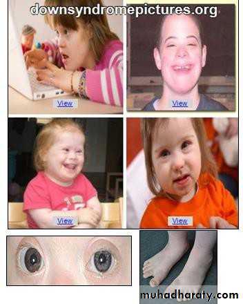
Structural chromosomal abnormalities
In structural chromosomal abnormalities, the cell has a normal number of 46 chromosomes but they are structurally abnormal.All chromosomes in a normal human karyotype have a long lower arm (known as the P arm), so that the centromere is located at different levels in between.
Deletion:
Involves loss of a piece of a chromosome, could be eitherTerminal deletion. In which, there is one break in the chromosome and the portion distal to this break is lost. This lost piece could be carrying important genes, result in signs and symptoms related to the lost genes products
Interstitial deletion. Where the piece of a chromosome between two breaks is lost resulting in a chromosome that is shorter than the original with the same consequences e.g. Ci du chat (loss of short arm of chromosome 5)
Sometimes, the piece lost carries no gene and therefore it cause no abnormality.
This is due to the fact that the genes are not situated on the chromosome one besides the other all along the chromosomes but there are parts of the chromosome carry no genes.
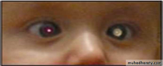
Retinoblastoma
Is rapidly developing cancer that develops from the cells of a retina,Caused by loss of retinoblastoma gene by interstitial deletion of the long arm of chromosome 13.
It is important to screen the eyes of such individuals regularly so that tumors can be treated early and sight preserved.
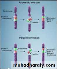
2. inversion
This abnormality result from 2 breaks in the chromosome and the piece between the two breaks will rotate 180 degree and is fixed back again in its rotated position.The breaks may involve either the short arm or the long arm and the inversion is called paracentric (the breaks are on one side of the centromere), or pericentric (the breaks may involve both arms and the centromere is involved in the inversion, which id more sever.)
3. translocation
It is define as an exchange of segments of chromosomes between chromosomes.Regular (reciprocal) translocation that takes place between any two chromosome is one of the main pathological changes seen in hematological malignancies.
These translocations may translocate some genes from their normal position to a new one where they are induced to function in an uncontrolled manner leading to override of the body’s regulatory control mechanism leading to malignancy.
Chronic myeloid leukemia
We the first malignancy to be linked to a chromosomal translocation known as the Philadelphia chromosome.
In this translocation, parts of two chromosomes (the 9th and 22nd) switch places.
As a result, part of the BCR (breakpoint cluster region) gene from chromosome 22 to is fused with the ABL gene on chromosome 9.
The abnormal fusion gene generates the BCR-ABL protein that inhibits DNA repair, causing genomic instability and matching the cell more susceptible to developing further genetic abnormalities.
Ring chromosome
This abnormality result from aberrant division of the centromere in a horizontal way rather than the perpendicular natural way during mitosis to separate the two sister chromatids into individual chromosomes.The resulting two chromosomes are imbalanced, one formed entirely of 2 short arm and the other 2 long arms.
These isochromosomes may be seen in some cases of DOWN’S or TURNER’S SYNDROMES (that is not due to trisomy or monosomy).