Diagnosis of genetic diseases:
Cytogenetics:The human geneome is composed of 23 pairs of chromosomes,which contain approximately 30,000 gene so examination of genetic material involve :
1-conventional cytogenetics analysis: this involve examination of entire chromosome by karyotype which's a photographic representation of a stained metaphase spread in which the chromomes are arranged in order of decreasing length .a variety of techniques for staining chromosomes used as Giemsa stain technique.
2. DIAGNOSIS OF GENETIC DISEASES
Diagnosis of genetic diseases requires the classical sequence of getting information about the patient like any medical disorder i.e. by history taking; clinical examination of the patient plus doing some additional. laboratory (hematological, biochemical, serological, hormonal, etc.) or radiological (plain X--ray, CT-scan, MRI, Echocardiogram, etc.) tests when indicated. If a provisional diagnosis is made or a list of few differential diagnoses is thought of, a confirmatory test should be sought for to confirm or rule out that diagnosis. A specific diagnosis for the genetic disorders may require a chromosomal study (a cytogenetic study to diagnose a numerical or structural. chromosomal abnormality), but many genetic diseases are caused -by subtle changes in individual genes that cannot be detected by karyotyping. Sometimes, the defect on the chromosome is more subtle (e.g. even deletion of the largest gene in the whole human DNA i.e. the dystrophin gene of a size of 2.4 M.B., is beyond the capability of the light microscope to detect). In this case, in situ hybridization is used to detect the mutated gene.Hybridization: This is a procedure used in the diagnosis of genetic and other pathologies as well as in the diagnosis of cancer. The procedure needs the availability of a probe for the gene you are testing whether the gene is abnormal,- mutated or absent. The latter is another type of mutation called gene deletion
How to prepare a probe? if you could extract the mRNA for the gene to be tested from tissues rich in that mRNA, then a labeled probe could be prepared. e.g. the hemoglobin polypeptide α and β. Their formation takes place in the developing marrow it is stored as mRNA which will form polypeptide; that will combine in duplicate, i.e. α2β2 to form the Hb molecule during their circulation in the peripheral circulationl Reticulocytes which are the young RBC contain very high amount of α-mRNA and β-mRNA( that could be extracted from them. Thalassemia is a blood disease caused by deletion of β-gene (β-thalassemia) or α-gene, (α-thalassemia). If you extract the reticulocytes from the blood of patients with β-thalassemia you can get α-mRNA and vice versa. Once you have your mRNA you could mix it in a system with an enzyme known as reverse transcriptase that could make DNA from mRNA and you add a solution containing the four basic nucleotide bases(Adenine, thymine,guanine and (cytosine). One of these bases is being labeled with a dye, so that it could label the newly formed DNA when it is incorporated in its formation. The resulting piece of DNA of gene that is labeled is known as a probe or marker. Once you have obtained your probe for any gene, you could proceed to hybridization. The first step is to extract the DNA from the patient you want to diagnose of whether he or she is suffering from a genetic disease. DNA could be extracted from any sample containing nucleated cells you could obtain from the body. For a prenatal diagnosis of a genetic disease, a sample of amniotic fluid cells or placental biopsy is required at early stage in pregnancy. The DNA obtained from the sample is subjected to the effect of RE to cut it in multiple small pieces; one of these pieces is normally the carrier of the gene we are testing for. Next is to separate the pieces of DNA according to their size by a process of gel electrophoresis (Figs 7-45). Then, we transfer the bands of DNA from the gel to a nitrocellulose filter paper by placing the paper over the gel in a buffer system. This process of transfer is known as Southern Blotting.
These obtained DNA bands are double stranded that could be converted into single strands by heating the nitrocellulose paper. Next, we pour the probe solution we have prepared previously over the filter paper and allow time for the reaction to take place; then we wash the excess probe and examine the paper for the presence of the probe on any band of the DNA. If the probe shows, it means that hybridization of the probe with an existing gene over one of the bands of our unknown DNA has taken place. This is because single stranded DNA sticks. to its complementary sequences when encountering it. If this occurs then the gene is present and the fetus is not suffering from gene deletion or genetic disease. Conversely, if no probe is detected on any of the otherDNA bands, then the gene is absent i.e. deleted and the fetus is suffering from a genetic disease.
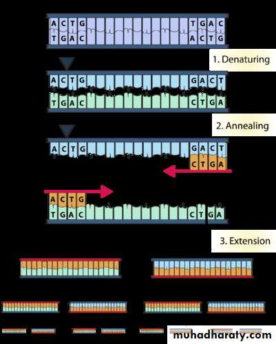
3. polymerase chain reaction (PCR)
PCR is the most frequently used molecular technique in a molecular pathology laboratory. Using a pair of priming complementary sequences (oligonucleotide primers) flanking a location of interest, together with unique heat-resistant polymerases (DNA copying enzymes), multiple copies of a targeted chimeric gene can be obtained Each PCR cycle involves 3 basic steps: denaturing, annealing, and polymerization. During denaturing, the 2 strands of the helix of the target genetic material are unwound and separated by heating at 90° to 95°C. During annealing, or hybridization, oligonucleotide primers bind to their complementary bases on the single-stranded DNA. This step requires a much cooler temperature, 55°C. Finally, during polymerization (at 75°C), the polymerase reads the template strand and quickly matches it with the appropriate nucleotides, resulting in 2 new helixes consisting of part of the original strand and the complementary strand that was just assembled.
The process is repeated 30 to 40 times, each cycle doubling the amount of the targeted genetic material. At the end of the PCR procedure, millions of identical copies of the original specific DNA sequence have been generated.
Since these copies are identical in electrical charge as well as molecular weight, they are expected to migrate simultaneously, forming a single band, when applied to an electrophoretic gel
PCR permits diagnosis of genetic mutations as well as malignant diseases .