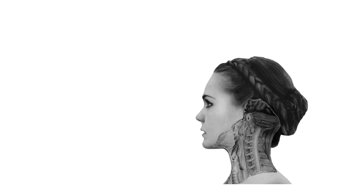
The Neck
Dr Firas Al-Hameed
M.B.Ch.B C.A.B.S MRCS (ENT) (England)
Thi-Qar Medical School

Fascial Layers of the Neck
• Fascia is an internal connective tissue which forms bands or sheets
that surround and support muscles, vessels and nerves in the body.
• There are two fascias in the neck – the superficial cervical fascia and
the deep cervical fascia.

Superficial Cervical Fascia
• It is a thin layer of subcutaneous connective tissue that lies between
the dermis of the skin and the deep cervical fascia. It contains
numerous structures:
• Neurovascular supply to the skin
• Superficial veins (e.g. the external jugular vein)
• Superficial lymph nodes
• Fat
• Platysma muscle
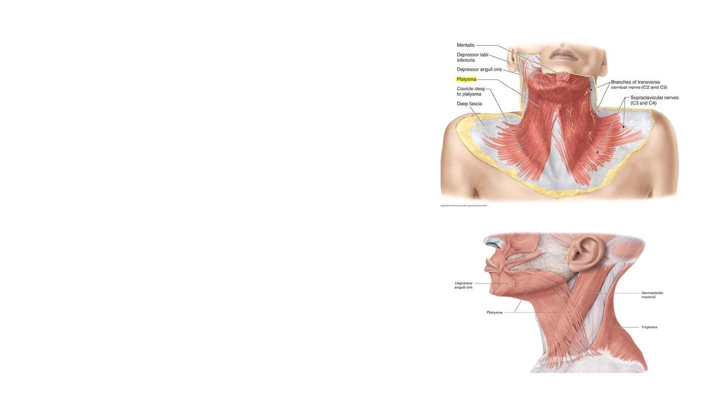
The platysma
• The platysma is a broad superficial muscle which
lies anteriorly in the neck.
• The superficial cervical fascia blends with the
‘paper thin’ platysma muscle.
• Originate
from the fascia of the pectoralis major
and deltoid.
• The fibres cross the clavicle, and meet in the
midline, fusing with the muscles of the face.
• Insertion
: inferior border of the mandible and skin
of lower face and lip
• Action
– releases pressure of skin on the subjacent
veins, depress mandible, pulls angle of mouth
downwards.
• Innervation
: cervical branch of the facial nerve
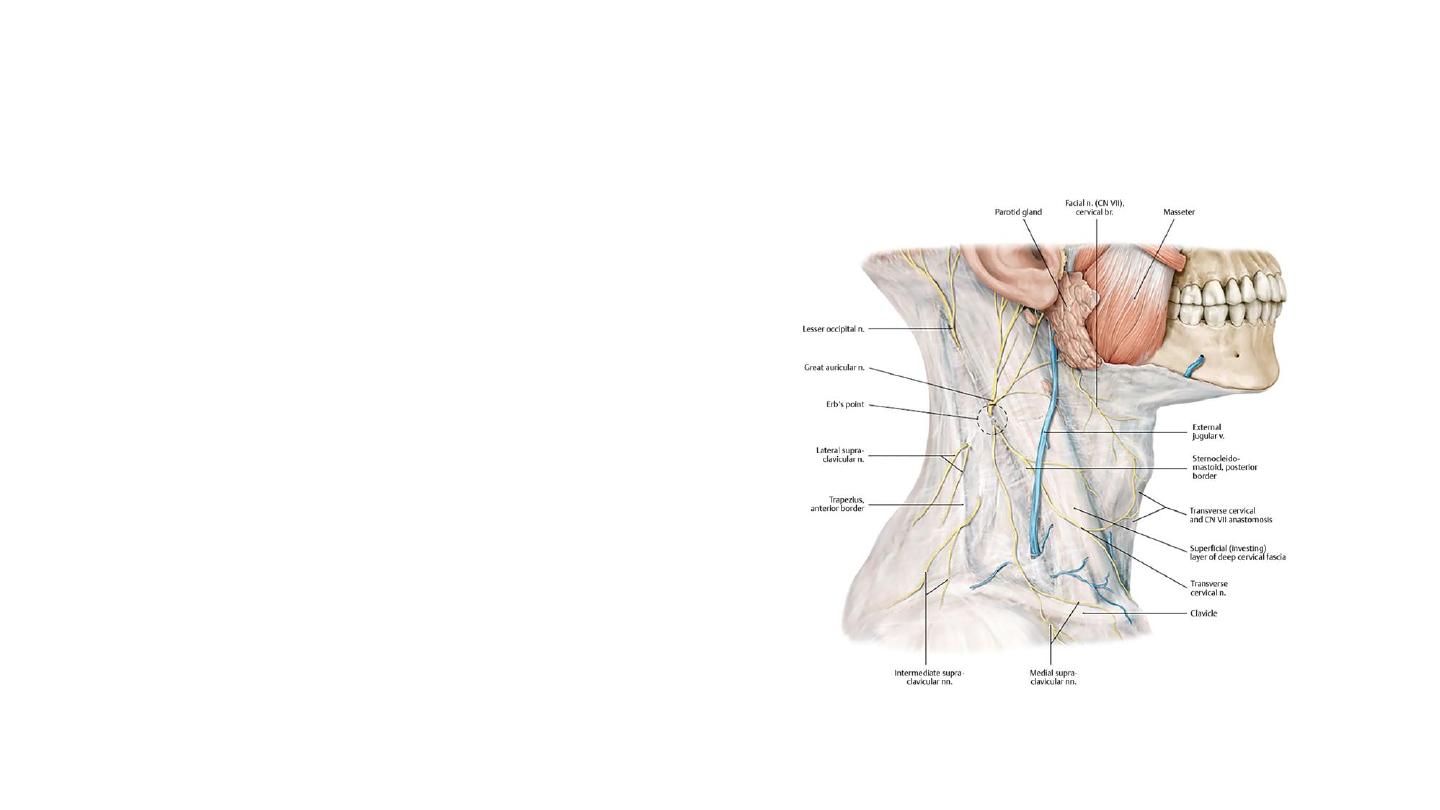
Neurovascular supply to the skin
• Lesser occipital nerve: from anterior
Rami of C2, C3
• Supply upper part of skin of inner surface
of auricle & adjacent part of skin of scalp
• Greet auricular nerve: from anterior
Rami of C2,C3
• Supply lower part of skin of inner surface
of auricle , skin over parotid gland & skin
over angle of mandible
• Transverse cervical nerve: C2,C3
• Supply of skin of anterior triangle
• Supraclavicular nerves: C3, C4
• Supply skin of posterior Triangle , skin of tip
of shoulder& skin of chest down to second
rib
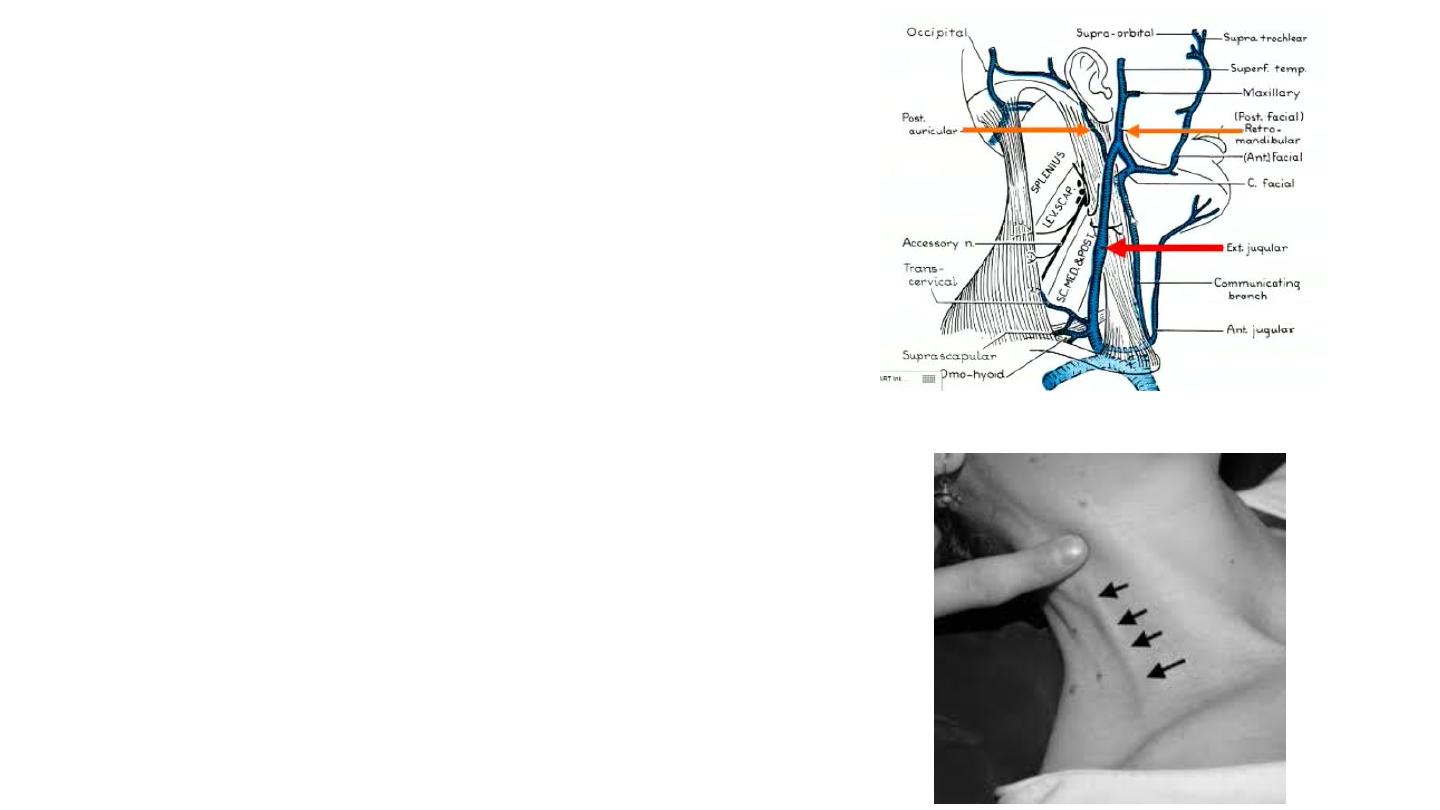
External Jugular Vein
• Begins just behind the angle of the mandible
by the union of the posterior auricular vein
with
the
posterior
division
of
the
retromandibular vein.
• It
descends
obliquely
across
the
sternocleidomastoid muscle
• Within the posterior triangle, the external
jugular vein pierces the investing layer of
fascia
• Drains into the subclavian vein
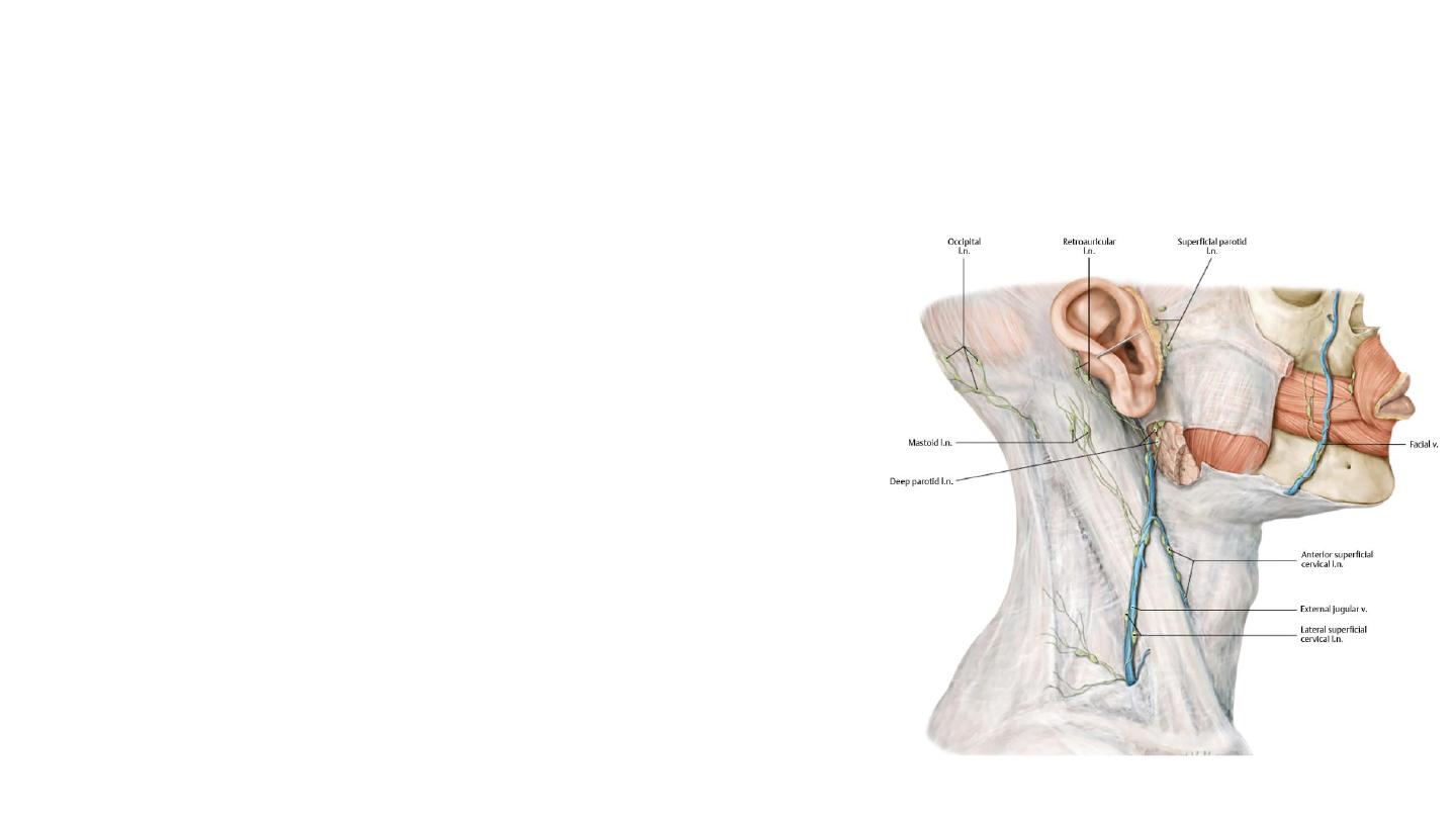
Superficial lymph nodes
• Lies along the external jugular vein
superficial to the sternocleidomastoid muscle
• Receive lymph vessels from the occipital and
mastoid lymph nodes
• Drain into the deep cervical lymph nodes
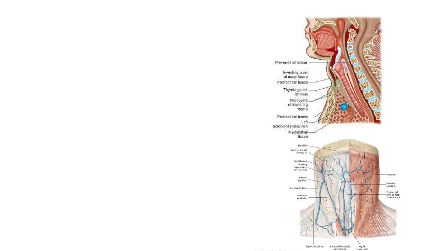
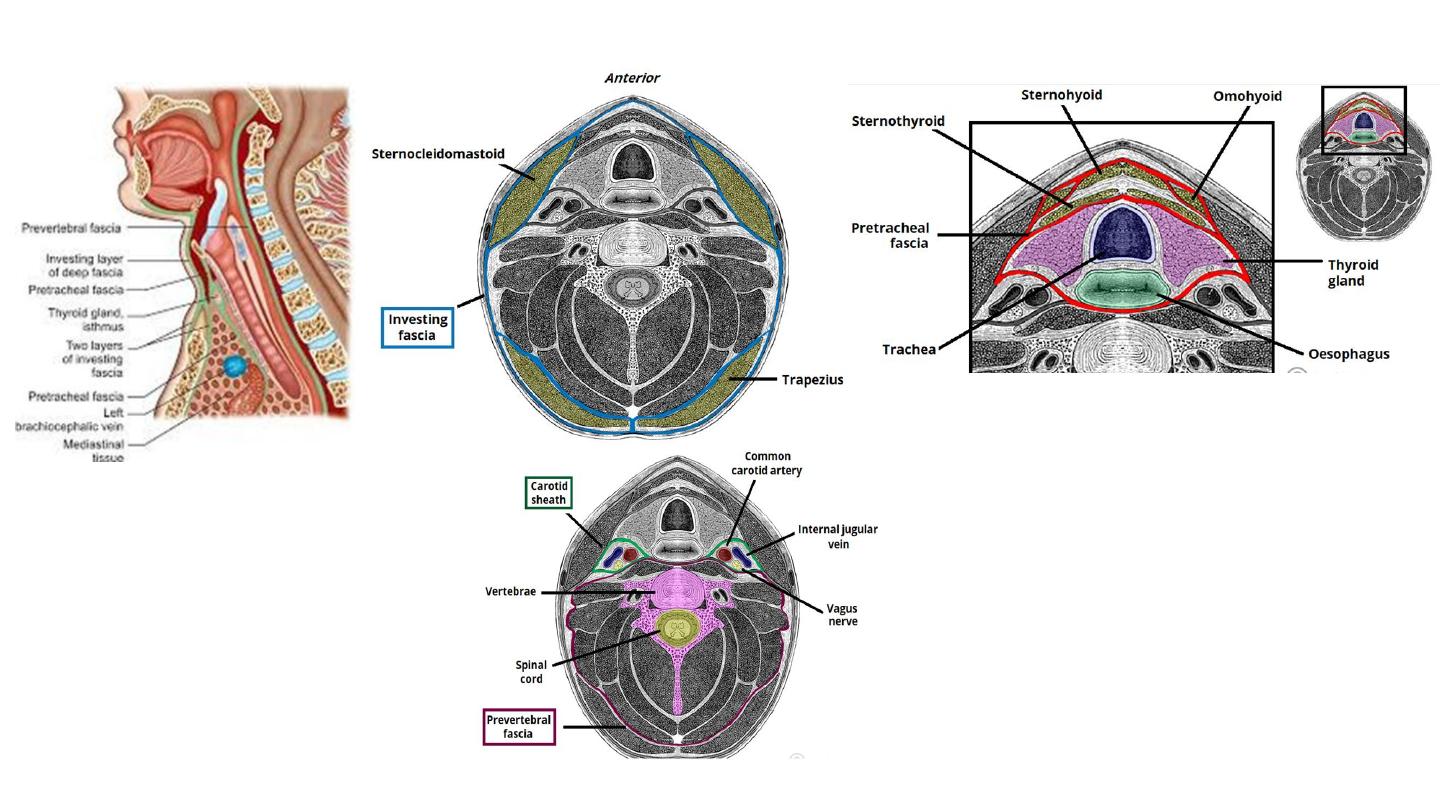
Deep Cervical Fascia
• Investing Layer
• The pretracheal layer
• Prevertebral Layer
• Carotid sheaths
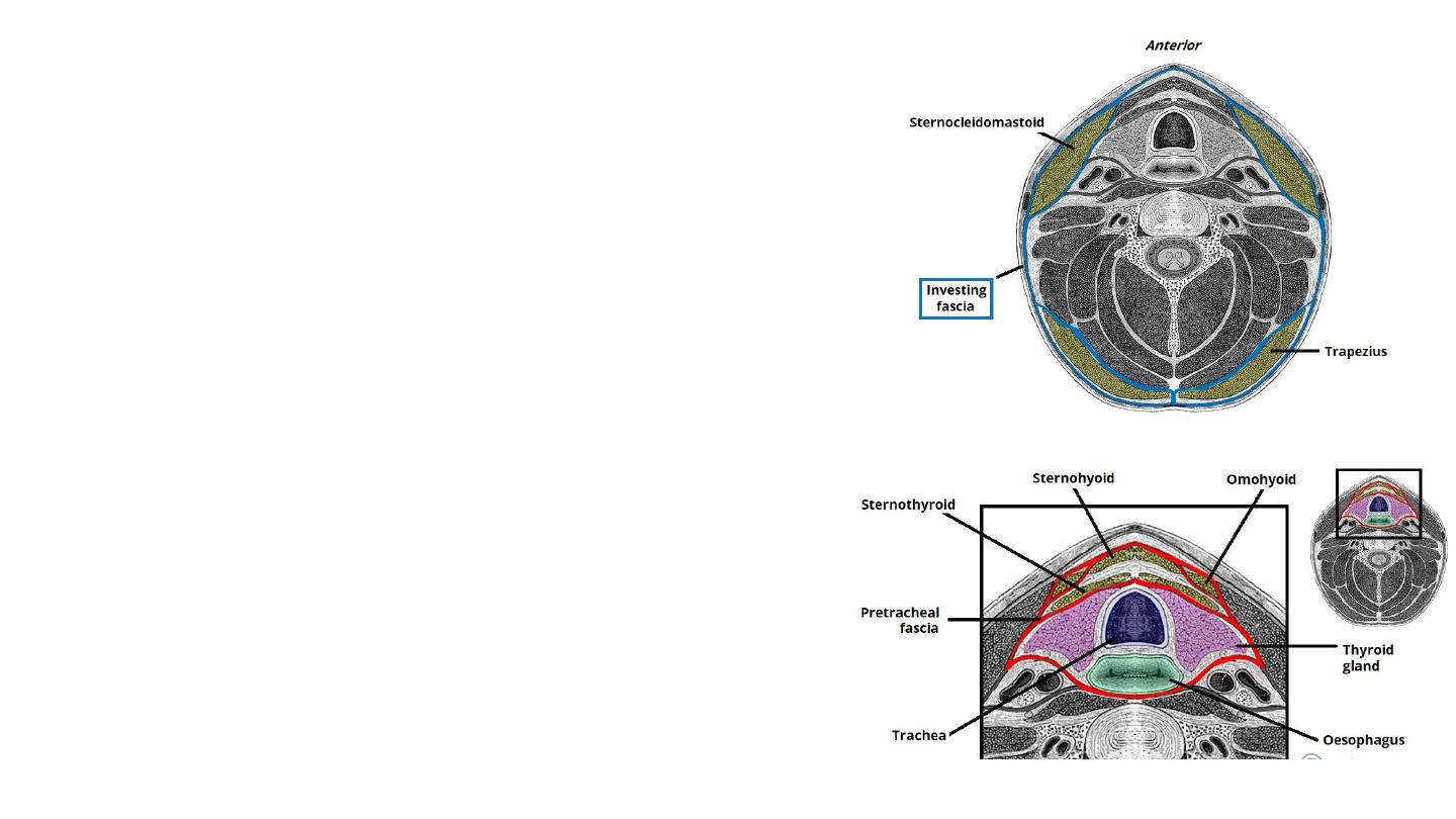
The investing layer
• The most superficial of the deep cervical fascia.
• It surrounds all the structures in the neck.
• Where it meets the trapezius and
sternocleidomastoid muscles, it splits into two,
completely surrounding them.
• Situated in the anterior neck.
• It spans between the hyoid bone superiorly and the
thorax inferiorly (where it fuses with the
pericardium).
• Surrounds the trachea, oesophagus, thyroid gland,
pharynx , larynx and infrahyoid muscles.
Pretracheal Layer
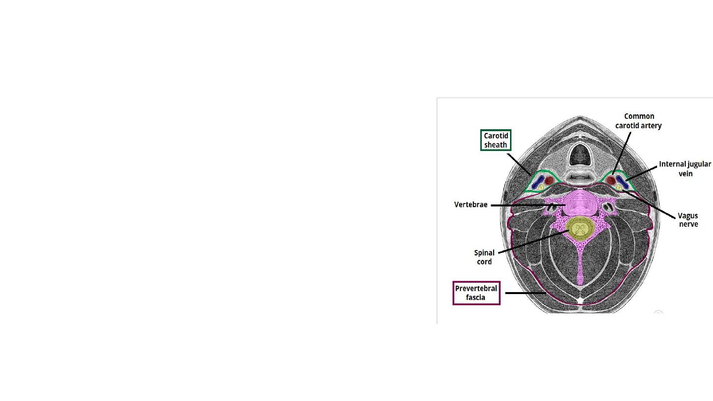
Prevertebral Layer
• Surrounds the vertebral column and its
associated muscles.
• Forms the floor of the posterior triangle of the
neck.
• Surrounds the brachial plexus and subclavian
artery

Carotid Sheath
• Paired structures on either side of the neck.
• The contents of the carotid sheath are:
• Common carotid artery
• Internal jugular vein.
• Vagus nerve.
• Accompanying cervical lymph nodes.
• The fascia of the carotid sheath is formed by
contributions from the pretracheal, prevertebral, and
investing fascia layers.
• Runs between the base of the skull to the thoracic
mediastinum. This is of clinical importance as a
pathway for the spread of infection.
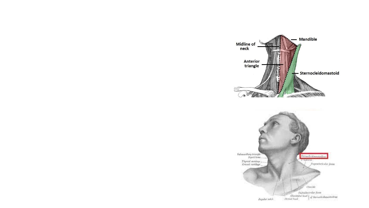
The Anterior Triangle of the Neck
• Located at the front of the neck.
Borders
• Superiorly – inferior border of the mandible (jawbone).
• Laterally – anterior border of the sternocleidomastoid.
• Medially – sagittal line down the midline of the neck.
• Sternocleidomastoid Muscle
• Origin: 2 heads Manubrium sterni & medial third of
clavicle
• Insertion: Mastoid process of temporal bone and occipital
bone
• Function :
• Rotate the head to the opposite side
• Flexes the neck
• When both sides of the muscle act together, it flexes
the neck and extends the head.
• Innervation: Spinal part of accessory nerve and C2 and 3,
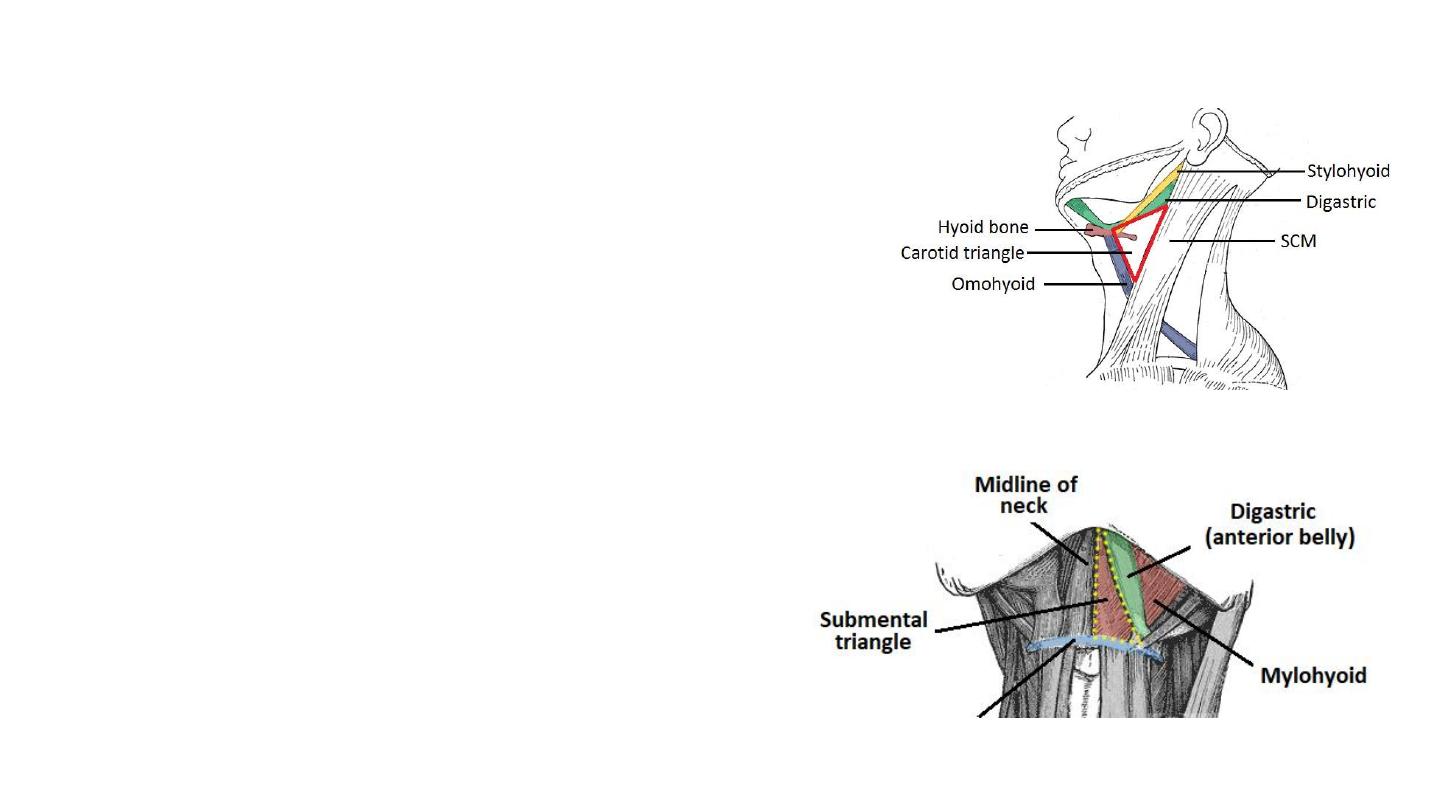
Subdivisions of anterior triangle
Carotid Triangle
• Superior – posterior belly of the digastric muscle.
• Lateral – medial border of the sternocleidomastoid muscle.
• Inferior – superior belly of the omohyoid muscle.
• Contents
• Common carotid artery
• Internal jugular vein
• Hypoglossal
• Vagus nerves
Submental Triangle
• Situated underneath the chin.
• Inferiorly – hyoid bone.
• Medially – midline of the neck.
• Laterally – anterior belly of the digastric
• The floor of the submental triangle is formed by the
mylohyoid muscle
Contents:
Submental lymph nodes
Hyoid bone
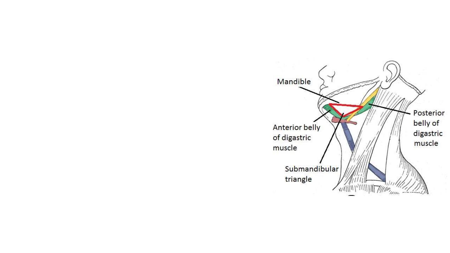
Submandibular Triangle
• The submandibular triangle is located underneath the
body of the mandible.
• Superiorly – body of the mandible.
• Anteriorly – anterior belly of the digastric muscle.
• Posteriorly – posterior belly of the digastric muscle.
• Contents:
• The submandibular salivary gland
• Lymph nodes.
• The facial artery and vein.
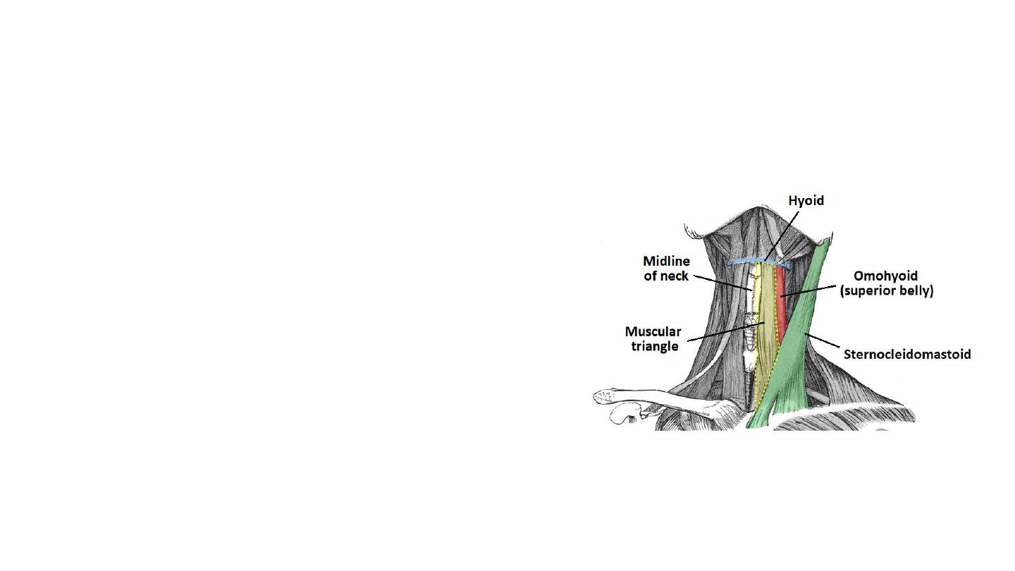
• Muscular Triangle
• Situated more inferiorly than the subdivisions.
It is a slightly ‘dubious’ triangle, in reality
having four boundaries.
• Superiorly – hyoid bone.
• Medially – imaginary midline of the neck.
• Supero-laterally – superior belly of the omohyoid
muscle.
• Infero-laterally – inferior portion of the
sternocleidomastoid muscle.
• Contents:
• The infrahyoid muscles
• Organs- the pharynx, oesophagus, larynx, trachea,
and the thyroid, parathyroid glands.
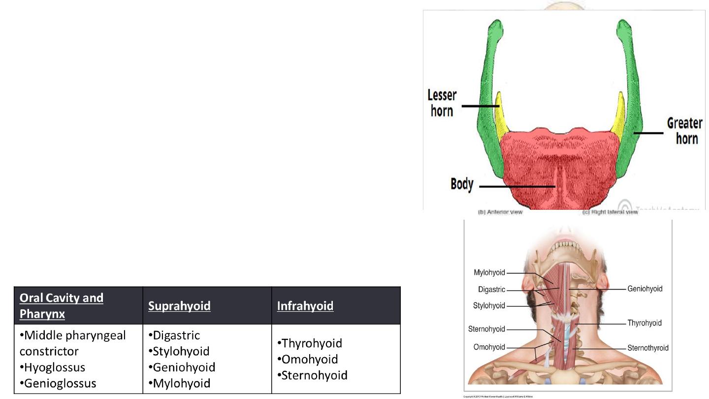
The Hyoid bone
• ‘U’ shaped structure
• Situated in the anterior midline of the neck
between the chin and the thyroid cartilage.
• At rest, it lies at the level of the base of the
mandible in the front and the third cervical
vertebra (C3) behind
• Does not articulate with any other bones, and is
suspended in place by the muscles and
ligaments that attach to it.
Muscular Attachments