
Neck
Dr Firas Al-Hameed
M.B.Ch.B C.A.B.S MRCS (ENT) (England)
Thi-Qar Medical School
Posterior triangle and suprahyoid muscles
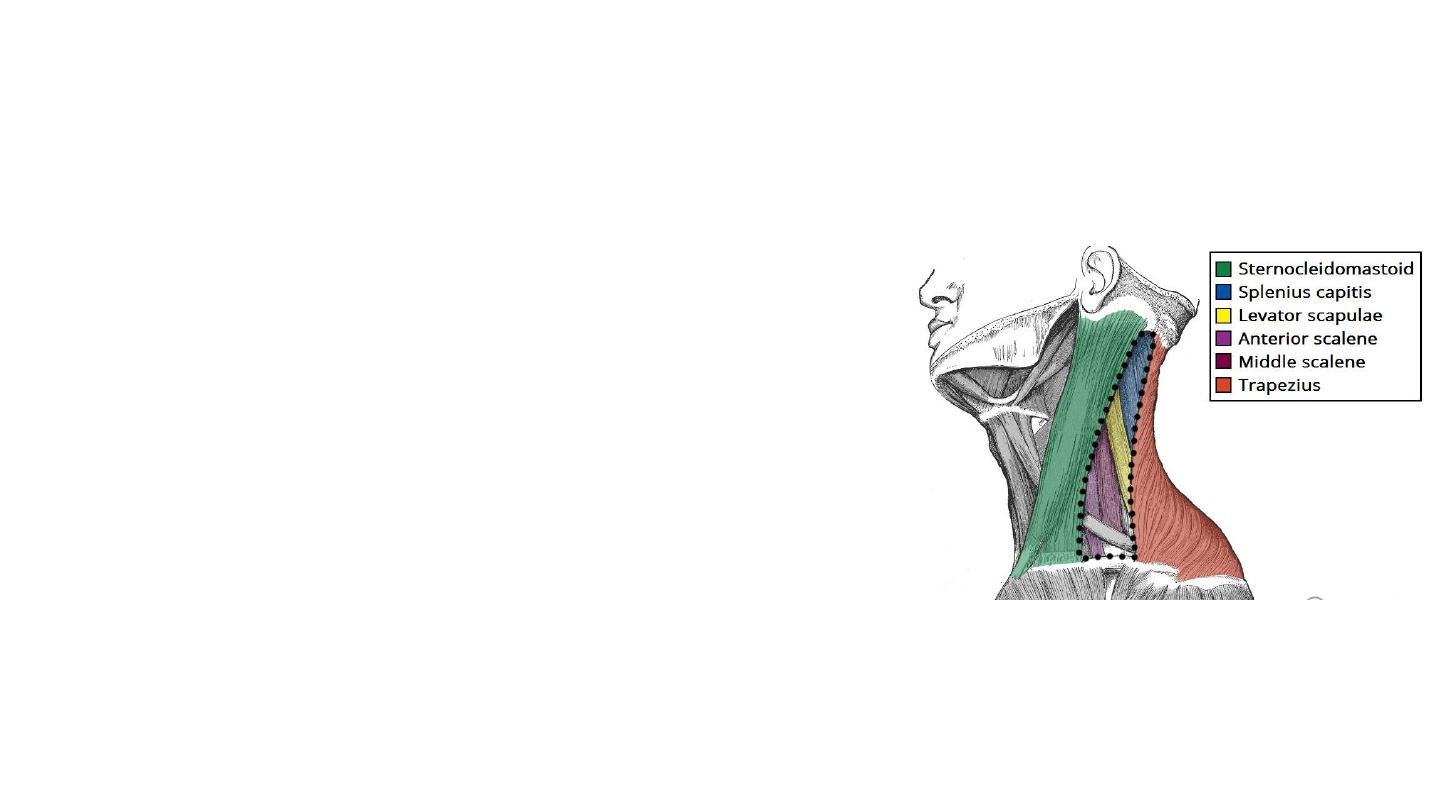
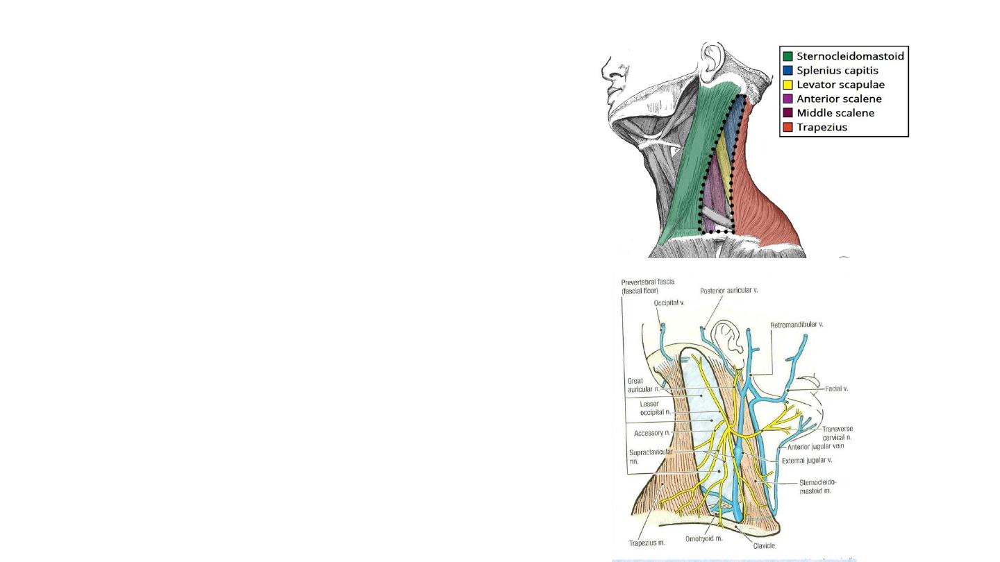
The posterior triangle of the neck
The posterior triangle of the neck is an anatomical
area located in the lateral aspect of the neck.
Borders
• Anterior – posterior border of the sternocleidomastoid.
• Posterior – anterior border of the trapezius muscle.
• Inferior – middle 1/3 of the clavicle.
• The posterior triangle of the neck is covered by the
investing layer of fascia, and the floor is formed by
the prevertebral fascia.
Contents
• Muscles:
• Inferior belly of the Omohyoid
• Vertebral muscles: splenius capitis, Levator
scapulae and anterior, middle and posterior
scalenes.
• Vasculature:
• External jugular vein, subclavian artery and
vein, transverse cervical artery and vein and
suprascapular a. & v.
• Nerves:
• Accessary nerve (CN XI), branches of cervical
plexus, phrenic nerve and brachial plexus
• Lymph nodes:
• Level v: Occipital and Supraclavicular
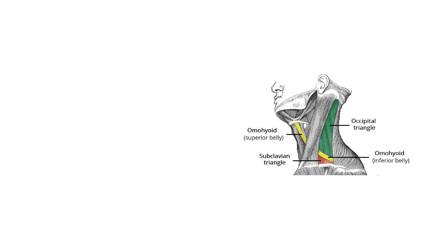
Subdivisions
• The omohyoid muscle splits the
posterior triangle of the neck into two:
• The larger, superior part is termed the
occipital triangle.
• The inferior triangle is known as the
subclavian triangle and contains the
distal portion of the subclavian artery.
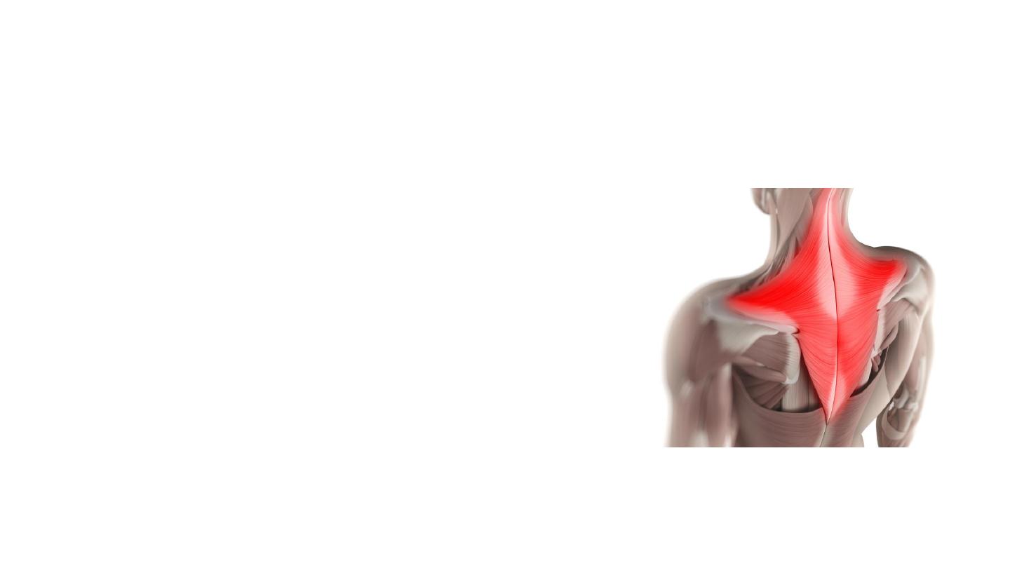
Trapezius Muscle
• Flat diamond-shaped muscle at back of
neck and chest.
• Origin: occipital bone and spines of C7 –
T12
• Insertion: Lateral 1/3 of clavicle, acromion,
spine of scapula
• Functions:
• Its main function is to stabilize and move the
scapula( elevation, depression, retraction).
• Assists in abduction of shoulder above 90
degrees.
• Extends the neck if both muscles contract and
the scapulae are stable.
• Nerve supply: accessory nerve (CN11)
• Dropped scapula/ winged scapula
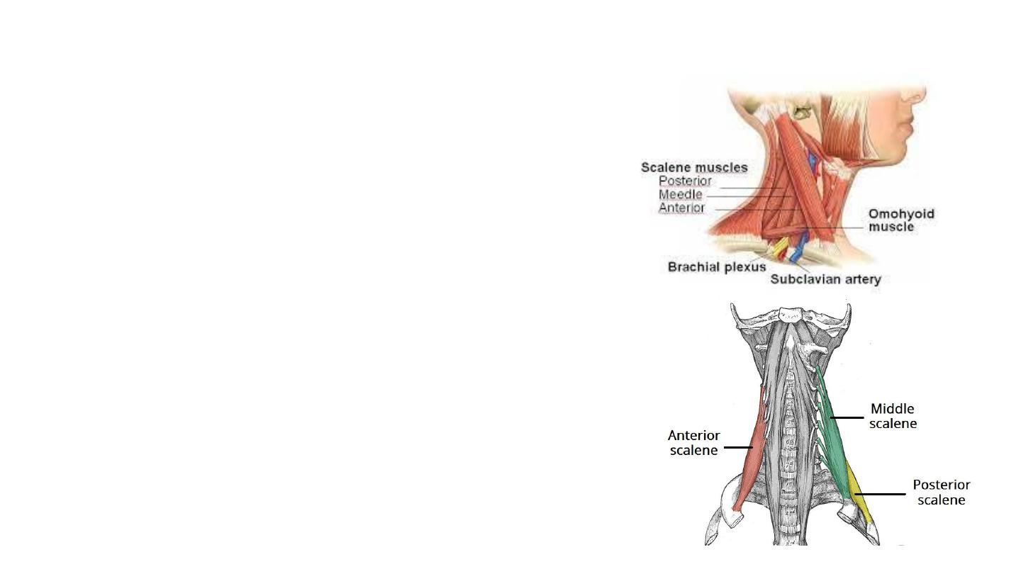
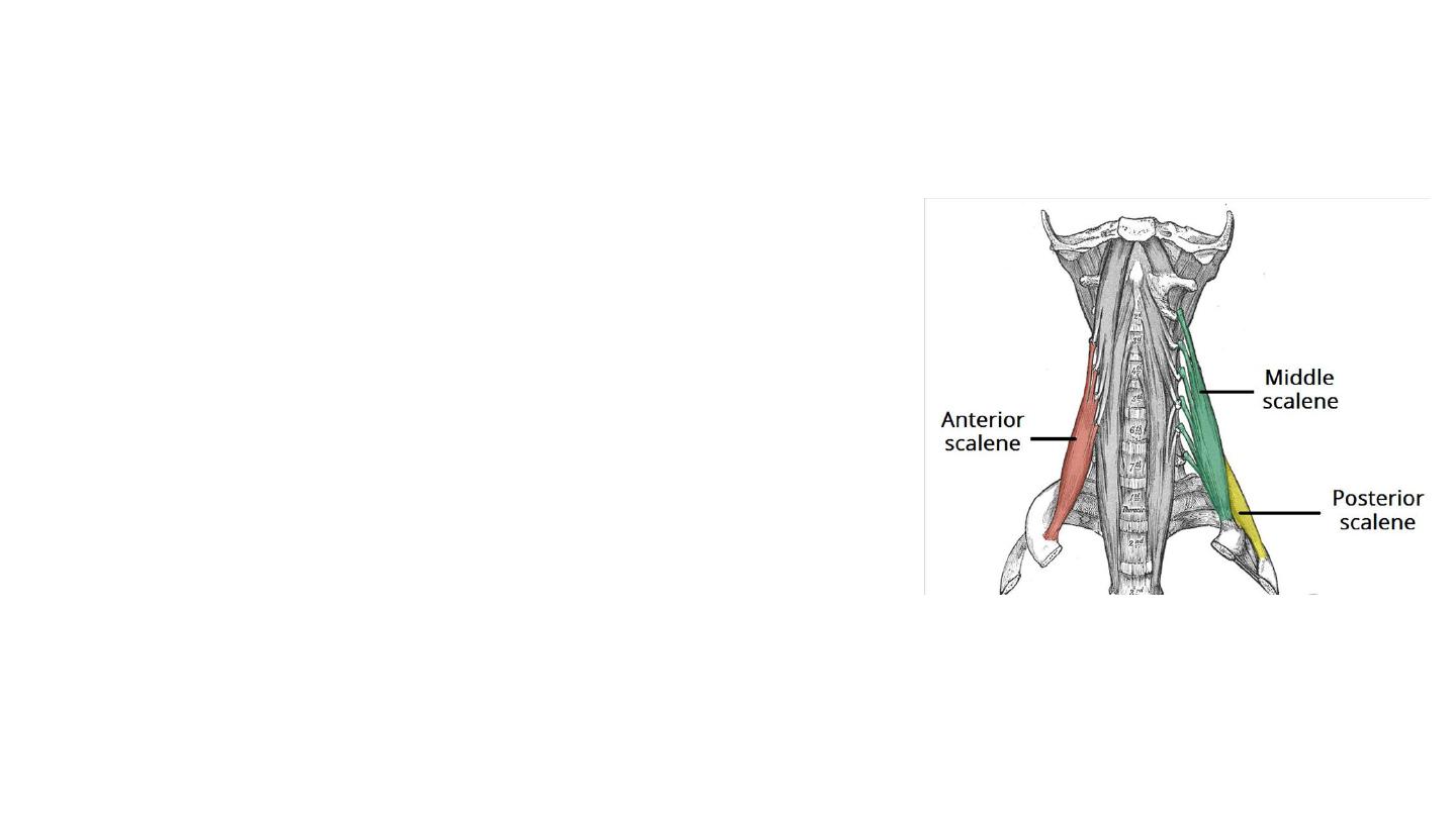
The scalene muscles
• Three paired muscles (anterior, middle and posterior),
located in the lateral aspect of the neck.
• The scalenes act as accessory muscles of respiration,
and perform flexion at the neck.
Anterior Scalene
• It lies on the lateral aspect of the neck, deep to the
prominent sternocleidomastoid muscle.
• Attachments: Originates from the anterior tubercles of
the transverse processes of C3-C6, and attaches onto
the scalene tubercle, on the inner border of the first rib.
• Function: Elevation of the first rib. Ipsilateral contraction
causes ipsilateral lateral flexion of the neck, and bilateral
contraction causes anterior flexion of the neck.
• Innervation: Anterior rami of C5-C6.
Middle Scalene
• It is the largest and longest of the three scalene muscles.
• It has several long, thin muscles bellies arising from the
cervical spine, which converge into one large belly that
inserts into the first rib.
• Attachments: Originates from the posterior tubercles of
the transverse processes of C2-C7, and attaches to the
scalene tubercle of the first rib.
• Function: Elevation of the first rib. Ipsilateral contraction
causes ipsilateral lateral flexion of the neck.
• Innervation: Anterior rami of C3-C8.
Posterior Scalene
• It is the smallest and deepest of the scalene muscles.
• Attachments: Originates from the posterior tubercles of
the transverse processes of C5-C7, and attaches into the
second rib
.
• Function: Elevation of the second rib, and ipsilateral lateral
flexion of the neck.
• Innervation: Anterior rami of C6-C8.
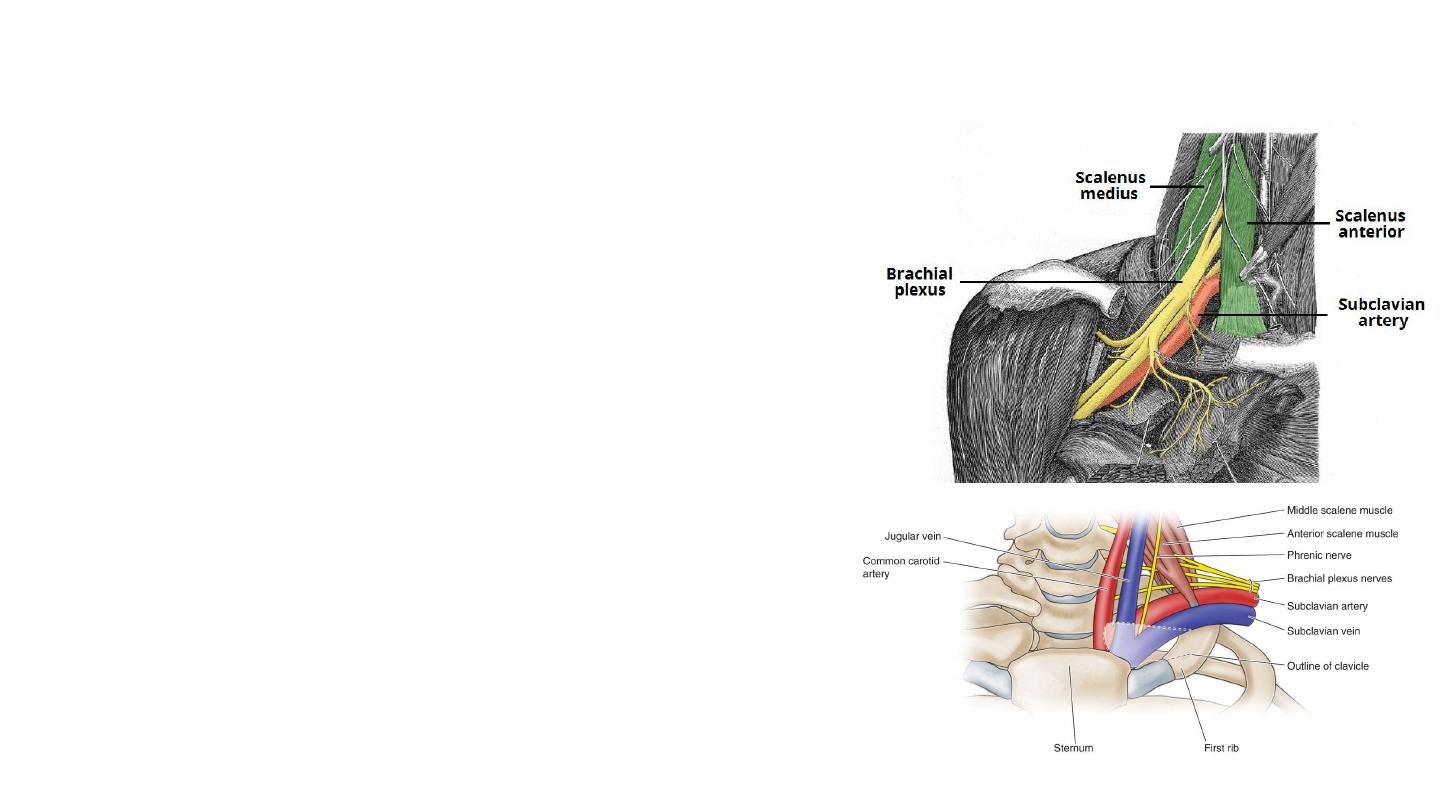
Anatomical Relationships of scalene muscles
• The brachial plexus and subclavian artery pass between the
anterior and middle scalene muscles.
• The subclavian artery is located
posterior to the anterior
scalene
.
• The subclavian vein and phrenic nerve pass
anteriorly to the
anterior scalene
– the subclavian vein courses horizontally
across it, while the phrenic nerve runs vertically down the
muscle.
• The brachial plexus is a network of nerve fibres that supplies
the skin and musculature of the upper limb. It begins in the
root of the neck, passes through the axilla, and runs through
the entire upper extremity.
• C5, C6, C7 and C8, and the first thoracic spinal nerve, T1.
• Phrenic nerve : arises from the anterior divisions of spinal
nerves C3-C5. It descends down the neck, within the
prevertebral fascia, to innervate the diaphragm.
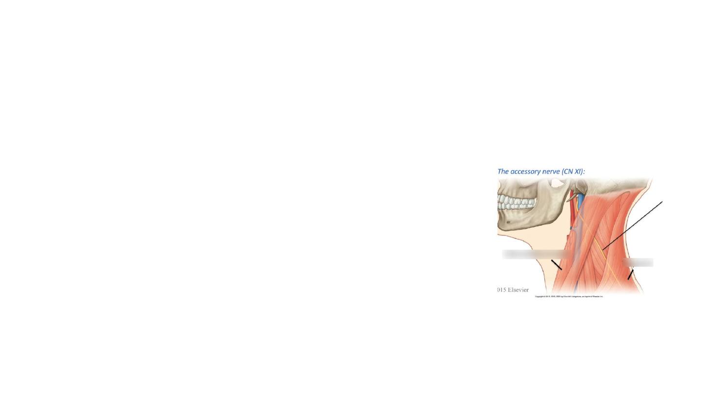
• The accessory nerve (CN XI) innervates sternocleidomastoid
and enters the posterior triangle. It crosses the posterior
triangle in an oblique, inferoposterior direction, within the
investing layer of fascia. It lies relatively superficial in the
posterior triangle, leaving it vulnerable to injury.
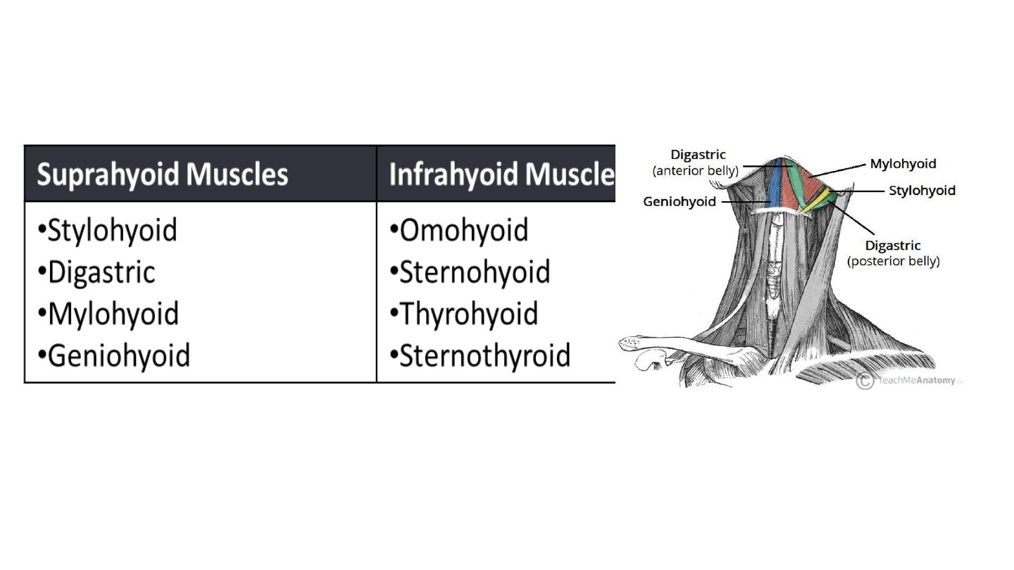
Suprahyoid and infrahyoid muscles
Suprahyoid muscles
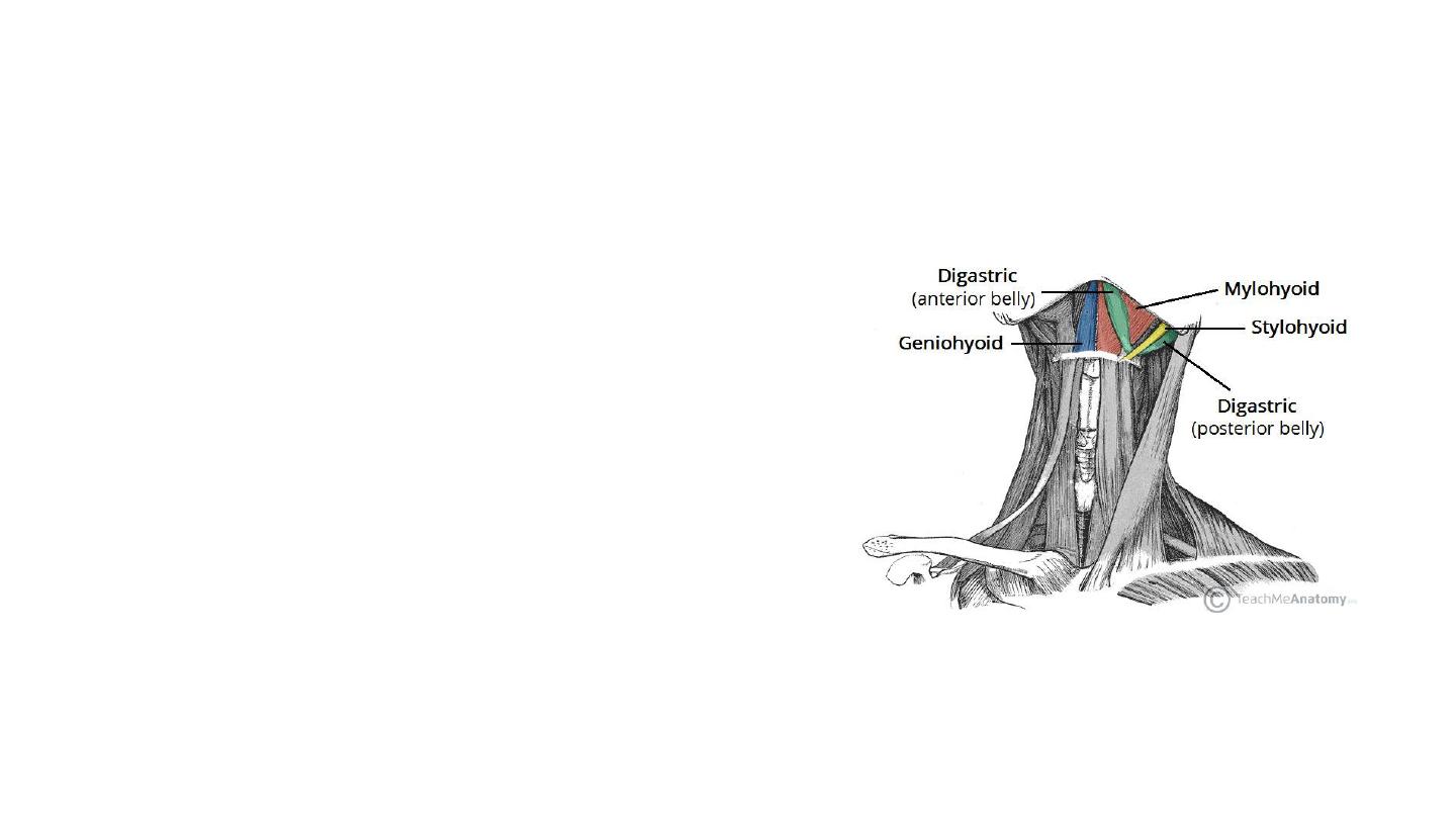
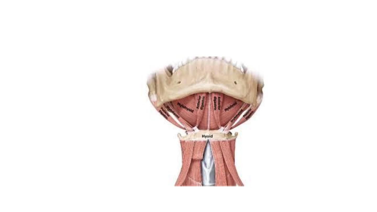
Stylohyoid
• The stylohyoid muscle is a thin muscular strip,
which is located superiorly to the posterior
belly of the digastric muscle.
• Attachments: Arises from the styloid process of
the temporal bone and attaches to the lateral
aspect of the hyoid bone.
• Actions: Initiates a swallowing action by pulling
the hyoid bone in a posterior and superior
direction.
• Innervation: Stylohyoid branch of the facial
nerve (CN VII).
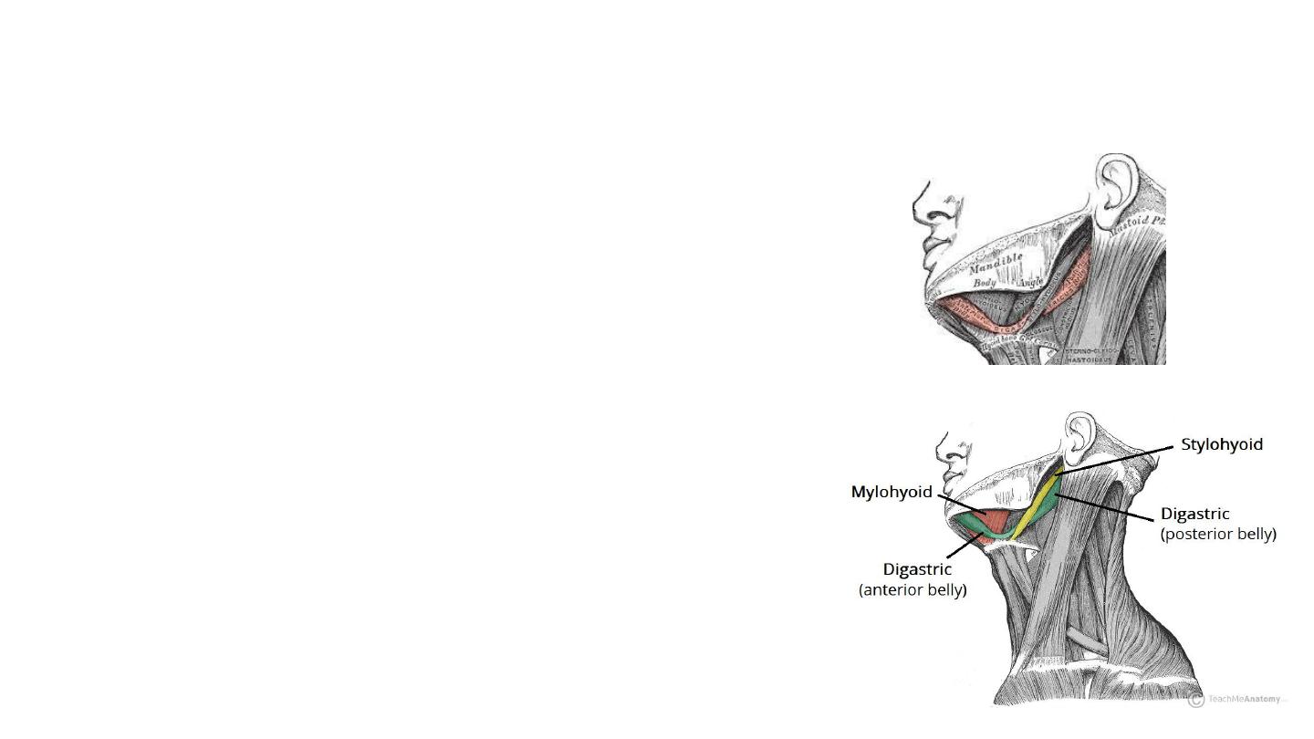
Digastric
• The digastric is comprised of two muscular bellies, which are
connected by a tendon
• Attachments:
• The anterior belly arises from the digastric fossa of the mandible.
• The posterior belly arises from the mastoid process of the temporal
bone.
• The two bellies are connected by an intermediate tendon, which is
attached to the hyoid bone via a fibrous sling.
• Actions: Depresses the mandible and elevates the hyoid
bone.
• Innervation:
• The anterior belly is innervated by the inferior alveolar
nerve, a branch of the mandibular nerve (which is derived
from the trigeminal nerve, CN V).
• The posterior belly is innervated by the digastric branch of
the facial nerve.
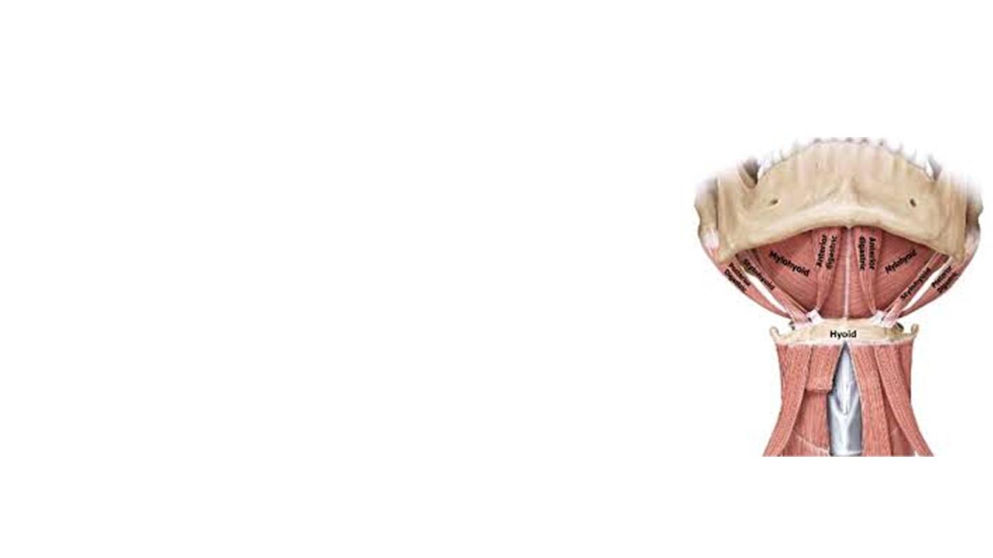
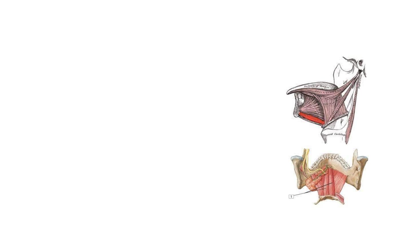
Mylohyoid
• The mylohyoid is a broad, triangular shaped
muscle. It forms the floor of the oral cavity and
supports the floor of the mouth.
• Attachments: Originates from the mylohyoid line of
the mandible, and attaches onto the hyoid bone.
• Actions: Elevates the hyoid bone and the floor of
the mouth.
• Innervation: Inferior alveolar nerve, a branch of the
mandibular nerve (which is derived from the
trigeminal nerve).
Geniohyoid
• The geniohyoid is located close to the midline
of the neck, deep to the mylohyoid muscle.
• Attachments: Arises from the inferior mental
spine of the mandible. It then travels inferiorly
and posteriorly to attach to the hyoid bone.
• Actions: Depresses the mandible and elevates
the hyoid bone.
• Innervation: C1 nerve roots that run within the
hypoglossal nerve.

The arterial supply :
• Branches of the facial artery, occipital artery, and lingual artery.