Lecture -3-
Anatomy of stomach
Dr . Raya Abdul Ameer
MBCHB.CABHS-RAD

• The stomach is a hollow muscular organ and
the most dilated portion of the gastrointestinal
tract. It is located on the left upper quadrant of
the abdomen between the esophagus and
duodenum
• It occupies the LT hypochondrial region ,
epigastric and umbilical regions
• It is J shaped
• 15- 25 cm in length
• Capacity = 1.5-2 liters

External features of stomach
;
it has
1) two orifices cardiac and pyloric
2) two curvatures lesser and greater
curvature
3) two surfaces antero-superior and
postero –inferior surfaces

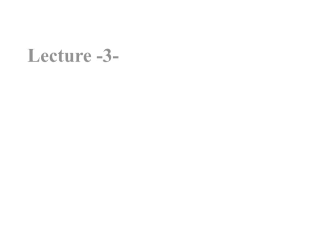
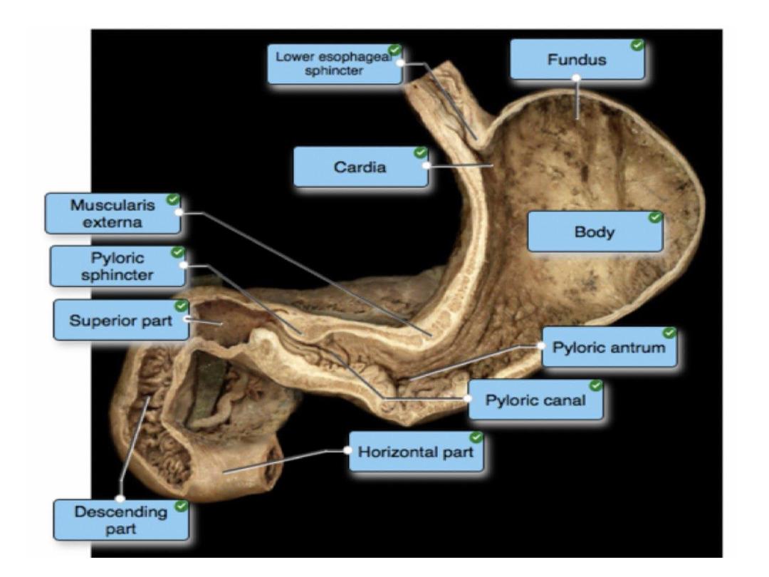
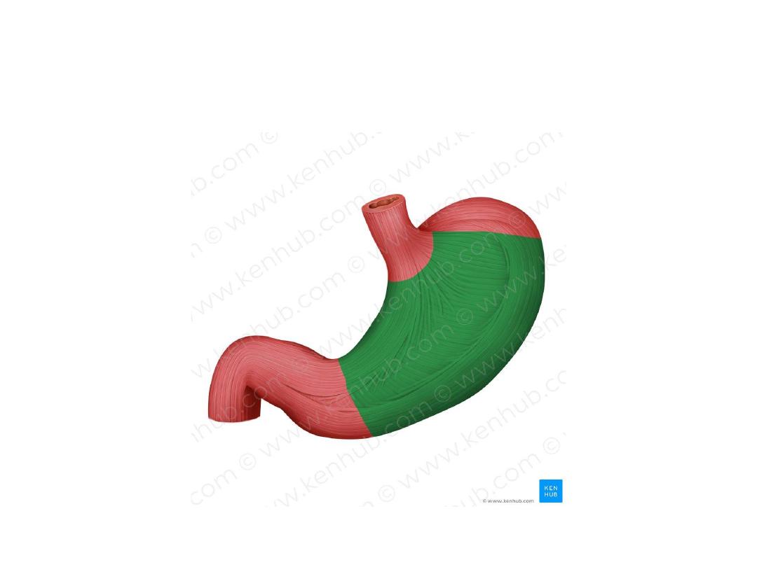
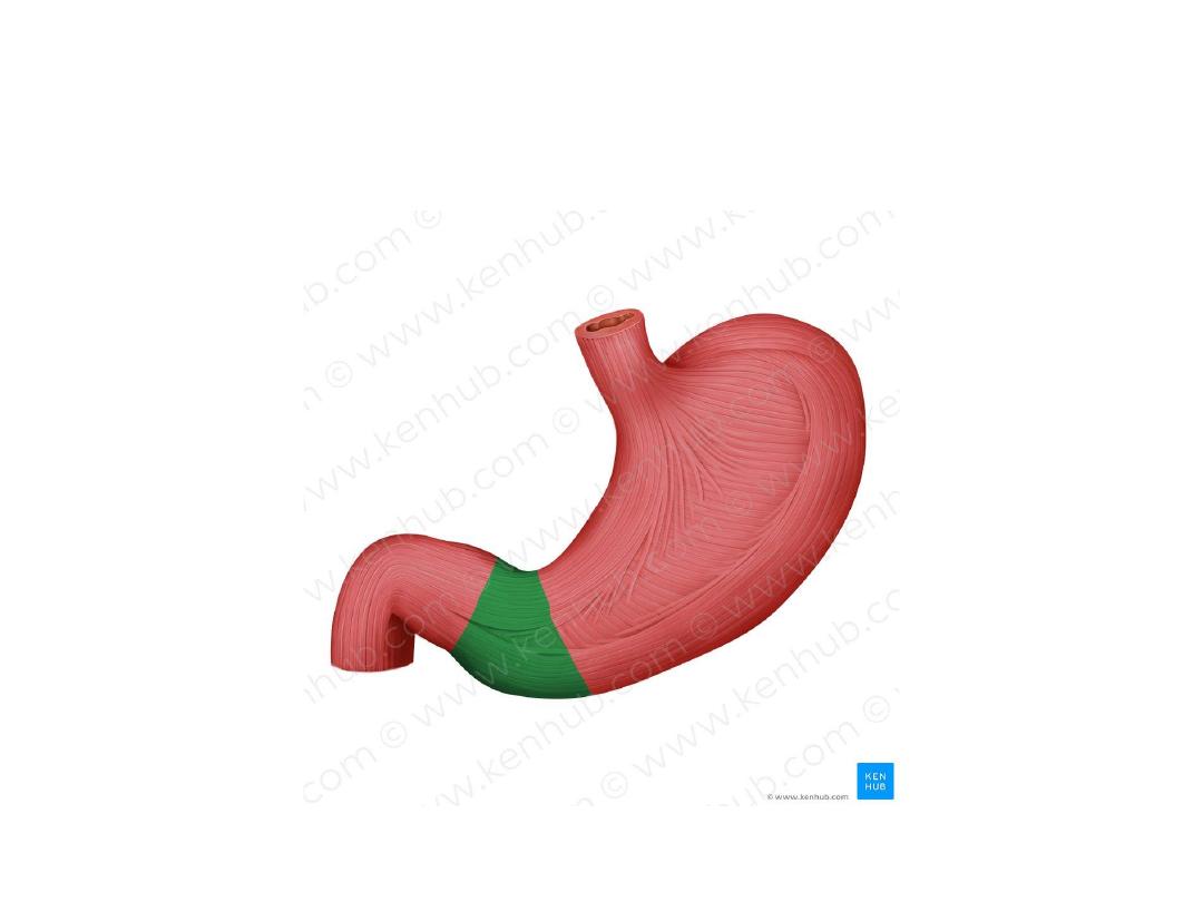
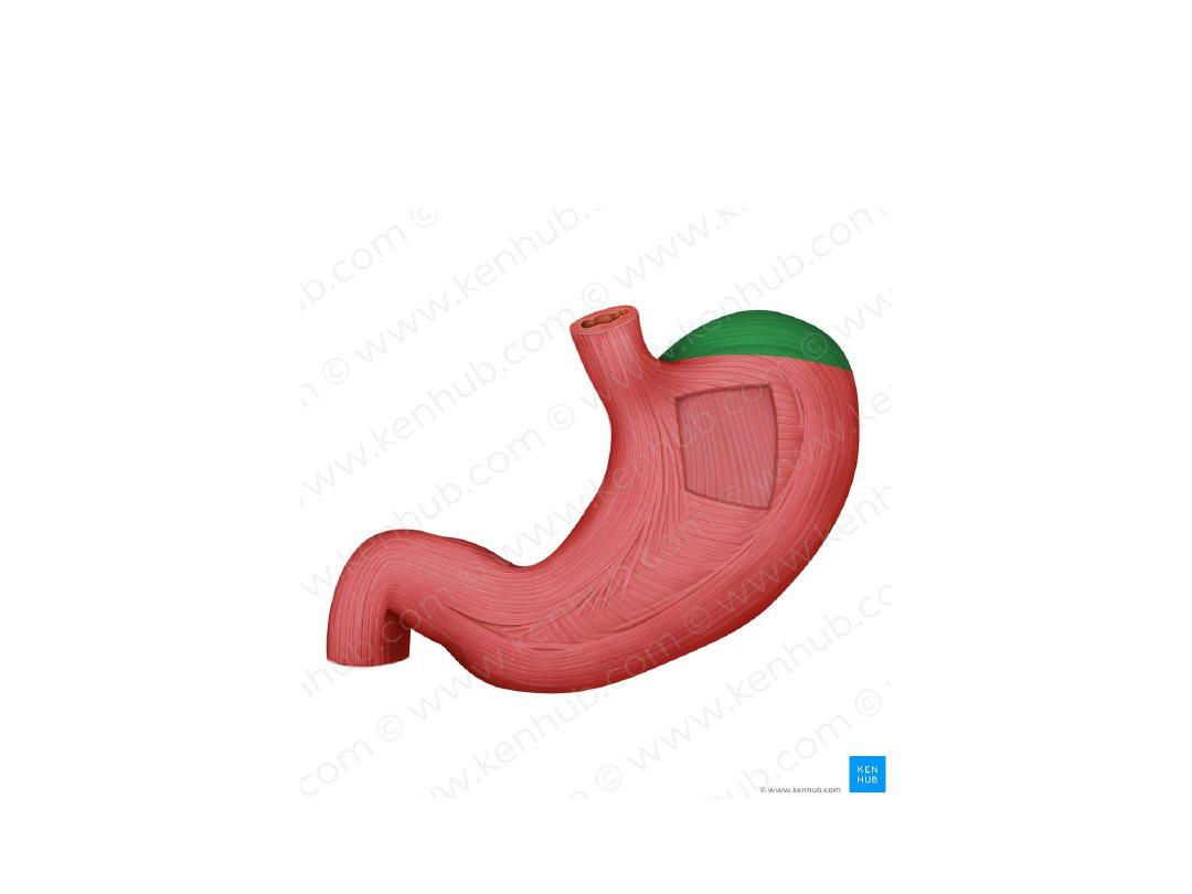
Parts of stomach
• The stomach has four main anatomical parts; the cardia, fundus,
body and pylorus:
• Cardia
– surrounds the superior opening of the stomach at the
T11 level.
• Fundus
– the rounded, often gas filled portion superior to and
left of the cardia.
• Body
– the large central portion inferior to the fundus.
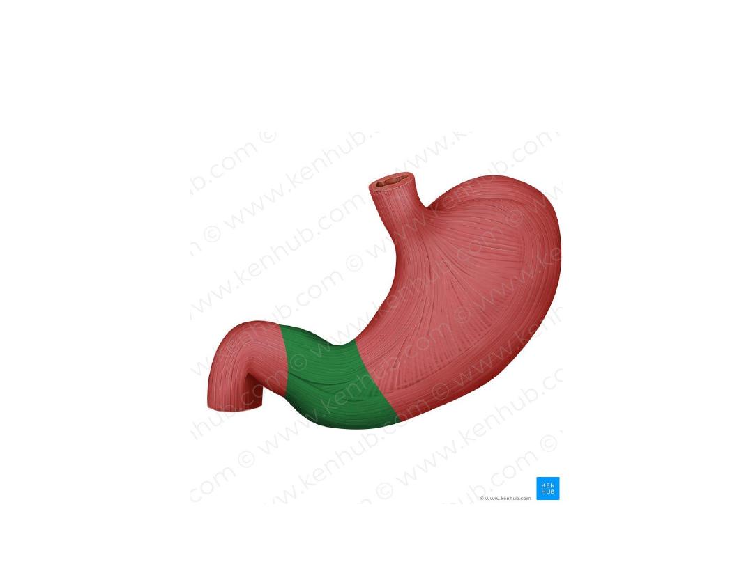
• Pylorus
– This area connects the stomach to the duodenum.
• It is divided into the
1)pyloric antrum,
2)pyloric canal and
3)pyloric
sphincter.
The
pyloric
sphincter
demarcates
the
transpyloric plane at the level of L1
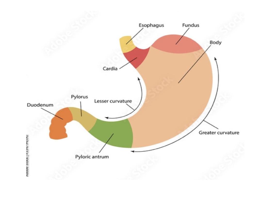

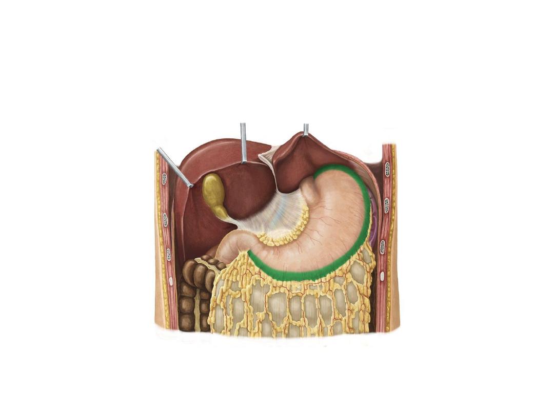
Curvatures of the stomach
The medial and lateral borders of the stomach are curved,
forming the lesser and greater curvatures:
• Greater curvature
– forms the long, convex, lateral border of
the stomach.
• Arising at the cardiac notch, it arches backwards and passes
inferiorly to the left.
• It curves to the right as it continues medially to reach
the pyloric antrum.
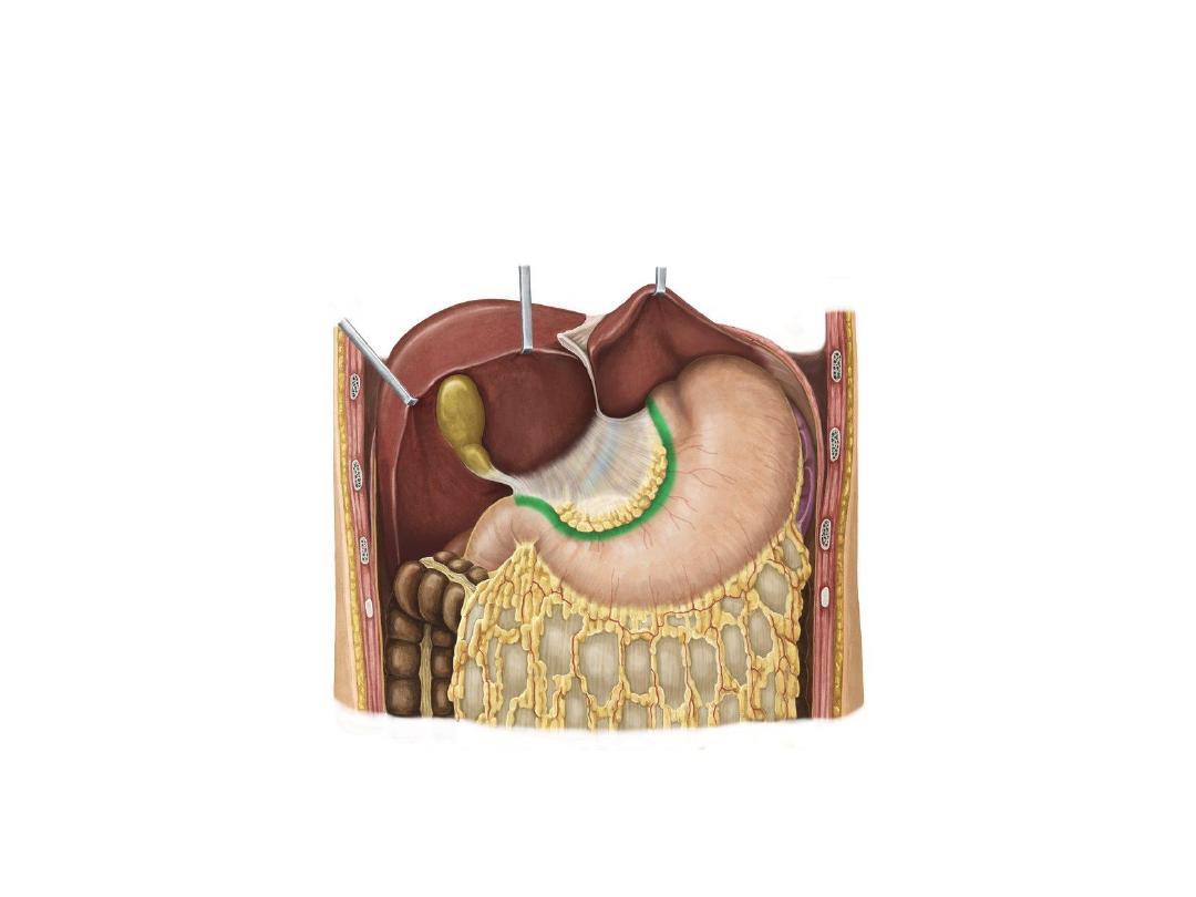
• Lesser curvature
– forms the shorter, concave, medial surface
of the stomach.
• The most inferior part of the lesser curvature, the angular
notch, indicates the junction of the body and pyloric
region
.
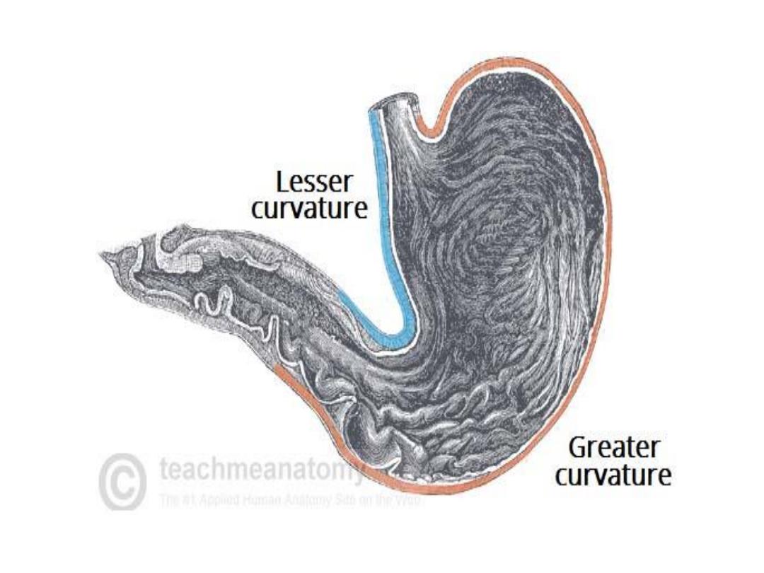

Sphincters of the Stomach
• There are two sphincters of the stomach,
located at each orifice. They control the
passage of material entering and exiting the
stomach

1)Gastroesophageal Oesophageal Sphincter
• Is a ring of smooth muscle fibers connect the
esophagus to the stomach , also called lower
esophgael sphinceter
2
) The pyloric sphincter
lies between the pylorus and the first part of
the duodenum
Site … trans pyloric plane at L1 level
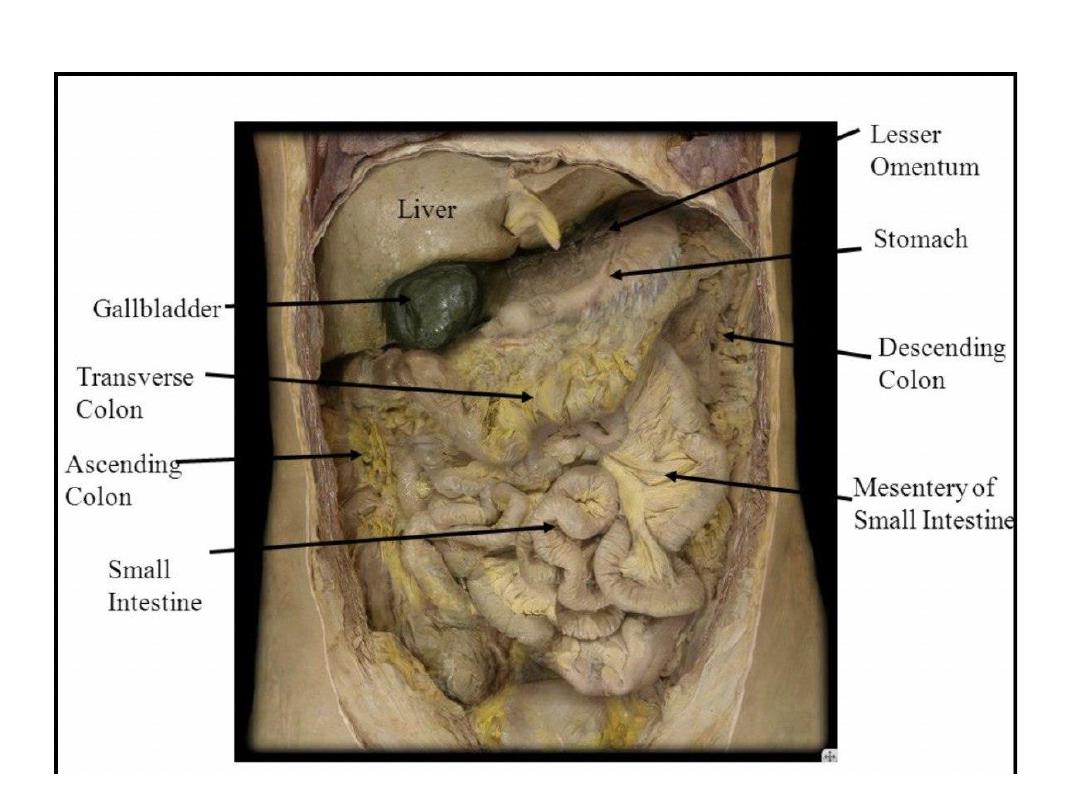

Anatomical relation of stomach
-Anterior
: Diaphragm, left lobe of liver,
and anterior abdominal wall
-
Posterior
: lesser sac, pancreas, left kidney
and adrenal gland, splenic artery and spleen
Superior
: Esophagus and LT dome
diaphragm
Inferior
: Transverse colon/mesocolon,
greater omentum

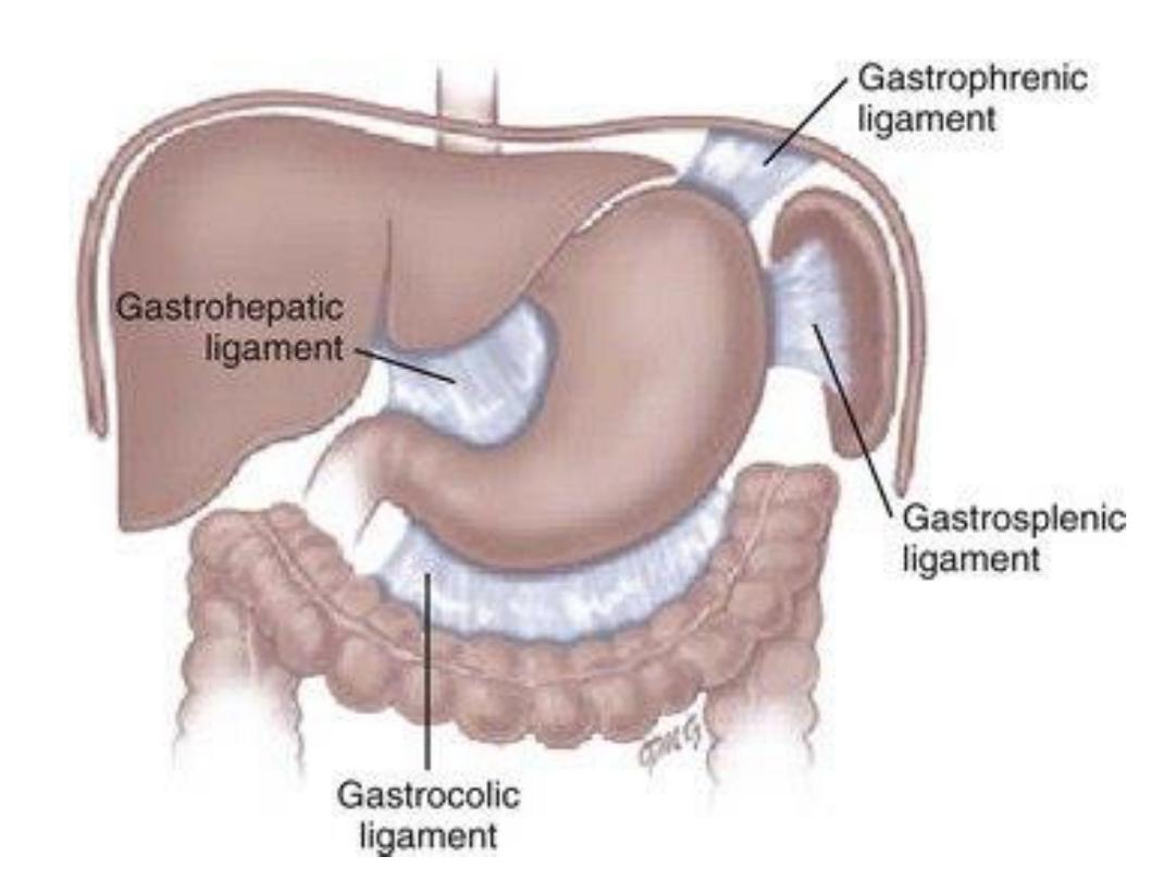
• Ligaments of the stomach
1)Hepato gastric ligament ..liver to the lesser
curvature of stomach
2)Gastrophrenic ligament :fundus to the
diaphragm
3 ) Gastrosplenic ligament : upper third of
greater curvature to spleen
4) Gastro Colic ligament :lower part of greater
curvature to the transverse colon

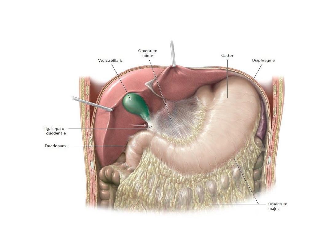
Omentum
The omenta are sheets of visceral peritoneum that extend from
the stomach and proximal part of the duodenum to other
abdominal organs
1) Greater omentum :
-
Consist of four layers of visceral peritoneum
-It descend from the greater curvature of the stomach
and proximal part of the duodenum then fold back up
and attached to the anterior surface of the transverse
colon
-
hanging from stomach like a curtain in front of small
intestine .
-It has a role in immunity and some times refer to as the
abdominal policeman , because it can migrate to the
infected viscera or to the site of surgical disturbance

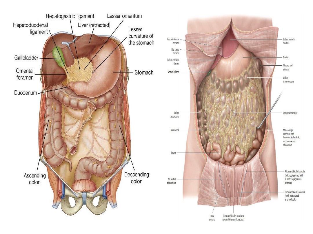
Lesser omentum :
• Is double layer of visceral peritoneum
and its smaller than greater omentum
• Attached from the lesser curvature of
stomach and proximal part of the
duodenum to the liver
• It consist of two parts ..hepto gastric
ligament and hepto duodenal ligament

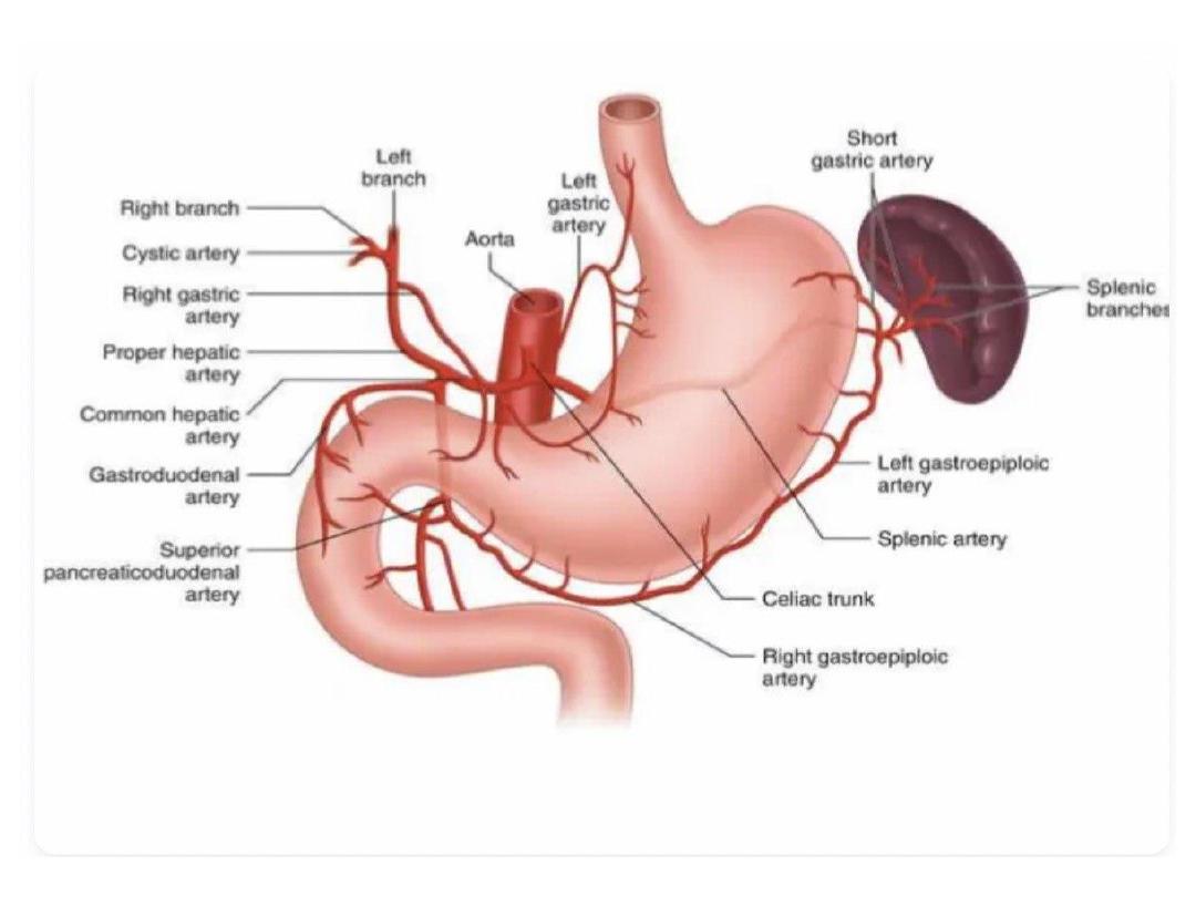
Blood supply to the stomach
1) Right gastric artery
: arises from hepatic artery,
forms anastomosis with left gastric artery; supplies
lower part lesser curvature, anterior and posterior
sides of the stomach
2) Left gastric artery
: arises from celiac artery ,
forms anastomosis with right gastric artery; supplies
upper part of lesser curvature, cardia, right upper
and posterior walls
3) Right gastro omental (gastro epiploic
)
artery: arises from gastro duodenal artery; forms
anastomosis with left gastro omental artery and
supplies inferior part of greater curvature

4
) Left gastro omental (gastro epiploic) artery
: arises
from splenic artery; forms anastomosis with right gastro
omental artery and supplies superior part of greater
curvature
5) Short gastric artery
: arise from splenic artery; supply
fundus and posterior wall of stomach
6 ) Gastroduodenal artery
: arises from common hepatic
artery; supplies pyloric part

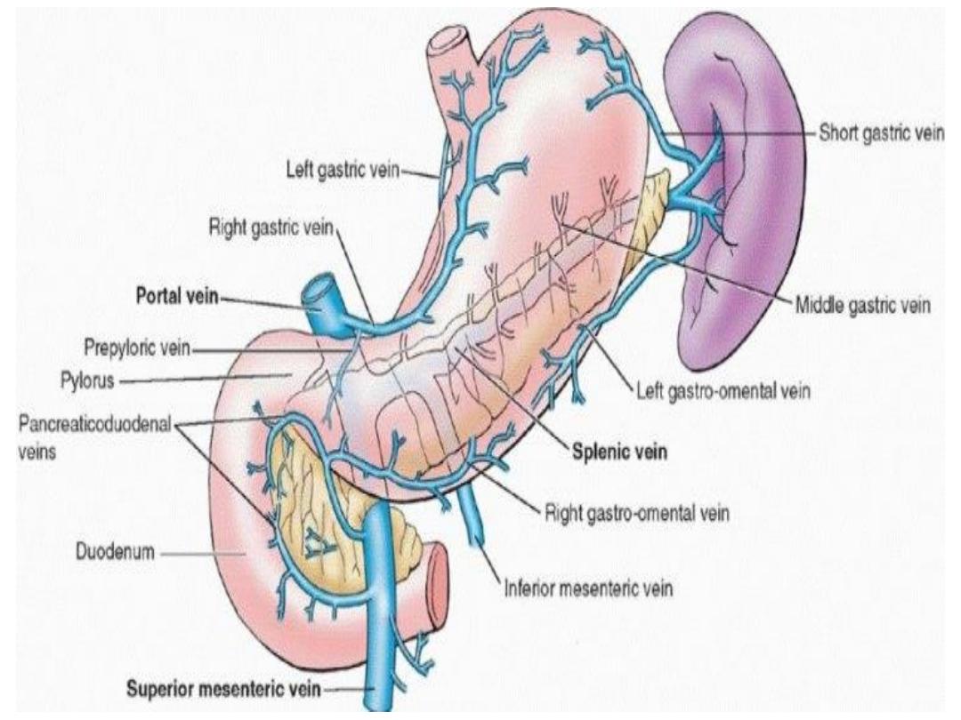
Venous drainage
1
) Right and Left gastric vein to the portal vein
2 ) Left gastro epiploic vein and short gastric
vein to splenic vein
3 ) Right gastro epiploic vein .to the superior
mesenteric vein

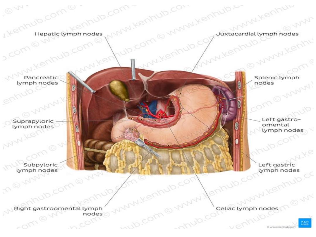
Lymphatic drainage of stomach
1) Cardia: Juxtacardial nodes
2) Fundus: Short gastric nodes
3 ) Lesser curvature: Right/left gastric nodes
4) Greater curvature: Right/left gastro omental
nodes
5 ) Pyloric part: Pyloric nodes (suprapyloric,
retropyloric, subpyloric nodes)
all are
Drain to celiac nodes → intestinal lymphatic
trunk → cisterna chyli → thoracic duct

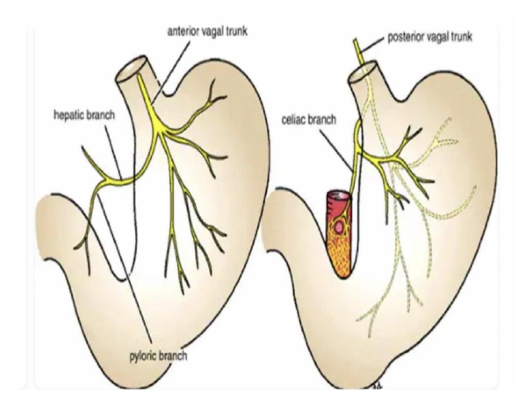
Nerve supply to the stomach
1) Parasympathetic nerve derived from the
vagus nerve
-Anterior . gastric nerve continuation of left
vagus supply anterior . surface of stomach
down to the pylorus
-Posterior . gastric nerve continuation of RT
vagus
Supply post surface of stomach except the
pylorus
.

• Sympathetic nerve supply
arises from the T6-T9 spinal cord segments
and passes to the coeliac plexus via the
greater splanchnic nerve


Lecture- 4
Anatomy of small intestine
Dr. Raya Abdul Ameer
MBCHB .CABHS –RAD

Small intestine
• It is the longest part of the digestive
system
• Its so called because its lumen diameter is
smaller than the large intestine although it
is longer than large intestine .
• It extend from the pylorus of stomach to
iliocecal valve where it joints the large
intestine
• Its about 6 meters ( 18-20 feet ) in length

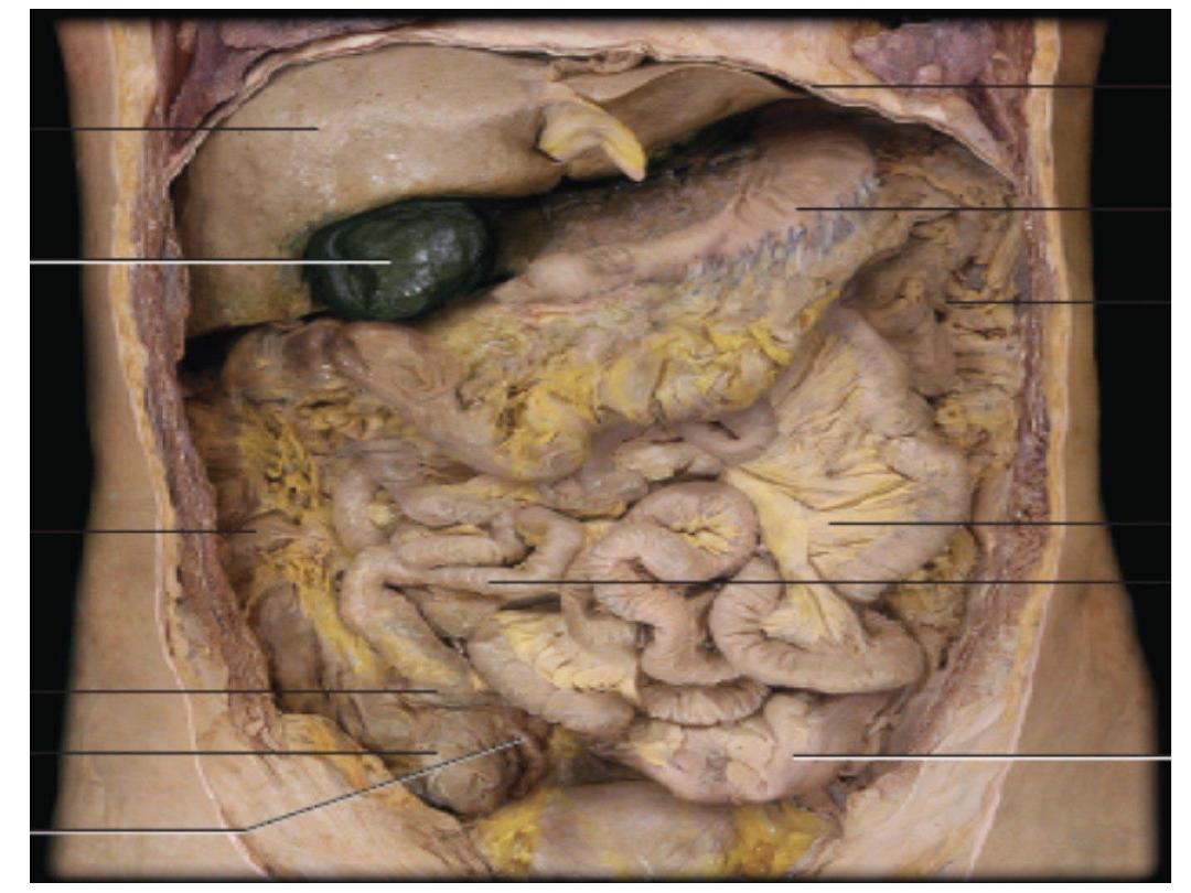
-
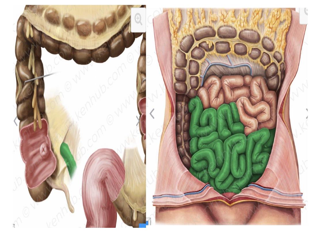
- It occupy the central & lower parts of the abdomen.
- It is surrounded by the curve of the large intestine &
covered anteriorly by the greater omentum & the anterior .
abdominal wall
-The main function is to complete digestion of food and
absorb nutrient
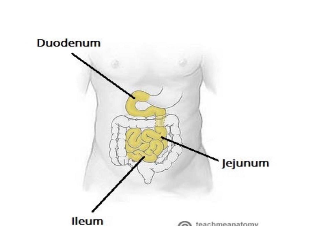
-Its divided into three parts
Duodenum ,jejunum and ileum
-all three parts are covered by greater momentum anteriorly
-the duodenum has both intraperitoneal and retroperitoneal
parts , while the jejunum and ileum are entirely
intraperitoneal

1.
Duodenum:
the first 10 inches & fixed to the posterior .
Abdominal wall.
2. Jejunum
: follows the duodenum. It is 8 feet long
and forms the proximal 2/5 of the small intestine.
3. Ileum
: next to the jejunum. It is 12 feet long and
forms the distal 3/5 of the small intestine. It ends by
joining the caecum at the ileocaecal
valve.
Note . both jejunum & ileum have a mesentery
attaching them to the posterior . Abdominal .
Wall.

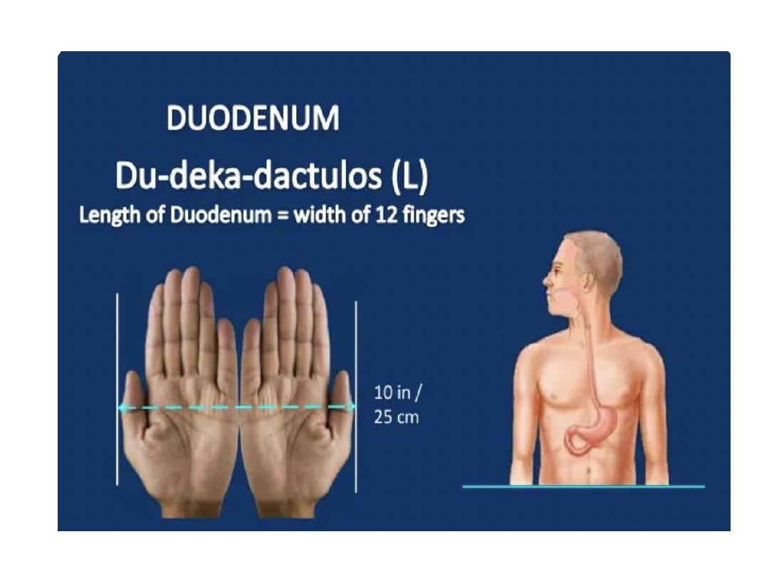
Duodenum
• It is the shortest , dilated and most fixed proximal
part of small intestine
• This portion of small intestine received its name
due to its length , in Latin duodenum mean 12
finger , which is the approximate length of the
organ
• 10 inches , 25 cm in length
• Its retroperitoneal and has devoiod of mesentery
except first segment
• It is fixed to posterior abdominal wall
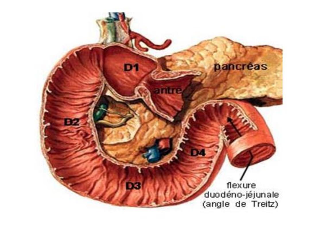
• C –shaped curvature curved around the head of
pancreas

• Lies above the umbilicus al level of L1,L2,L3
vertebra
• Receive bile duct and pancreatic ducts
Begins: at the pyloric end of the stomach ½
inch to the right of the median plane.
Ends: at the duodenojejunal flexure 1 inch to
the left of the median plane
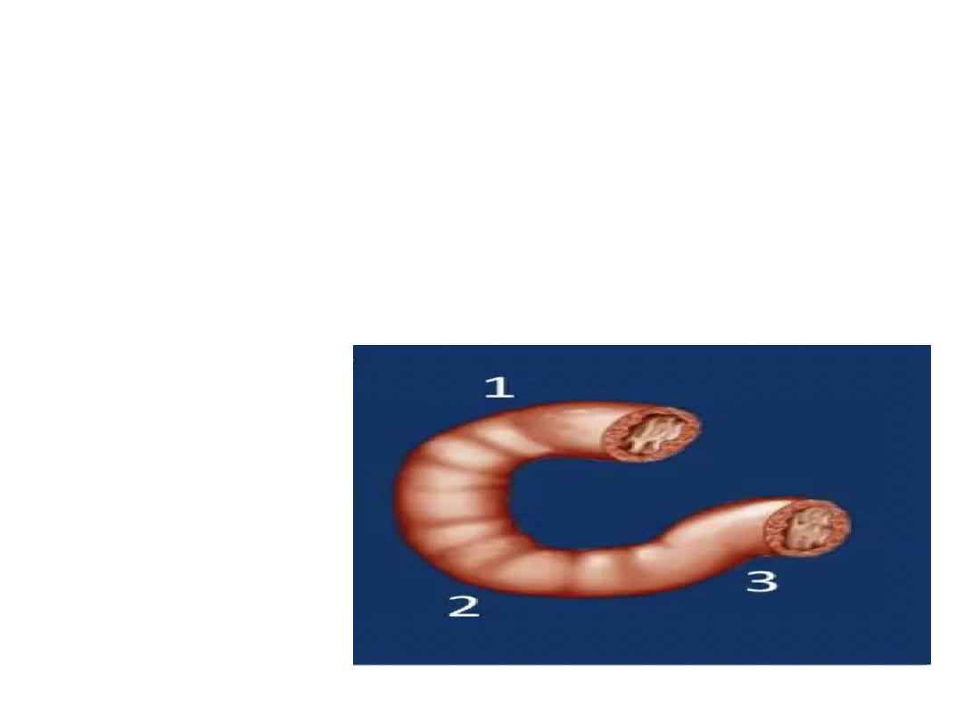
It has three flexures
1-superior duodenal flexure
2-inferior duodenal flexure
3-duodeno-jejunal flexure

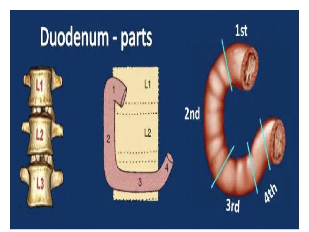
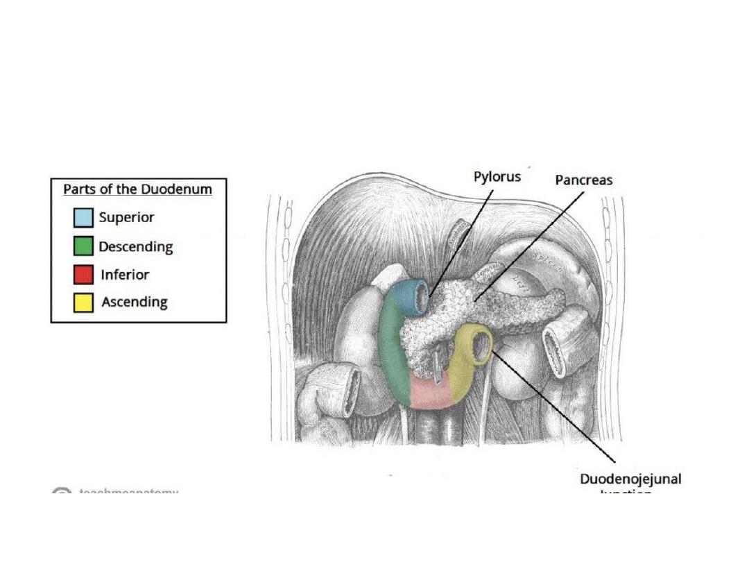
• The duodenum can be divided in to four parts
/segments , each part has a different anatomy
(shape) ,and perform a different based function
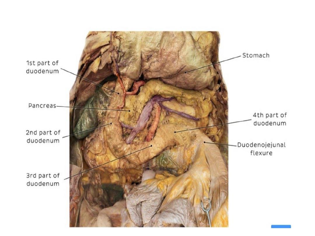
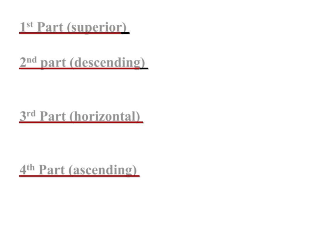
1
st
Part (superior
) 2 inches( 5 cm ) long & lies
opposite the 1
st
L. vertebra.
2
nd
part (descending
) 3 inches( 7.5 cm ) long
& extends from L1 to L3 vertebra.
3
rd
Part (horizontal)
4 inches ( 10 cm )& lies
at the level of L3 vertebra.
4
th
Part (ascending)
1 inch(2.5 cm ) & ascends
from the level of L3 to L2.

Peritoneal relations:
The duodenum is mostly retroperitoneal and
fixed to the posterior .abdominal .wall except
the following 2 mobile parts:
1. The proximal
1 inch which is suspended by :
a. lesser omentum: above
b. greater omentum: below
2. The distal end
which is attached to the right
crus of the diaphragm by a fibro muscular band
called the suspensory muscle of duodenum
First Part of Duodenum
Begins:
as a continuation of the pylorus, ½ inch to the
Rt. Of the median plane at the level of L1 (transpyloric
plane).
-Course:
it passes upwards, backwards & to the Rt.
undercover of the quadrate lobe of the liver.
Ends:
close to the neck of the gall bladder by curving
downwards to become the 2
nd
part
-
Length 5 cm
-Proximal 2.5 cm movable , attached to lesser omentum
above and greater omentum below
-Distal 2.5 cm fixed , retroperitoneal covered with
peritoneum only on anterior aspect

• Most common site of duodenal ulcer
Duodenal bulb /duodenal cup
-Is very first part of the duodenum which is
slightly dilated
-It is intraperitoneal
-2.5 cm in length
-It is mobile and has mesentery
-Smooth walled
-It is connected to the liver by hepatodudenal
ligament
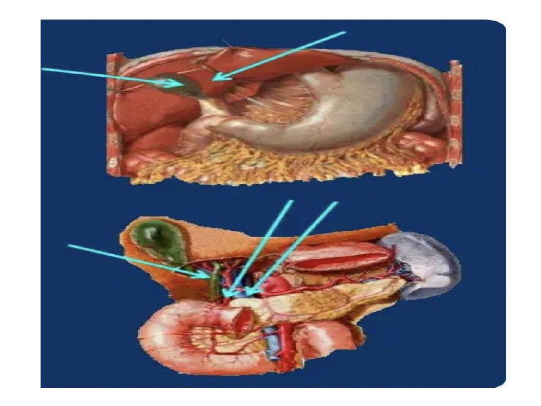
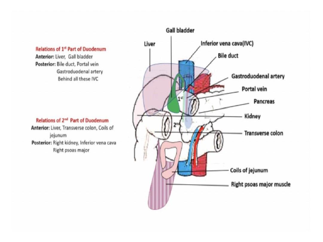
Relations of the 1
st
part
I. Anteriorly
1. Quadrate lobe of liver
2. Neck and body of gall bladder
II. Superiorly
Epiploic foramen
III. Inferiorly
Head and neck of pancreas
IV. Posteriorly
Common .Bile . duct, gastroduodenal artery . &
portal vein
2
nd
Part of Duodenum
Begins:
at the level of L1 as a continuation of
the 1
st
part.
Course:
it descends vertically downwards
infront of the hilum of the Rt. Kidney.
Ends:
at the level of L3 by curving to the left to
become the 3
rd
part.
Peritoneum: it is covered by peritoneum only
anteriorly except its middle part which is devoid
of peritoneum & is directly related to transverse
colon
Relation of 2
nd
part of duodenum:
(
A) Anteriorly
1-Rt. Lobe of liver
2. Transverse Colon
3. Coils of jejunum
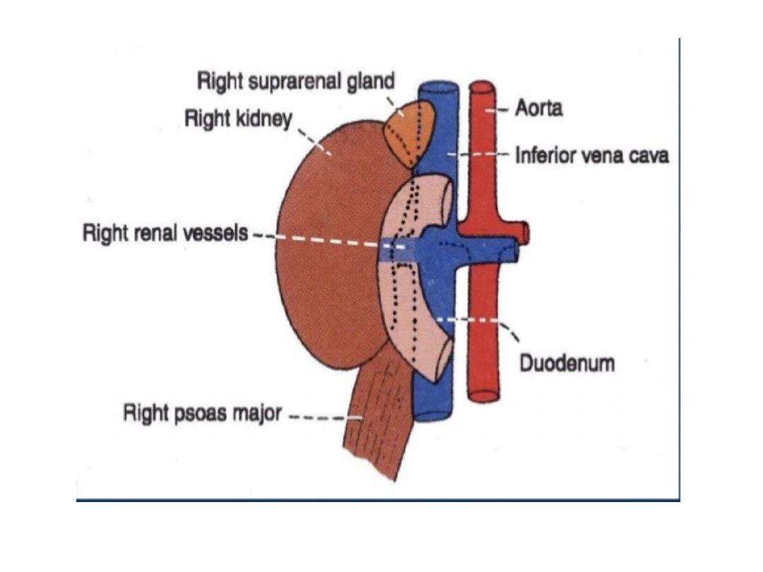
(B) Posteriorly
-1-The hilum of Rt. Kidney
2. Rt. Renal vessels
3-Rt. Psoas major muscle
4-Some times part of the RT supra renal gland
5-Rt edge of inferior vena cava
(C) Laterally
1. Fat infront of the right kidney
2. Rt. Colic flexure ( hepatic flexure )
3. ascending colon
D) Medially:
1. Head of pencreas
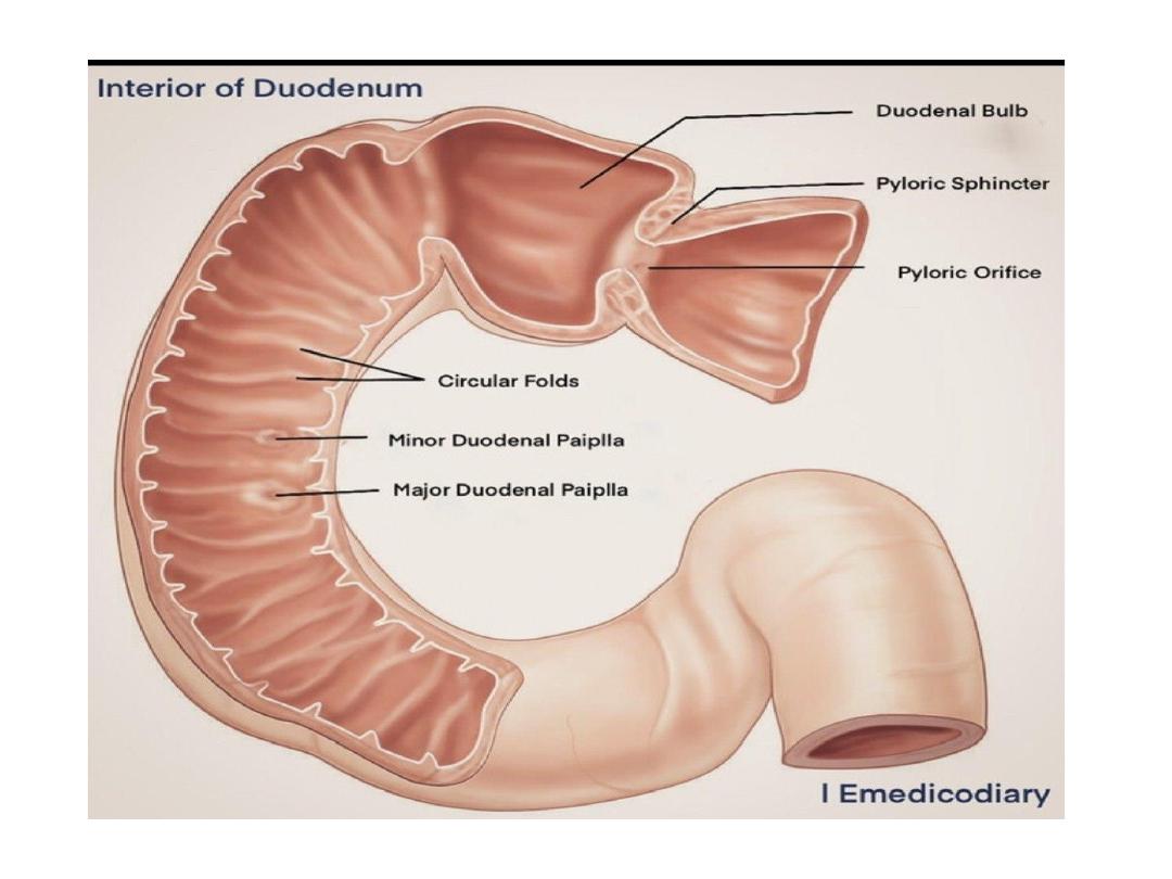
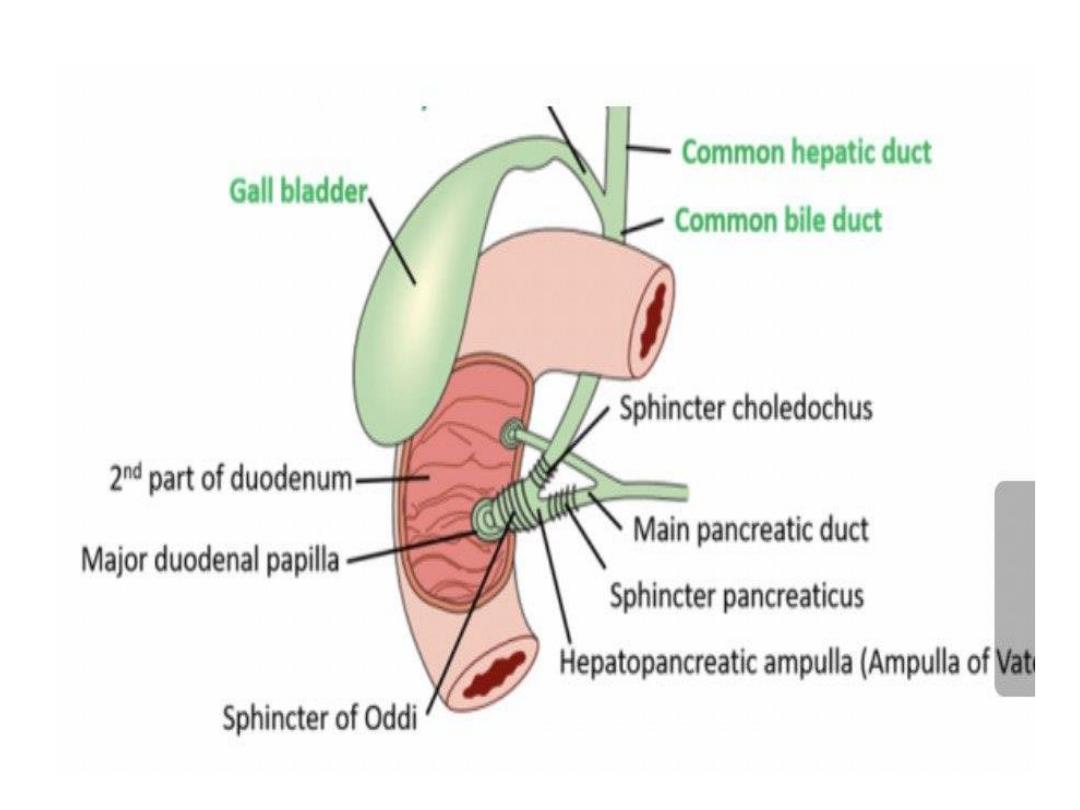
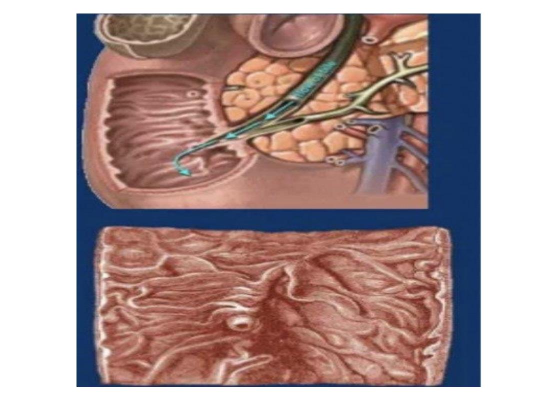
Interior of the second part of duodenum
Two elevations are presents along the posteromedial aspect
1_ major duodenal papilla
-It is conical elevation situated 8-10 cm from pyloric end of the
stomach
-Ampulla of Vater ( which formed by union of bile duct and main
pancreatic duct ) open to it , the opening is guarded by sphincter
of Oddi
2-minor duodenal papilla
is a small conical elevation about 2
cm above the major papilla and the accessory pancreatic duct
open to it .
Note : Sup. & Inf. Pancreatico-duodenal vessels anastomosis
in the groove between head of pancreas & 2
nd
part of
duodenum
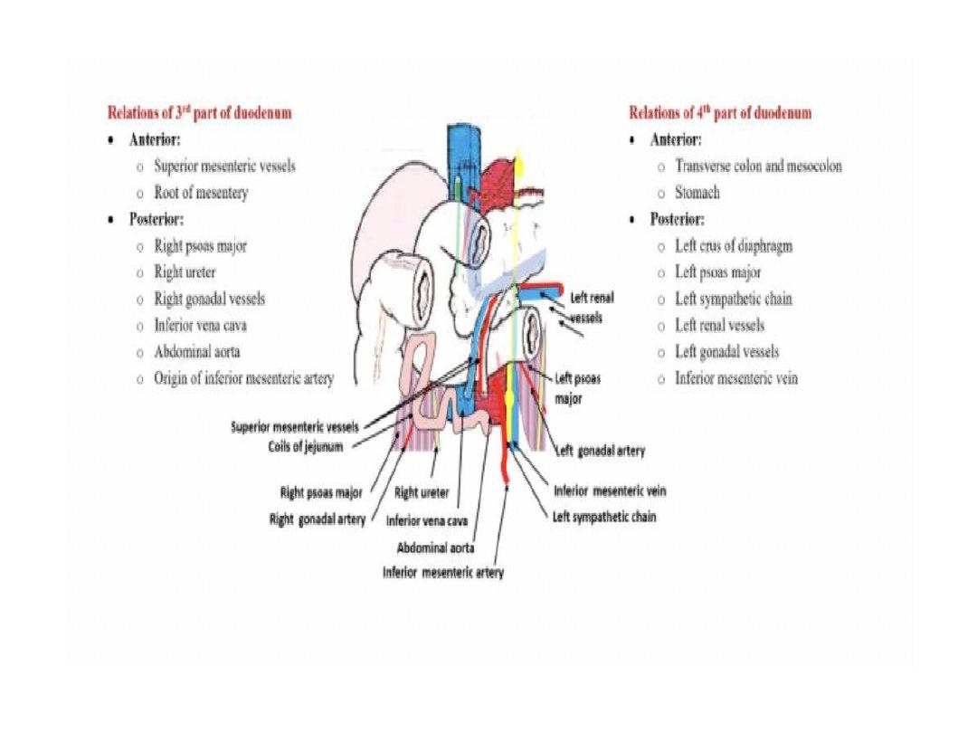
3
rd
Part of Duodenum
Length:
it is the longest part (4 inches long).
Course:
it passes horizontally form Rt. To Lt. at
the level of L3 vertebra.
Peritoneum:
it is covered by peritoneum
anteriorly and inferiorly
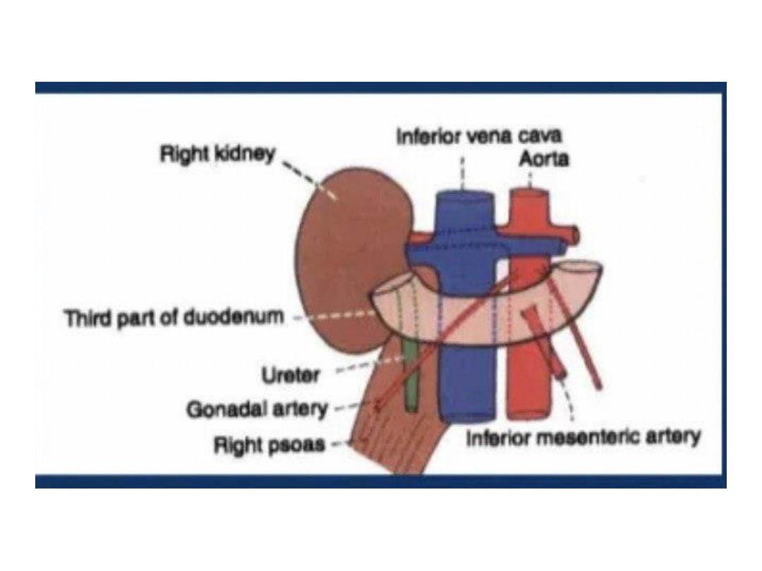
Relations:
A. Superiorly:
head of pancreas and uncinate
process
B. Inferiorly:
Coils of jejunum
C. Anteriorly:
1. Coils of jejunum
2. Root of mesentery
3. Superior Mesenteric vessels
D. Posteriorly (from right to left):
1-Rt. Psoas major muscel.
2-Rt. Ureter.
3. Inferior vena cava
4-Rt. Gonadal vessels
5. Abdominal aorta
6-the origin inferior . Mesenteric artery.
4
th
part of Duodenum
Length:
it is the shortest part (one inch long)
Course:
it ascends along the Lt. side of the
vertebral
column (from L3 to L2)
Extend from the front of the aorta to
duodenojujenal flexureon the Lt. side of L2
-kept in position by suspensory muscle of
duodenum .
Peritoneum:
it is covered by peritoneum
anteriorly & to the left
.
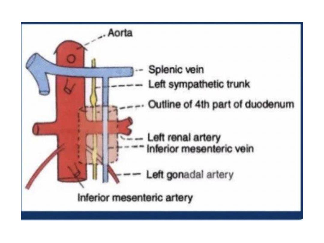
Relations of the 4
th
part ..
Anteriorly :
1-
transverse colon and mesocolon
2-posteroinferior surface of stomach .
(
Posteriorly:
1-LT crus of diaphragm
2-left. psoas major Muscle.
3. Lt. sympathetic trunk
4-Lt. renal vessels
5-Lt Gonadal vessels
6-Lt supra renal vein
7-Inferior mesentric vein

RT side
• Uncinate process of pancreas
LT side
• LT kidney and ureter
Superiorly
• Body of pancreas
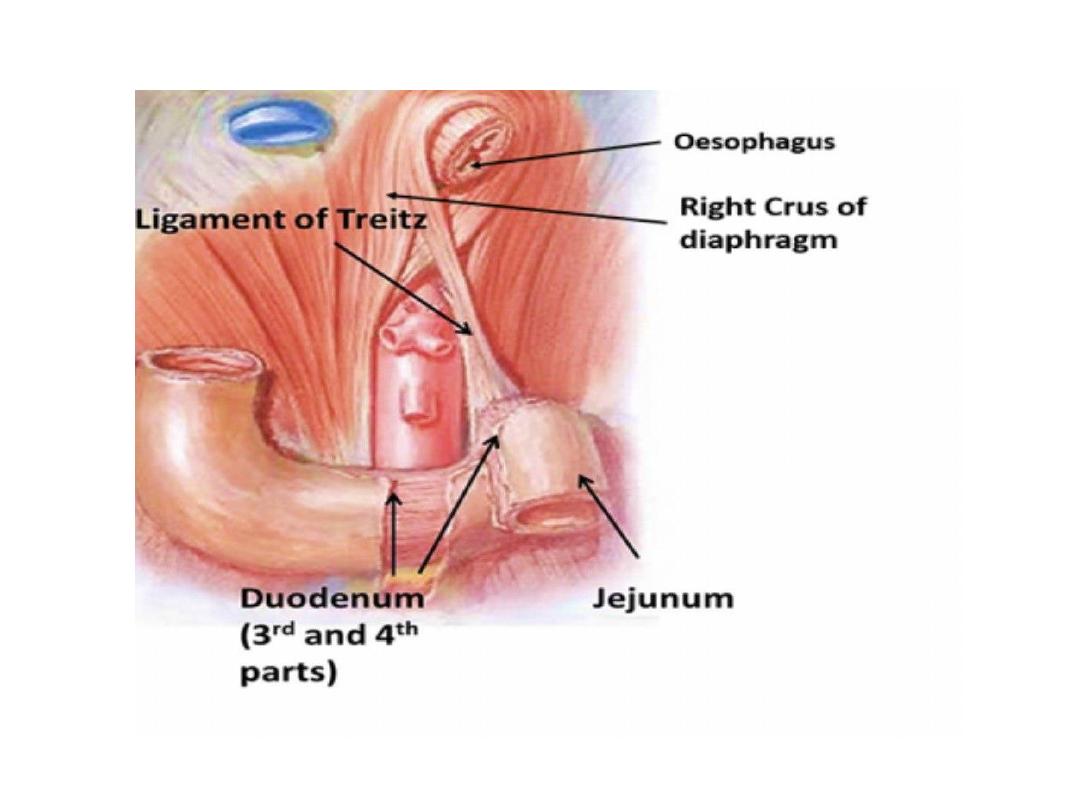
Suspensory muscle of duodenum
(Lig. Of Treitz)
- It is a fibromuscular band which suspends the
duodenojejunal flexure.
- It
arises
from the Rt. Crus of diaphragm close to
the RT side of the esophagus.
- It
descends
behind the pancreas to be attached to
posterior surface of the duodenojejunal flexure &
the 3
rd
& the 4
th
parts of duodenum.
- Upper third consist of striated muscle , middle
third of elastic fiber , and lower third of smooth
muscle

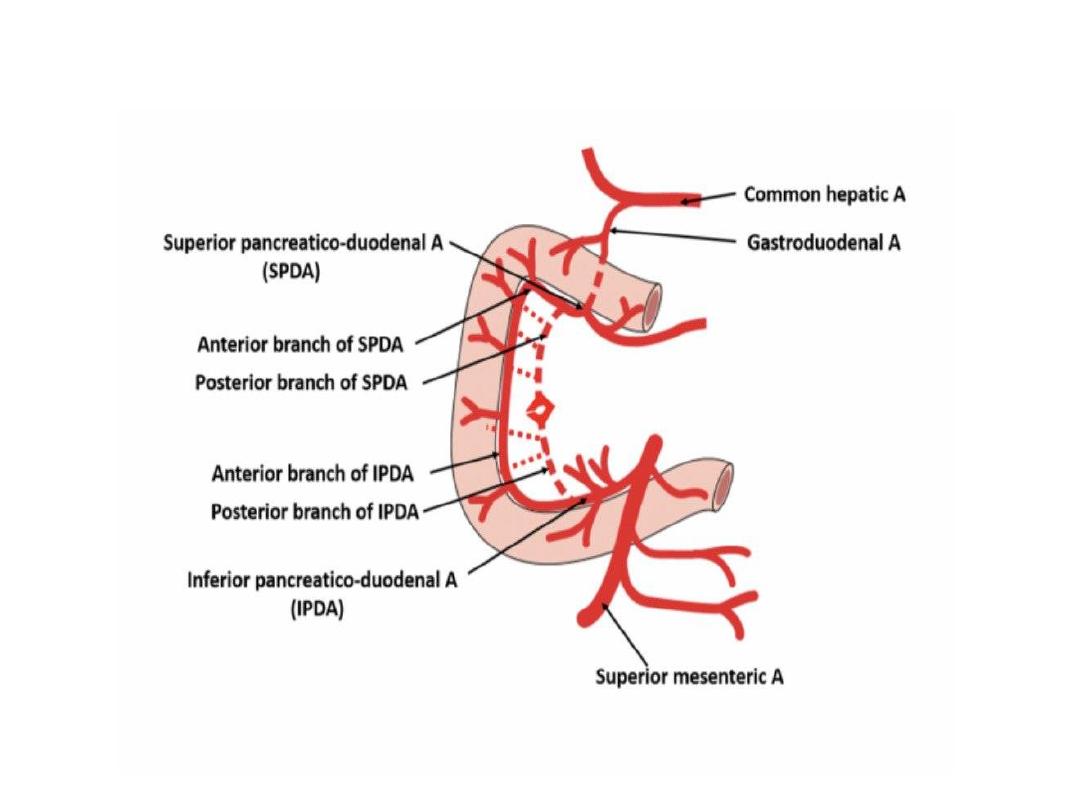
Arterial blood supply to the duodenum
1)
above the level of bile duct opening ( first part
and mid of second part )
• Supplied by anterior and posterior branches of
superior pancreaticoduodenal artery …branch
from gastroduodenal artery
2) below the level of bile duct opening ( from
mid D2 to ligament of treitz )
• Supplied by anterior and posterior branches of
inferior pancreaticoduodenal artery ..branch
from superior mesentric artery

The duodenal cup ( first 2.5 cm of the
duodenum ) ..receive additional supply
from:
1-RT gastric artery
2-RT gastro epiploic artery
3-Retero duodenal artery ( branch from
gastro duodenal artery )
4-Supra doudenal artery ( branch from
hepatic artery )

Venous drainage
1
) Proximal part
Superior pancreatico duodenal vein …drain to Rt
gastroepoploic vein and portal vein
2) Distal part
Inferior pancreatico duodenal vein ..drain to
superior mesenteric vein
Lymphatic drainage :
Pancreatrico dudenal lymph nodes …to
superior Mesenteric & hepatic lymph
nodes…to .celiac Lymph Node.
• Nerve supply :
• sympathetic nerves … via celiac plexus
• Parasympathetic anterior and posterior
vagal trunk

Applied anatomy
-First part of the duodenum is the commonest
site of peptic ulcer
-Commonest site of perforation is anterior
surface of first part
-Third part of the duodenum is vulnerable to
injury as it lies anterior to vertebral column

Lecture -5-
Anatomy of small intestine
( part II )
Dr. Raya AbdulAmeer
MBCHB,CABHS-RAD

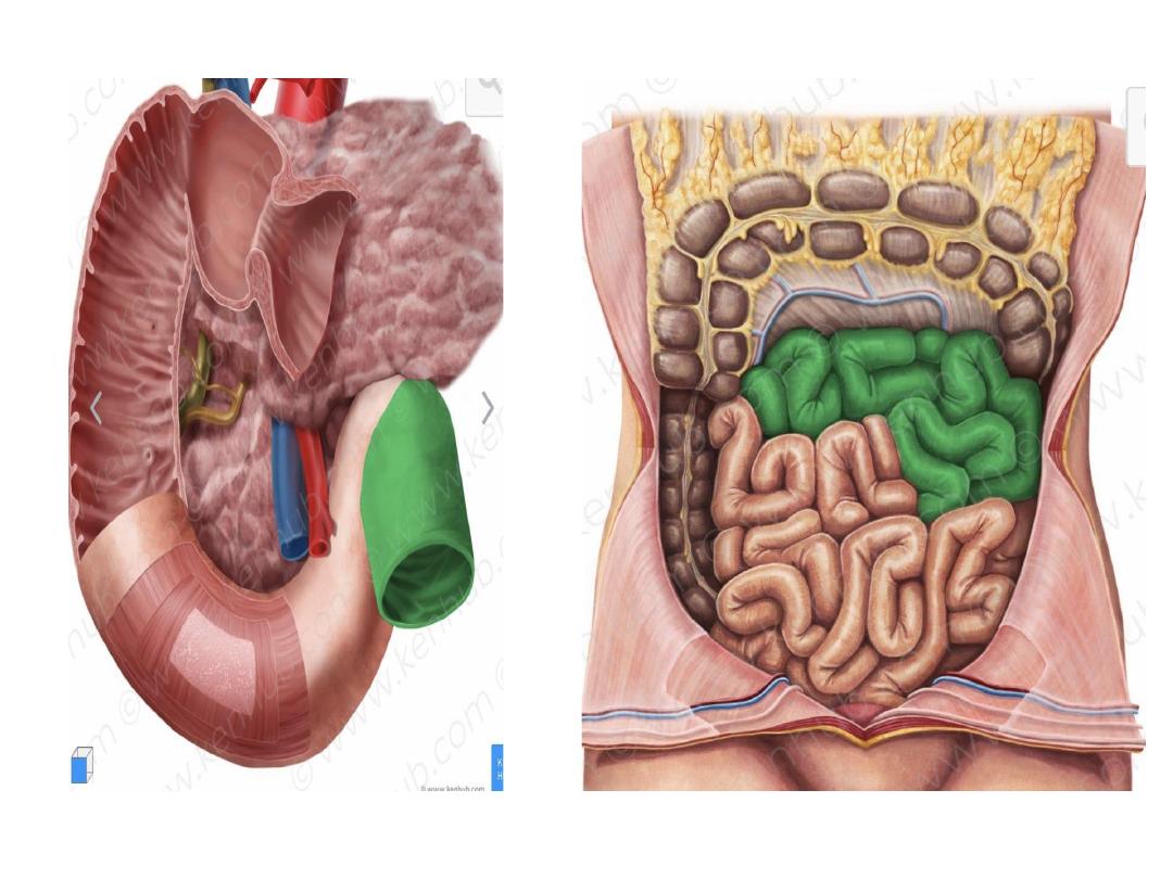
The jejunum
• The jejunum is the second part of the small
intestine.
• It begins at the duodenojejunal flexure and
ends at ileum -is found in the
• The jejunum is entirely intraperitoneal as the
mesentery proper attaches it to the posterior
abdominal wall.
• It is about 2/5 of total length of small intestine
( 1.5-3.5 meters )

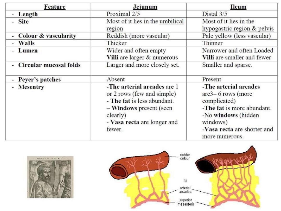
• There is no clear line of demarcation between
the jejunum and ileum, but there are some
anatomical and histological differences that
distinguish them:
• The jejunum represents the proximal two-fifths
of the jejunum-ileum continuum
• The wall of the jejunum is thicker and its lumen
is wider than in ileum
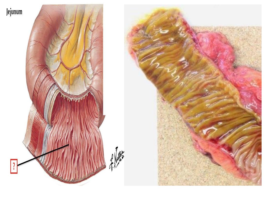
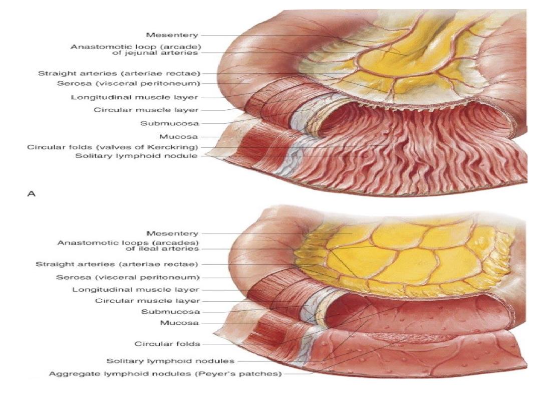
• The jejunum contains more prominent circular
folds in the mucosa called ( valve of
Kerckring) or (plica circularis ) or ( vulvulae
conniventes)

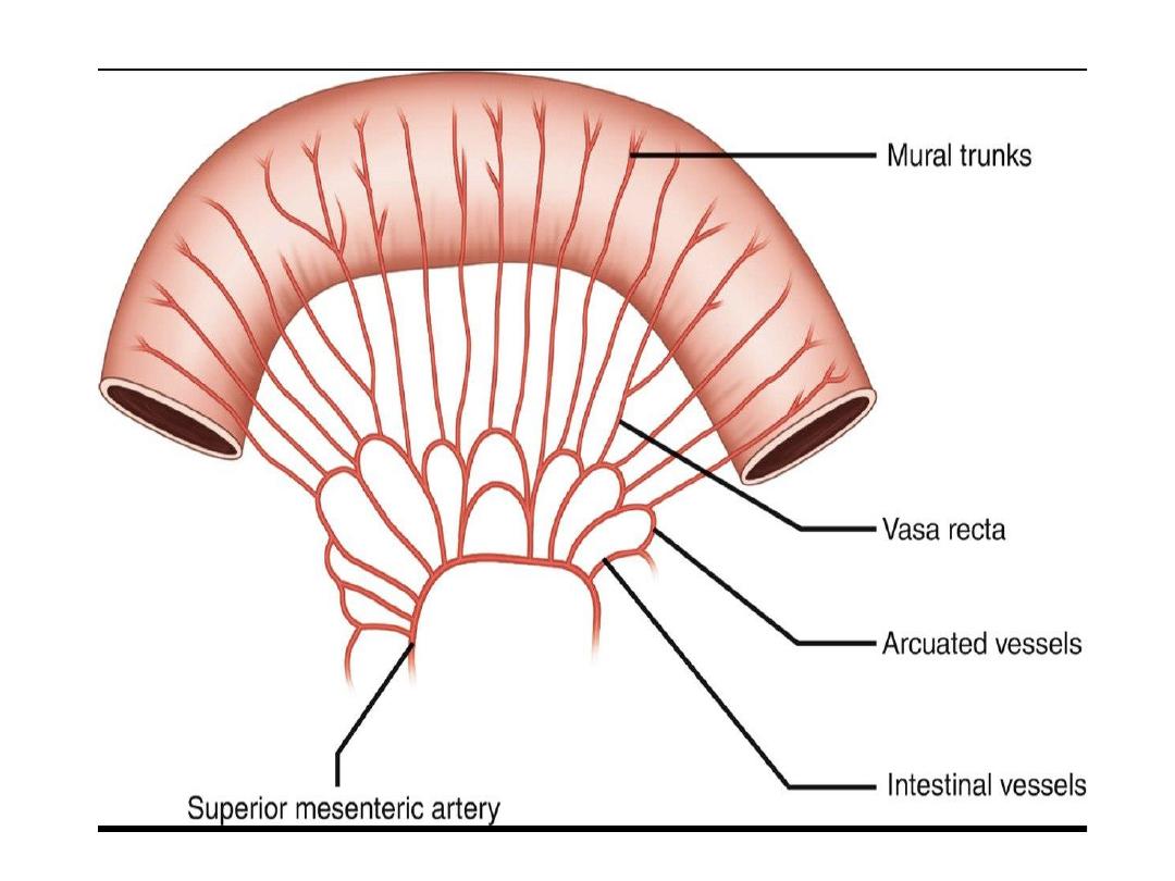
• Arterial supply
• Jejunal branches ( about five branches )
from superior mesenteric
artery
with
arterial blood. These form arcades with the
other arteries of the small intestin
e
.
Venous drainage
Corresponding to arteries and drain to
superior mesenteric vein
Lymphatic drainage
To superior mesenteric Lymph nodes

Nerve supply
• Sympathetic
Spinal segments T9-T10 , celiac plexus and
superior mesenteric plexus
• Para sympathetic
Vagus nerve

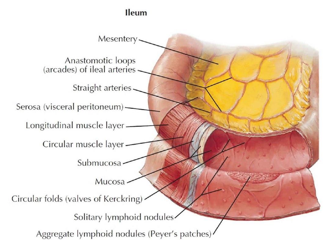
Anatomy of the ileum
• Last of the three parts of the small intestine, found
between the jejunum and large intestine
• Its completely intraperitoneal
• At the distal end, the ileum is separated from the
large intestine, into which it opens, by the
ileocecal valve.
• The ileum itself is very rich in lymphoid follicles
• and is attached to the posterior
abdominal
wall
by the

• The ileum makes up about 3/5 of the total
length of the small intestine (2.5 to 3.5
meters).
• Compared to the jejunum, the parallel
running circular folds in the mucosa
(valves of Kerckring) are less prominent.
• it is rich in lymphoid follicles( pyres
patch
)

Arterial supply of ilieum
• About twelve ileal arteries called straight
arteries (branches of the superior mesenteric artery )
supply the ileum with arterial blood. These form
arcades with the other arteries of the small intestine
.
• Venous drainage
• Superior mesenteric vein
Nerve supply :
• Sympathetis
… coeliac plexus and the superior
mesenteric plexus
• Para sympathetic
• vagus nerve (cranial nerve X
)
.
Lymphatic drainage
To superior mesenteric Lymph nodes
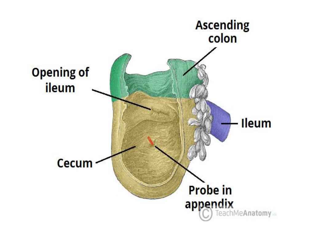
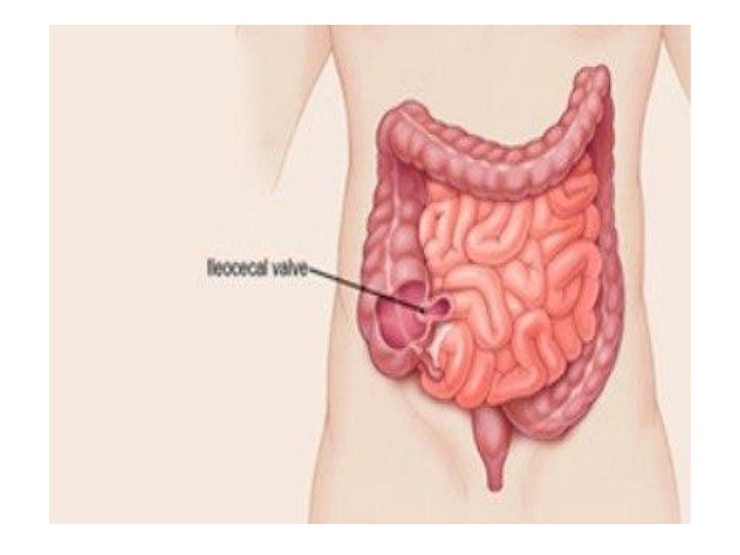
• It lies at the junction between the ileum
and the cecum
• a functional sphincter formed by the
circular muscle layers of both the ileum
and cecum.
Function: it regulates the passage of
ileal contents into the cecum & prevents
reflux from cecum to ileum.
Ileocecal valve

Pyers. Patches
• Are group of lymphoid follicles that found in
the mucous membrane of the small bowel (
ileum )
• Has important role in immune function
• One patch is around 2 to 5 centimeters long
and consists of about 300 aggregated lymphoid
follicles
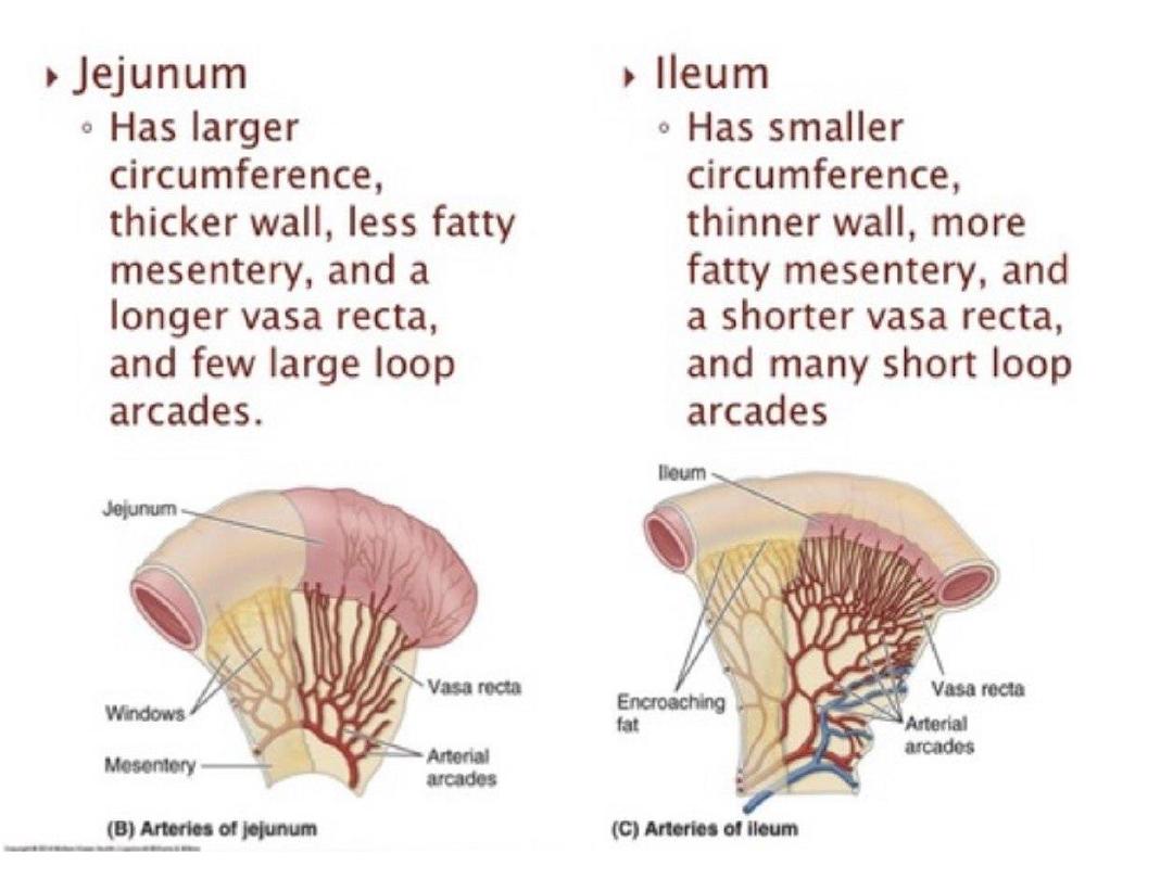

Mesentric window :
• Thin translucent membrane in between the
artery /vein pairs feeding the small intesti
ne

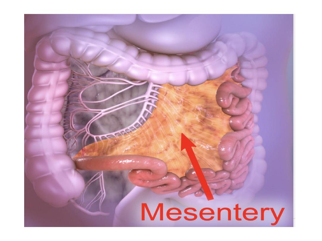
Anatomy of The mesentery
• It is a double fold of peritoneal tissue that
suspends the small intestine and large intestine
from the posterior abdominal wall
Function
The mesentery has several functions in the
abdomen:
1-Suspends the small and large intestine from the
posterior abdominal wall; anchoring them in place,
whilst still allowing some movement.
2-Provides a conduit for blood vessels, nerves
and lymphatic vessels

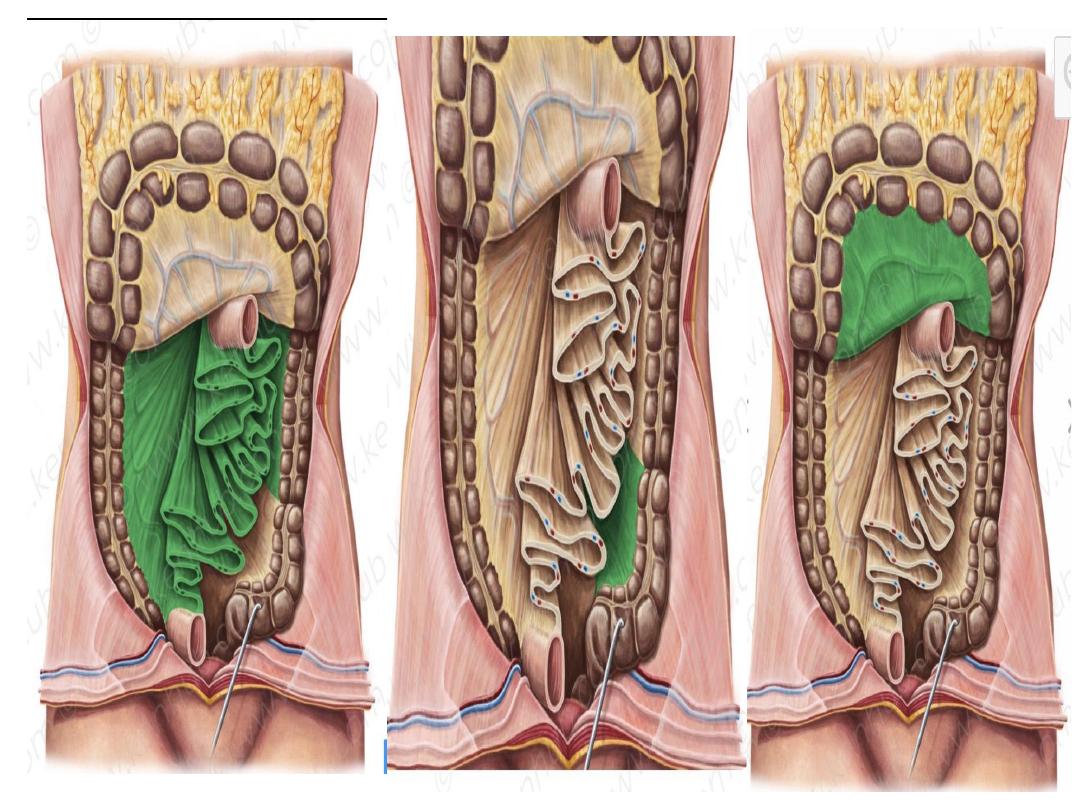
Three mesentries
1-Mesentery proper ( small bowel mesentery )
• From jejunum and ilium to the posterior
abdominal wall
2-Transverse mesocolon
• From transverse colon to posterior abdominal
wall
3-Sigmoid mesocolon
• From sigmoid colon to posterior abdominal
wall –pelvic wall

Mesentery of Small Intestine
It is a peritoneal fold enclosing the free part of the
small (jejunum & ileum) & connecting it to the post.
Abdominal wall.
Shape:
fan-shaped fold having broad free border &
narrow attached border:
(a) Free border
: is 6 meters (20feets) long & encloses
the jejunum & ileum.
(b) Attached border:
(Root of mesentery): 6 inches
long & 6 inches away from the free border
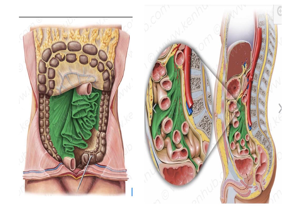

-It is attached to the posterior . Abdominal
wall extending from the duodenojejunal
flexure (on the left side of L2) to ileocecal
junction (above the Rt. Sacroiliac joint).-
-
-
The root crosses 6 structures on the
posterior . Abdominal . wall
.
-
1) two parts of duodenum( 3
rd
and 4
th
parts)
2) two large vessels ( abdominal aorta and
inferior vena cava )
3)two muscles ( RT iliacus muscle and RT
psoas major muscle )
:

Contents of the mesentery:
1.
Coils of jejunum & ileum in the free
margin of the mesentery.
2.
Superior mesenteric artery & its branches(
jejunal and ilial branches )
It runs downwards & to the Rt. In the root of
mesentery
3.
Superior . Mesenteric Vein. & its
tributaries:
- Runs in the root of mesentery on the Rt.
Side of the superior . Mesenteric artery

4.
Lymphatics & 3 raws of mesenteric
Lymph nodes .
(a) Small lymph nodes near the
intestine in the free border.
(b) Medium-sized L.Ns in the middle
of the mesentery
(c) Large L.Ns: lie along the superior
. Mesenteric vessels
5-
plexuses of autonomic nerve fibers
around the arteries.
6.
Extraperitoneal fatty tissue