Lecture -6-
Anatomy of large
intestine
Dr.Raya Abdul Ameer
MBCHB,CABHS-RAD

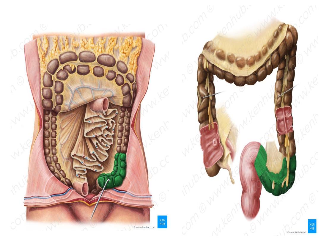
• The large intestine, also known as the large
bowel, represents the last part of the
gastrointestinal tract .,
• it has a length of approximately 1.5 meters,
• extending from the ileocecal junction to
the anus.
• Most of the large intestine is located inside the
abdominal cavity, with the last portion residing
within the pelvic cavity.
• Some parts of it are intraperitoneal while
others are retroperitoneal.

•
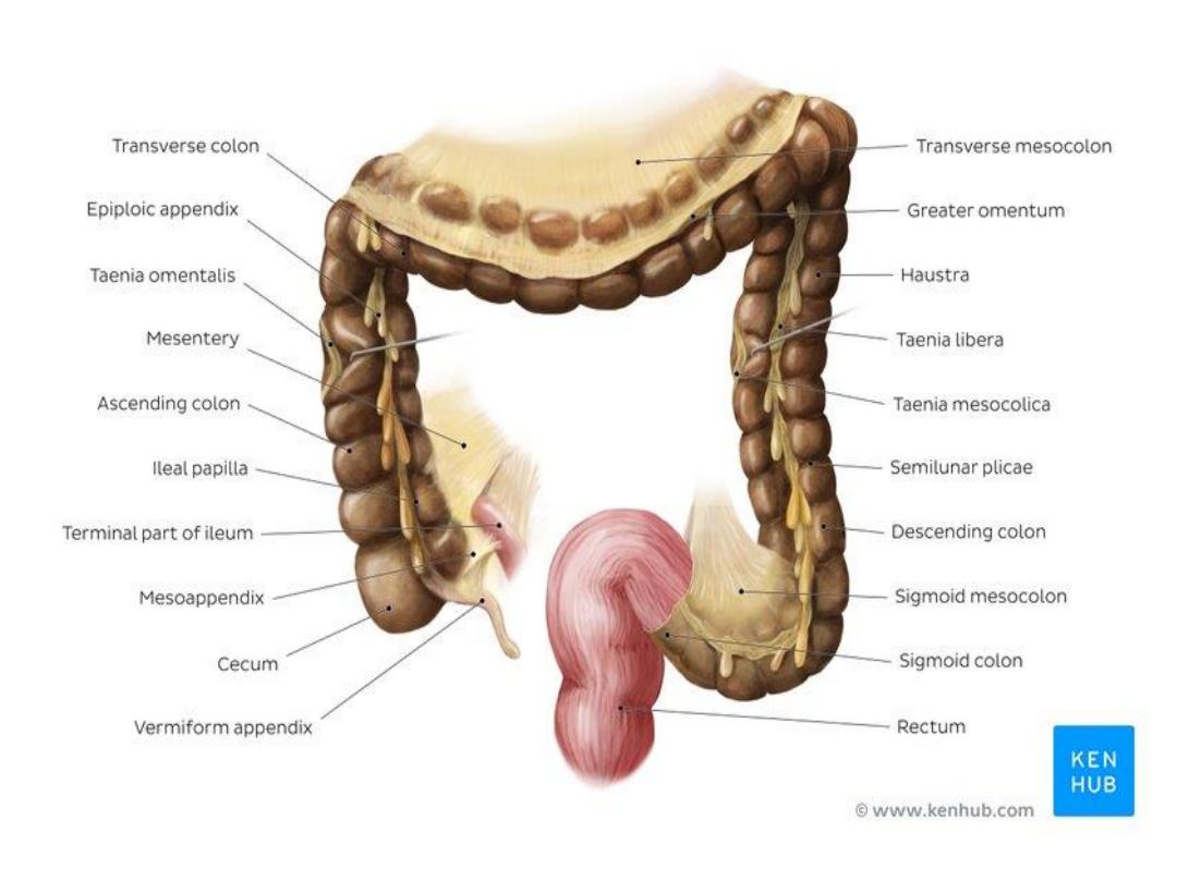
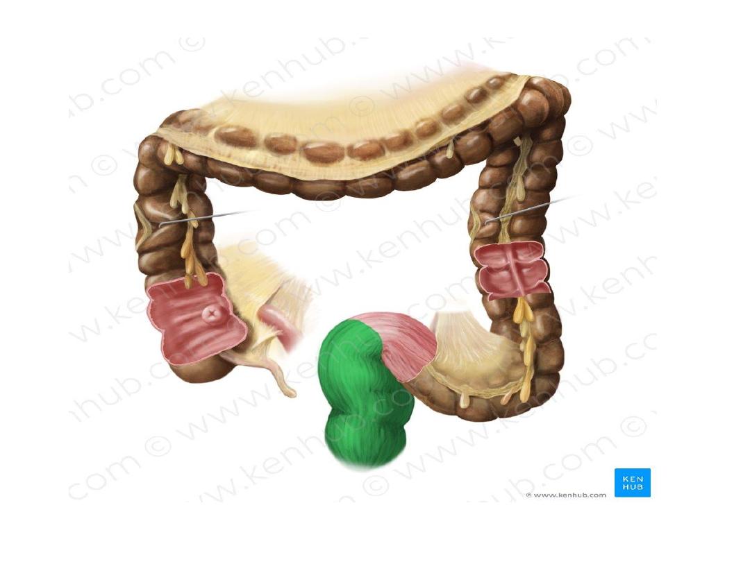
Features specific to the large intestine
1) Omental or epiploic appendages
are fat filled
pouches of peritonium that are attached externally
to the walls of the large intestine.
2) Teniae coli
are three longitudinal bands of smooth
muscel located underneath the peritoneum that
extend along certain sections of the large intestine.
Their contractions facilitate the peristaltic action of
the large intestine, propelling the fecal matter and
forming the haustra.
3)
Haustra are sacculations
that occur along the large
intestine, providing it with its characteristic ‘baggy’
aspect. They are created by semilunar folds on the
internal surface of the large intest
ine.

• The large intestine consists of eight parts;
the cecum , appendix , ascending colon,
transverse colon, descending colon, sigmoid
colon , rectum and anal canal
• The middle four sections (ascending to
sigmoid parts) form the
.

Cecum
• The cecum is the first part of the large
intestine, lying in the right iliac fossa of
the abdomen
• Shape
blind pouch 3 inch in length
• The cecum is intraperitoneal with various folds
and pockets (retrocecal peritoneal recesses)
surrounding it
• The terminal ileum joins the cecum at
the ileocolic junction.
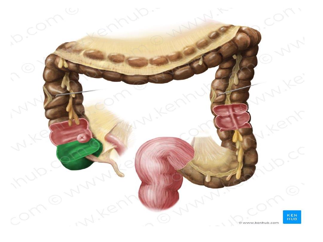

Appendix
• The vermiform appendix is a blind lymphoid
pouch located in the right iliac fossa which arises
from the cecum.
•
Size
½
-9 inch average 4 inch it’s the narrowest part
of GIT
• connected by the meso-appendix.
• The appendix has a role in the maintenance of
gut flora and mucosal immunity.
• It open in to the cecum at posteromedial aspect of
cecum , 1inch below iliocecal junction
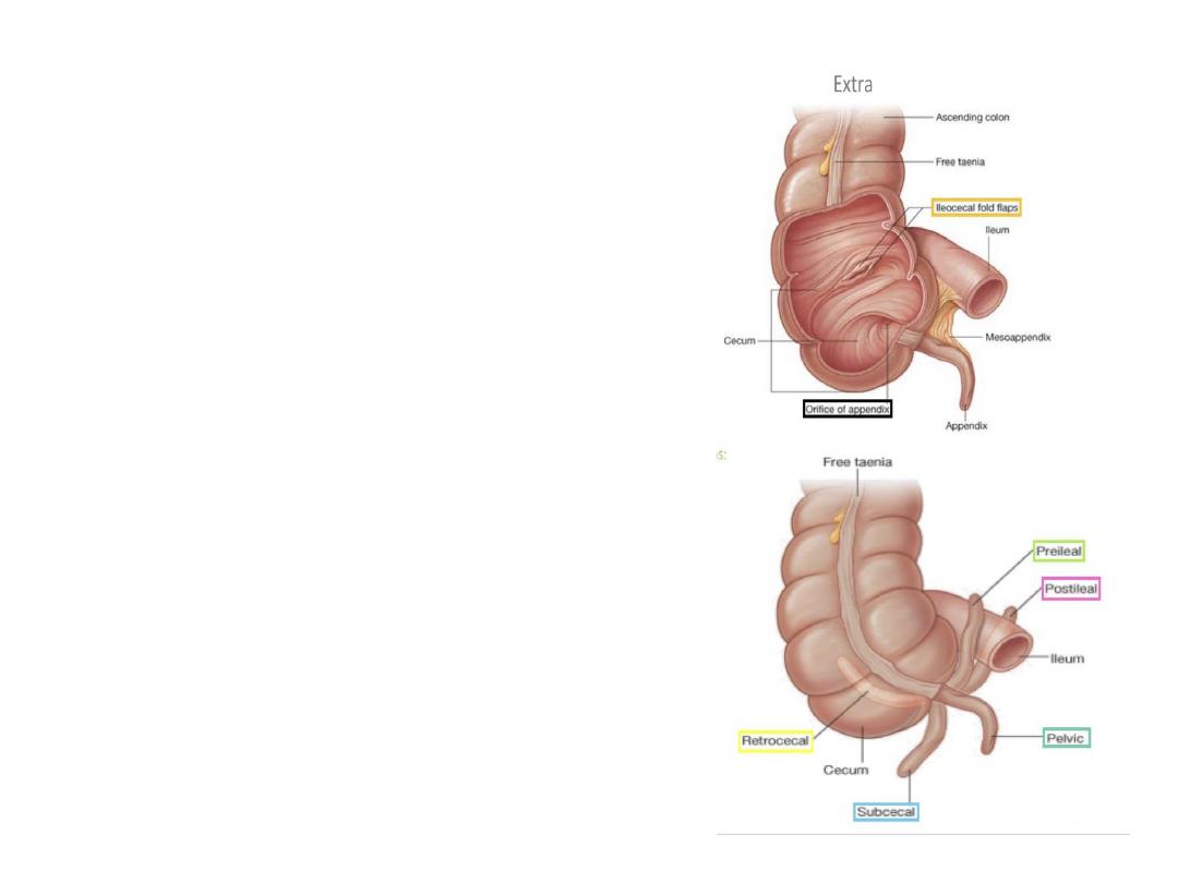
• Posiotions of appendix
• 1-retrocecal ..most common
• 2-pelvic
• 3-subcecal
• 4-perilial
• 5-post ilial ..least common
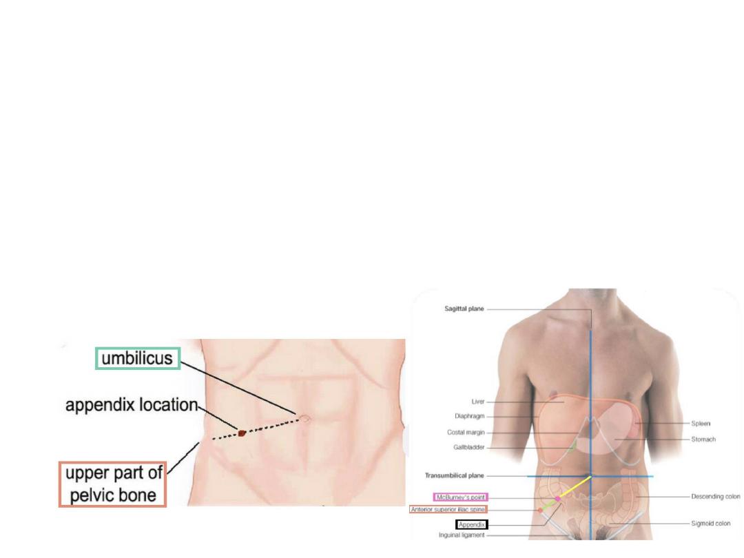
Surface anatomy
• The base of appendix is marked by
McBurneys
point
: a point at the junction of lateral 1/3 and medial 2/3
of a line traced from RT anterior superior iliac spine
to umbilicus
• Importance
:tenderness and rebound at this point is
suggestive of appendi
citis

Ascending colon
• The portion of the large intestine located
between the cecum and rectum is termed
the colon.
It consists of four parts; ascending,
transverse, descending, and sigmoid
.
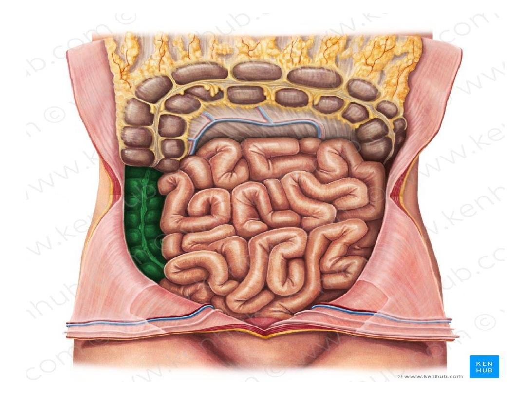

• The ascending colon travels through the right
iliac fossa, Right flank , and Right
hypochondrial region
• It ends at the right colic (hepatic) flexure.
• The ascending colon is retroperitoneal and it is
connected to the posterior abdominal wall by
the Toldt’s fascia
.
• A deep vertical groove or recess (
right
paracolic gutter)
lies between the ascending
colon and the lateral abdominal wall.
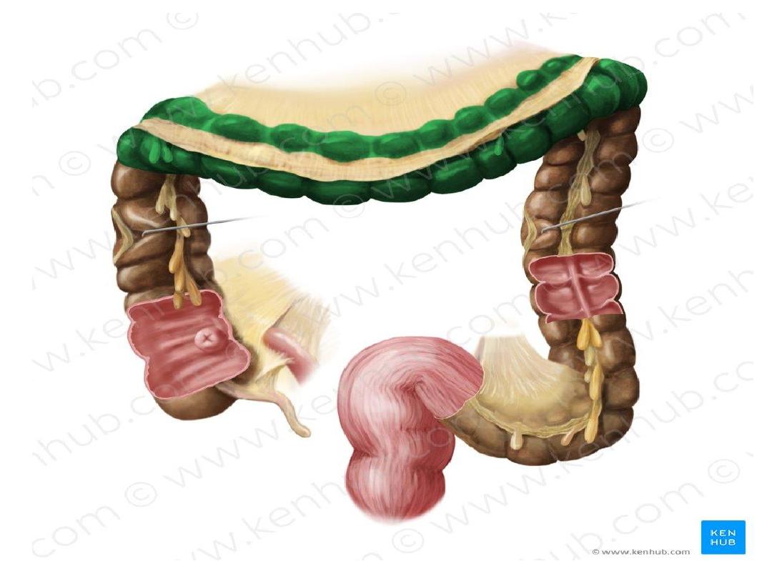
Transverse colon
• The transverse colon is the second major part of the
colon.
• It extends between the right and left colic (splenic)
flexures, spanning the right hypochondriac
, epigastric and left hypochondriac regions of the
abdomen.
• The greater curvature of
the stomach and gastrocolic ligamnet are superior
to the transverse colon, while the greater
omentum hangs over and extends inferiorly to it.
• The transverse colon is intraperitoneal. A peritoneal
mesentery (transverse mesocolon) attaches it to the
posterior wall

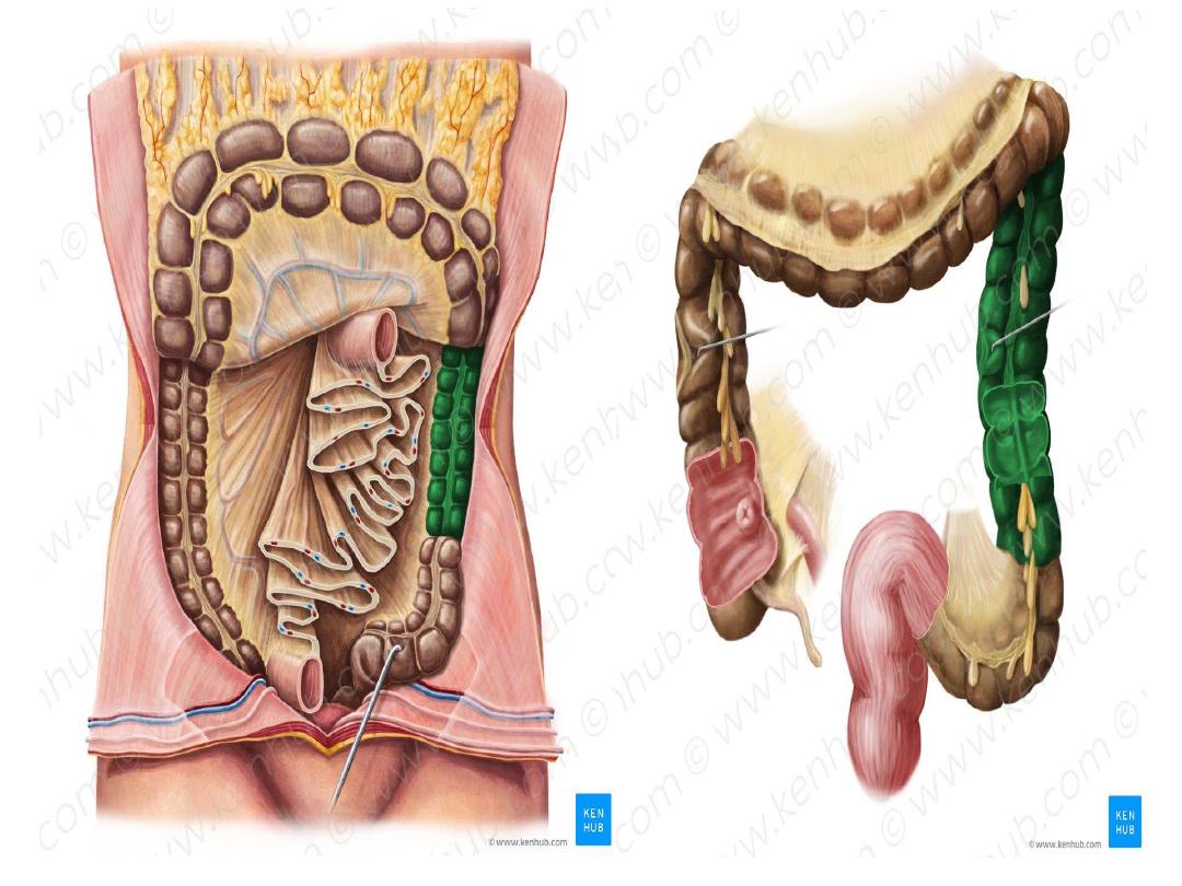
Descending colon
• The descending colon extends between the left
colic flexure and sigmoid colon.
• It travels through the left hypochondriac
region, left flank and left iliac fossa.
• The left paracolic gutter is located between the
descending colon and the lateral abdominal
wall.
• This part of the colon is retroperitoneal
. Toldt’s fascia fixes the descending colon to
the posterior abdominal wall.

Sigmoid colon
• The S-shaped sigmoid travels from the left
iliac fossa until the third sacral vertebra
(rectosigmoid junction).
• This part of the colon is intraperitoneal. It is
connected to the pelvic wall by the sigmoid
mesocolon.

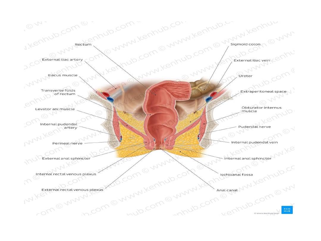
Rectum
The rectum stretches between the rectosigmoid
junction and the anal canal. The typical
characteristics of the large intestine (taenia coli,
haustra, epiploic appendages) change or even
terminate at the rectum.
The rectum has a characteristic S-shape marked by
several bends or turns; sacral, anorectal and lateral
flexures. The latter correspond with three infoldings
called transverse rectal folds.
The rectum ends at a dilated ampulla
.

• The rectum is partially intraperitoneal since the
inferior third is subperitoneal.
• The peritoneum reflects from the rectum
towards the bladder in males (rectovesical
pouch) and the vaginal fornix in females
(recto-uterine pouch or pouch of Douglas).
• The spaces around the rectum are potential
spaces for infections, abscess formation, and
many other pathologies
.

Anal canal
The anal canal forms the terminal part of the
gastrointestinal tract.
It extends from the anorectal junction to the anus.
The anus represents the external orifice of the
entire digestive system.
.
the pectinate line / dentate makes the distinction
between the superior and inferior parts of the anal
canal. They differ in terms of neurovascular
supply and lymphatic drainage
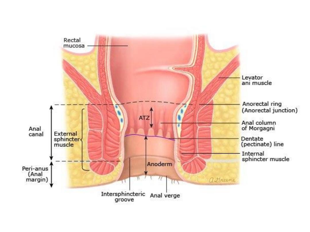

Respectively, they involuntarily and
voluntarily control the release of stool. Both
sphincters are tonically contracted to prevent
the uncontrolled release of fecal matter or
flatus
.

Peritoneal covering
Parts with mesentery :
1-transverse colon
2-sigmoid colon
3-appendix
4-cecum
retroperitoneal parts
1-ascending colon
2-descending colon
3-upper 2/3 of rectum

Parts devoid of peritoneal covering :
• 1-lower 1/3 of the rectum
• 2-anal canal

Relations of cecum , ascending and decsending
colons
Anterior
1-greater omentum
2-coils of small intestine
3-anteror abdominal wall

Posterior relations
• Cecum
..psoas major and iliacus
• Ascending colon
..iliacus ,quadratous
lumborium and Right kidney
• Descending colon
…Left kidney , quadratous
lumborium , iliacus , psoas major

Relations of transverse colon
Anterio
r ..greater omentum and anterior
abdominal wall
Posterior
..second part of duedenum , pancreas,
superior mesentric vesseles
Superior
..liver ,gall bladder ,stomach
Inferior
…coils of small intestine

Relations of rectum
Anteriorly
..
• Male …seminal vesicle , posterior surface of
urinary bladder and prostate gland
• Female ..posaterior wall of the vagina
Posteriorly In both male and female
• Sacrum ,sacral plexus and coccyx

ArterialBlood supply
The large intestine receives arterial blood
predominantly from the
The SMA supplies the midgut derivatives, such
as the cecum, appendix, ascending colon and
the proximal two-thirds of the transverse colon
via three main branches: ileocolic,
,
and middle colic arteries

• The inferior mesenteric artery supplies
the hindgut derivates, namely the posterior
third of the transverse colon, descending colon,
sigmoid, colon, rectum and the upper part of
the anal canal via three branches:
,
sigmoid, and
.
• The middle and
, which
stem from the
, also supply
hindgut derivates

Cecum
Iliocolic artery
Appendix
Appendicular artery
Ascending colon
Ileocolic and right colic arteries
Transverse colon
Middle colic artery and LT colic artery
Descending colon
Left colic artery
Sigmoid colon
Sigmoid artery
Rectum
Superior part: superior rectal artery
Middle & inferior parts: middle rectal artery
Anal canal
Superior to pectineal line: superior rectal
artery
Inferior to pectineal line: inferior rectal artery

Venous drainage
• The midgut derivates drain first into the colic
veins, which in turn empty into the
• Hindgut derivates flow directly into the
inferior mesenteric vein.
• The rectum has a special venous
drainage. Middle rectal and inferior rectal
veins drain this part of the gastrointestinal tract
into the internal iliac and internal pudendal
veins, respectively.

Cecum
Ileocolic vein
Appendix
Ileocolic vein
Ascending colon
Right colic vein
Transverse colon
Superior mesenteric vein
Descending colon
Inferior mesenteric vein
Sigmoid colon
Inferior mesenteric vein
Rectum
Superior, middle, inferior rectal veins
Anal canal
Superior to pectinate line: superior rectal vein
Pectinate line: internal rectal venous plexus
Inferior to pectinate line: inferior rectal vein

Nerve supply
Parasympathetic innervation
• The
provide parasympathetic
innervation to the large intestines.
• The
(S2-4) also
contribute to the large intestines’ parasympathetic
supply.
• The vagus nerve fulfils this role in the gut to the
point of the transverse colon, while the pelvic
splanchnic nerves carry on this function from the
left colic flexure down wards.

Sympathetic innervation
• The T10-L2 thoracolumbar outflow of
fibers
They form synapses at the superior and inferior
mesenteric, and the inferior hypogastric plexuses.
•
The superior mesenteric plexus provides sympathetic
innervation to the cecum, appendix, ascending and
transverse colon (near to the left colic flexure),
• while the inferior mesenteric plexus innervates the colon
from the left colic flexure to the
.
• The inferior hypogastric plexus also innervates the rec
tum

Lymphatic drainage
• Throughout the large intestines, the lymph nodes are
arranged in four general groups.
• The first group
is epicolic
nodes that rest on the outer
surface of the intestinal wall.
• The second is
paracolic
nodes that lie along the
intestinal margin.
• Thirdly, there
are intermediate nodes
that lie along (and
conveniently take the names of) the arterial branches of
the superior and inferior mesenteric arteries.
• Finally, there are the
preaortic nodes
that are found at
the branching point of the coeliac trunk and the
superior and inferior mesenteric arteries.
• Clinical notes