• Immunological Techniques in Research and Clinical Medicine
• Philip L. Cohen, M.D. Chief of Rheumatology, LKSOM• Presented by
• Dr. Minen Al-Kafajy
• December-18-2021
• Antibodies – Remarkable Tools for Research and Diagnosis
• You can make an antibody to practically any immunogen.• Monoclonal antibodies have a single specificity, so the immunogen need not be pure (e.g., whole cells or lysates)
• Antibodies are stable (decades at ‐20C!)
• They can be covalently coupled to enzymes, chromophores, biotin, and many other things..
Albert Coons, M.D.
• 1912‐1978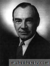
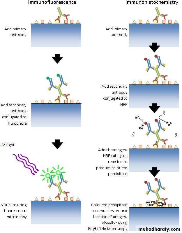
• Coons’ Insight
• Antibodies can be “tagged” with small fluorescent molecules and still retain their binding specificity• These “tagged” antibodies can be used as probes to visualize specific molecules in tissues, cells, or anywhere
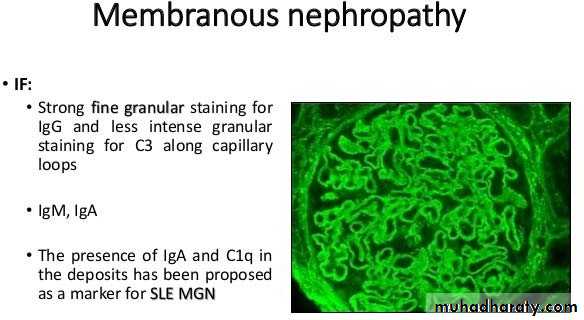
SLE Systemic lupus erythrematosus
MGN membranous glomerulonephritis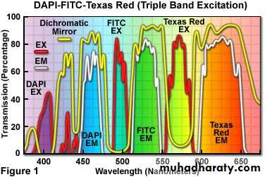
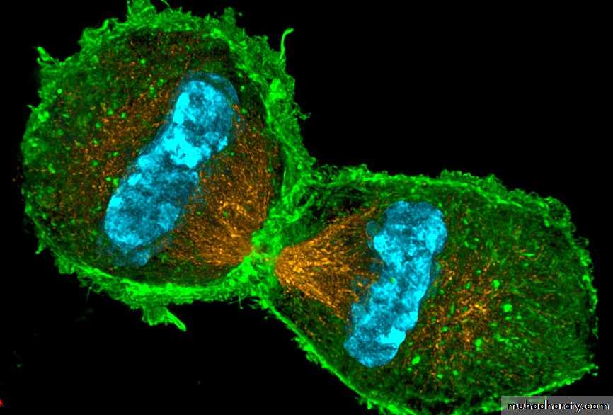
• Multiple Color Immunofluorescence
للاطلاع• Some Considerations for Fluorescence Staining
• Usually requires freshly snap‐frozen tissue• Conjugated antibodies are less stable than native molecules, are light‐sensitive
• For fixed tissues, immunohistochemistry is preferred
• Can be quantitated using laser capture fluorescence microscopy
• Confocal and dual photon microscopy
• Methods for Diagnosis
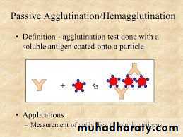
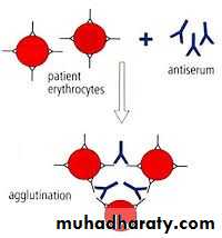
• Immunodiffusion (Ouchterlony)• Hemagglutination and latex agglutination
• Complement fixation
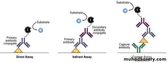
• ELISA
• Western Blot and other electrophoretic methods
• Antibodies as Tools for Quantitating Proteins and Almost Everything Else
• Monoclonal abs are exquisitely specific• Essentially any molecule can be quantitated
• It helps to have two monoclonals with different specificity (sandwich assays)
• Examples of what’s measured: hormones, drugs, cytokines, tumor‐derived proteins (e.g. PSA)
• Sensitivity to ng levels at least
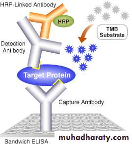
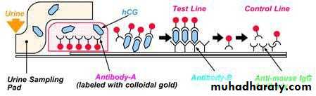
• Principle of the Sandwich ELISA
• ELISA Plate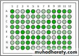
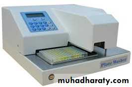
• ELISA Plate Reader
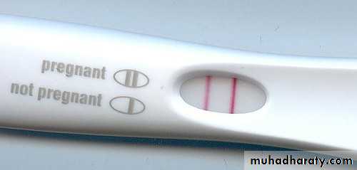
• An Important ELISA Application
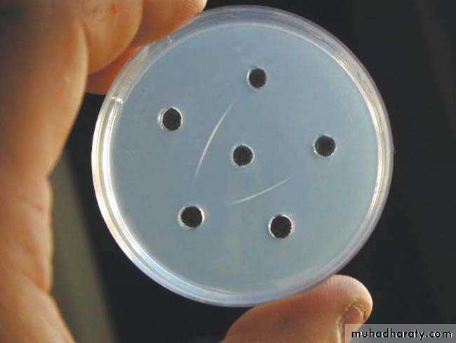
• Immunodiffusion Detection of Antibodies
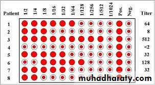
• Hemagglutination on a Microtiter Plate
• Direct Coombs Test
• ELISA Principles
• Some Points About ELISAs
• Understand O.D.s (optical densities)• How to make ELISAs quantitative
• Issues of Specificity and Stickiness
• Western Blot
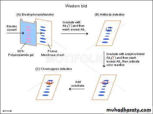
• Typical Western Blot
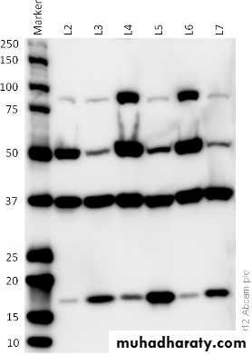
• Uses of Western Blots
• For determining antibody specificity when the antigen is complex• WBs are at best semi‐quantitative
• Some antibodies “don’t blot”
• Need for probity in data presentation
• Assays of Cellular Immunology
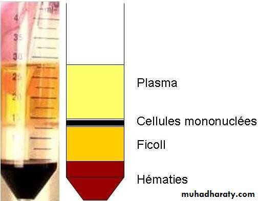
• DTH – “red bump in the skin”• Most cellular assays depend on density separation of “PBM” – peripheral blood mononuclear cells
• Ficoll Hypaque Separation of PBM
• Using Isolated PBM to Study Immunity
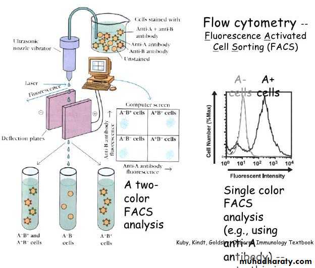
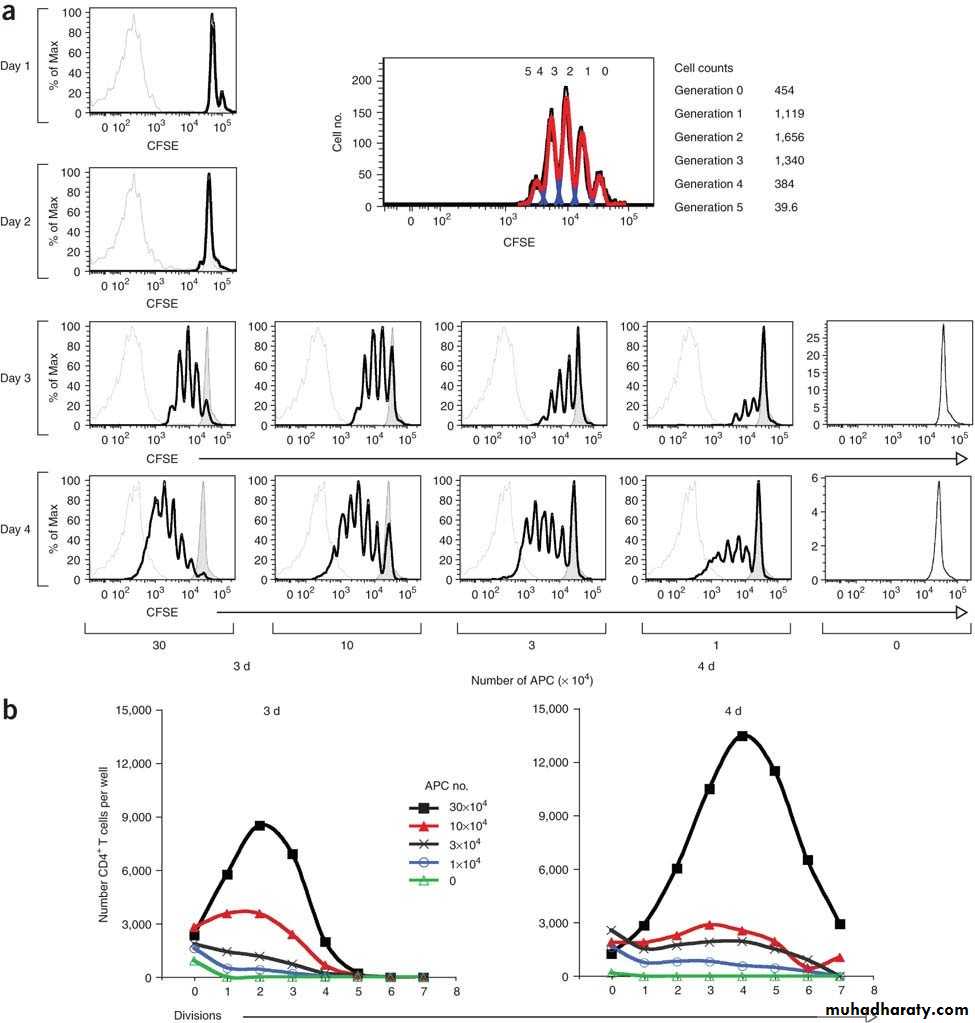
• Counting cells via flow cytometry• T‐cell functional assays: antigen, aCD3, or mitogen‐induced proliferation (CFSE preferred these days, formerly 3H thymidine incorporation Into DNA)
• Cytotoxicity
• Cytokine production
• Cell mixing and culture expts.
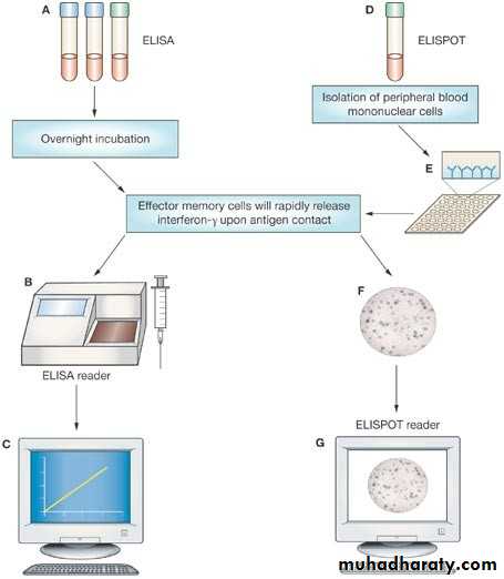
• Quantiferon Gold – a Cytokine Assay
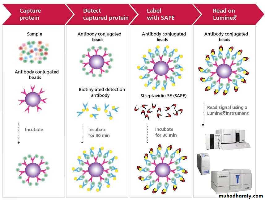
• A Multiplex Cytokine Assay
• Proliferation Measured by CFSE Dilution
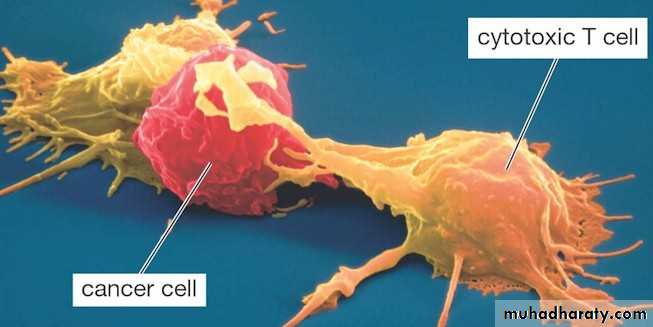
• Cytotoxic T Cells Destroying a Cancer Cell