Parasitology
Notes…
1
Parasitology Lecture.4 NON PATHOGENIC AMOEBA
1- E.coli. 2- E.gingivalis . 3- Dientamoeba fraglis.
4- Endolimax nana . 5- Iodoamoeba butschlii
6-E.hartmanni 7-E.dispar
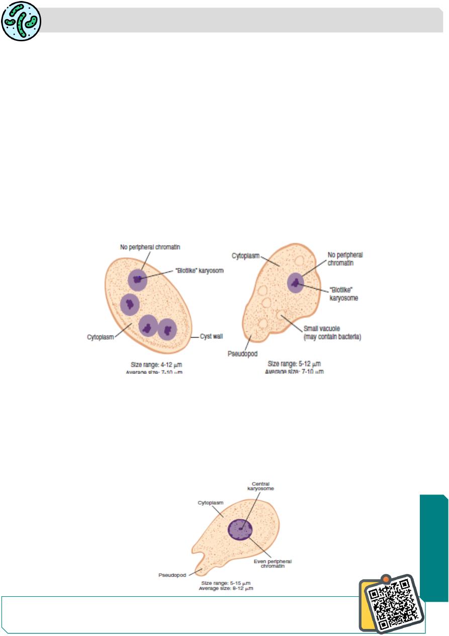
Endolimax Nana
This common commensal amoeba is widely distributed.
Habitat in the human large intestine.
The trophozoite is small (nana: small), less than 10 μm in size with a sluggish motility .
The nucleus small contains a large blot-like( karyosome)
There is little or no peripheral chromatin
The cyst is small 5 μm size, oval in shape , and quadrinucleate with glyocgen mass and
chromidial bars, which are inconspicuous orabsent .
It is non-pathogenic
Entamoeba Hartmanni
It is ccurs wherever E. histolytica is found.
It is a separate species of non pathogenic commensal intestinal amoeba.
It is much smaller than E. histolytica, called (small race) of
E.histolytica
.
the trophozoite measuring 4–12 µm, the cyst 5–10 µm in size with 1-4nuclei.
Trophozoites do not ingest red cells and their motility is less vigorous.
The cyst resembles that of Endolimax nana.
N
eed S
om
e H
el
p?
Parasitology
Notes…
2
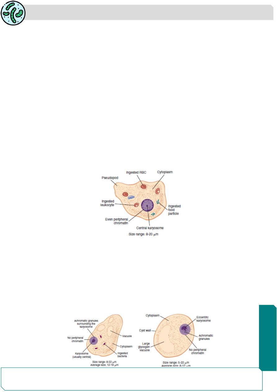
Entamoeba Gingivalis
It is global in distribution.
It is a commensal ,non pathogenic
Habitat in the mouth ( gingival tissues),tonsillar crypts and bronchial mucus.
the organism thrives in diseased gums, but is not considered a causal agent,It is
destroyed in stomach if swallowed.
Only the trophozoite is found; no cystic stage .
The trophozoite is about 10–20 μm, actively motile with multiple pseudopodia.
Cytoplasm contains food vacuoles with ingested bacteria, leuocytes, and epithelial cells.
the only species which ingests leucocytes
Nucleus is round with central karyosome lined by coarse chromatin granules
The amoeba lives in gingival tissues and is abundant in unhygienic mouths.
It is transmitted by direct oral contact ,kissing ,contact with fomites (drinking glasses,
eating utensils, etc.)
E. gingivalis have been found in bronchial washings and vaginal and cervical smears,
where it can be mistaken for E. histolytica
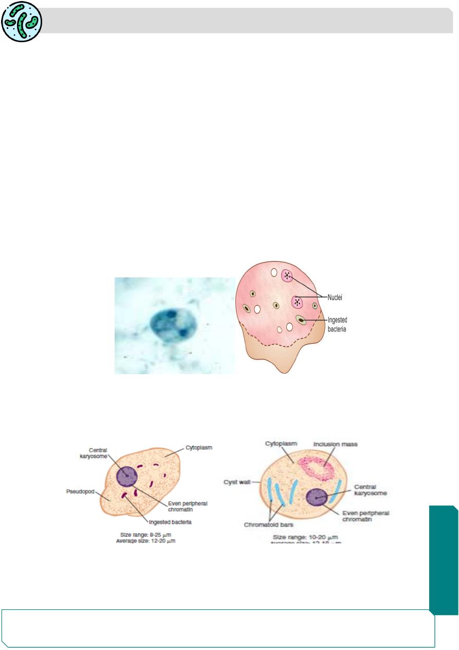
Iodamoeba butschlii
widely distributed, but less common than E. coli and Endolimax Nana.
The trophozoite is small, 6–12 µm, with single nucleus ,the prominent karyosome is half
the size of the nucleus having bull’s eye appearance,
The cyst is oval, uniucleate, and has a prominent iodine staining glycogen mass
(iodophilic body).
the cyst is often called the “iodine cyst” due to the presence of a large glycogen vacuole
which stains dark brown with iodine.
Parasitology
Notes…
3
Dientamoeba Fragilis
D. fragilis was previously considered as an amoeba but has now been reclassified as an
amoeboflagellate.
based on electron microscopic study and antigenic similarity toTrichomonas.
The name D.fragilis is derived from the binucleate nature of trophozoite (Dientamoeba)
and the fragmented appearance (fragilis) of its nuclear chromatin
It is worldwide infection . the most common intestinal protozoan in Canada.
It lives in colonic mucosal crypts, feeding on bacteria. It does not invade tissues, but may
rarely ingest RBCs
it has only trophozoite stage ,but no cyst stage.
The trophozoite is 7–12 μm in diameter. It is motile with broad hyaline leaflike
pseudopodia.
They have 1-4 nuclei; the binucleate form being the most common , with no peripheral
chromatin on the nuclear Membrane transmitted by the fecaloral route or by the eggs of
Enterobius vermicularis and other nematodes, which may serve as a vector
Entamoeba polecki
E. polecki is usually a parasite of pigs and monkeys.
rarly it occure in humans, non pathogenic ,distinguished from Entamoeba histolytica the
cysts of E.polecki have one nucleus
Parasitology
Notes…
4
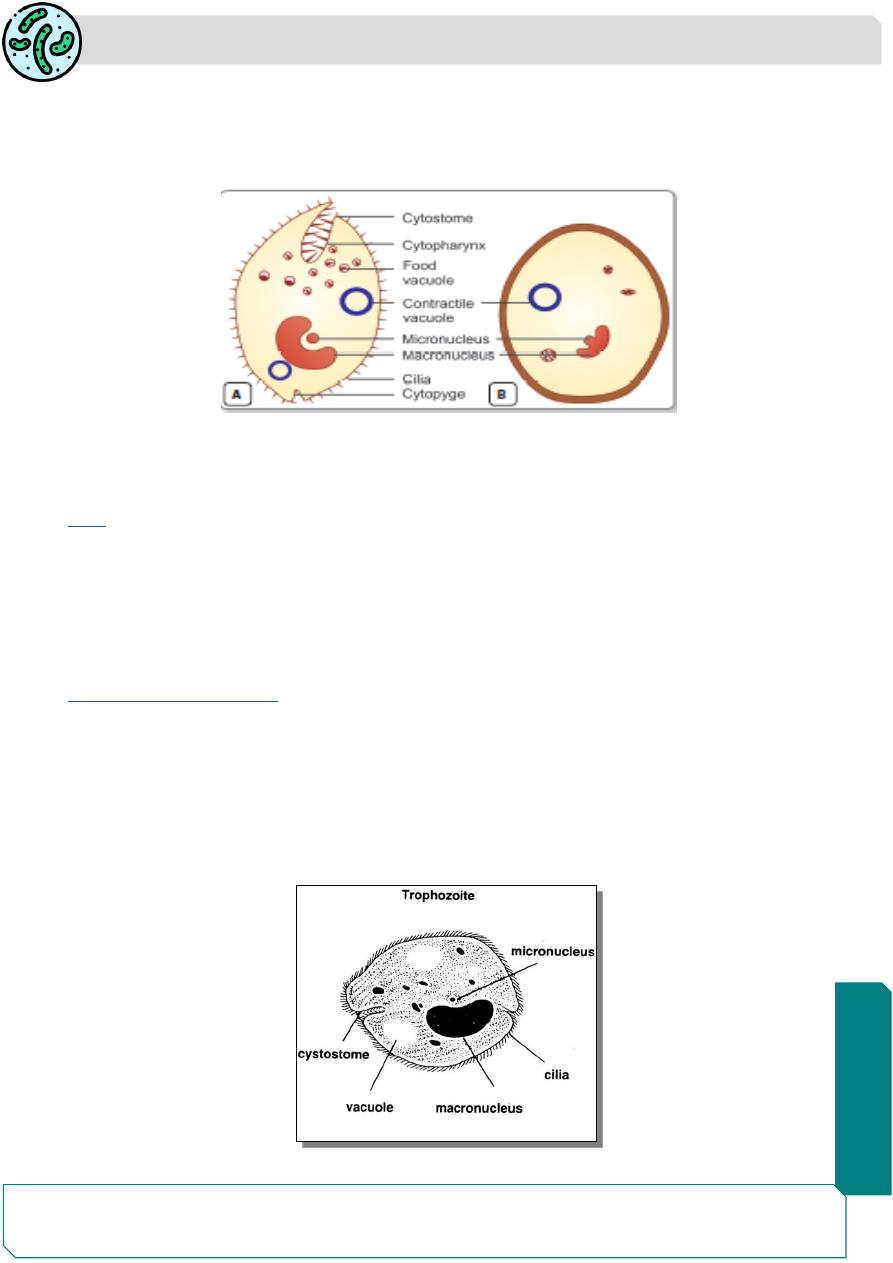
Balantidium coli .
Balantidium coli belongs to the Phylum Ciliophora and Family Balantididae.
the organism was named Balantidium, which means “little bag or sac like appearance
It is the Largest ciliate protozoan parasite of humans .
It is present worldwide, but the prevalence of the infection is very low.
The most endemic area is New Guinea, where there is a close association between man
and pigs .
Habitat
B. coli resides in the large intestine of man, pigs, and monkeys.
Morphology
Balantidium coli occurs in 2 stages: trophozoite, cyst
Trophozoite
The trophozoite lives in the large intestine, feeding on cell debris, bacteria, starch .
The trophozoite is actively motile and is invasive stage of the parasite found in dysenteric
stool.
It is a large ovoid cell, about 50-150 µm in length and 35–50 µm wide.
The motility of trophozoite is due to the presence of short delicate cilia over the surface
of the body.
The cilia around the mouth are longer then other
Parasitology
Notes…
5
Its anterior end is narrow and posterior end is broad.
At the anterior end, there is a groove (peristome)leading to the mouth (cytostome), and
a short funnel shaped gullet (cytopharynx).
Posteriorly, there is a small anal pore (cytopyge).
The trophozoite has 2 nuclei—a large kidney-shaped macronucleus and lying in its
concavity a small micro nucleus.
The cytoplasm has 1 or 2 contractile vacuoles and several food vacuoles.
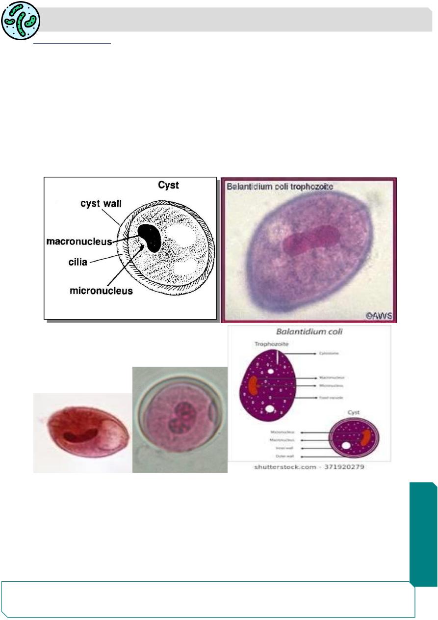
Cyst
The cyst is spherical in shape and measures 40–60 µm in diameter.
It is surrounded by a thick and transparent double layered wall.
The cytoplasm is granular. Macronucleus,micro nucleus, and vacuoles are also present
in the cyst. The cyst is the infective stage of B. coli.
It is found in chronic cases and carriers.
Trophozoite Morphology
▪ Cilliated parasite
▪ Oval shape
▪ Greenish yellow color size 50-150 µm
▪ Large Kidney or bean shape Macronucleus
▪ Small micronucleus
▪ Retractile food vacuole
Parasitology
Notes…
6
Cyst moraphology
▪ 40-60µm
▪ Spherical shape
▪ Cyst wall is thick consist
▪ of 1-2 layers
▪ No cytostome
▪ Macronucleus ,
▪ micronucleus
▪ Conractile vacules
▪ No cilia
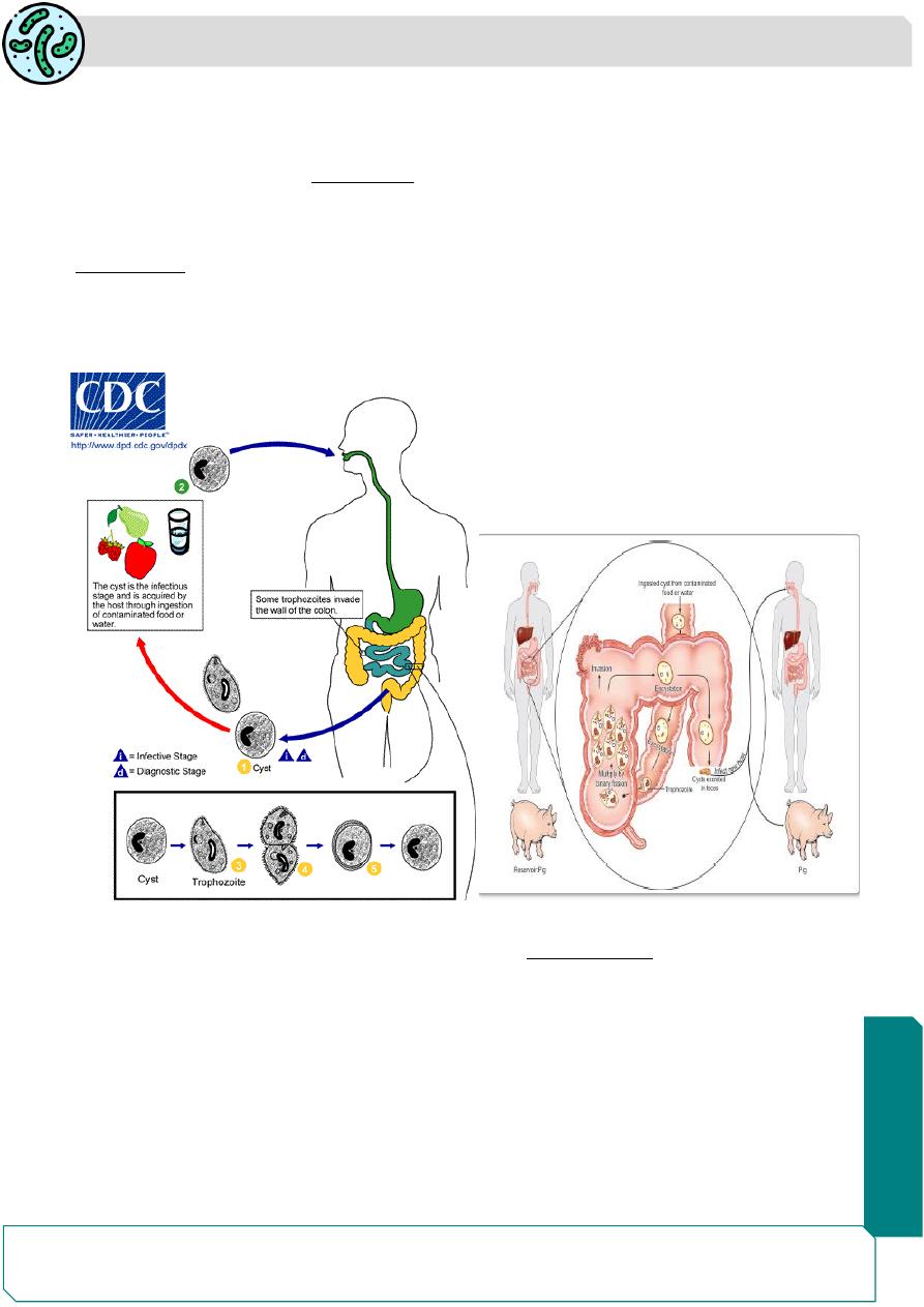
Life Cycle
Balantidium coli need one host only.
Natural host:Pig.
Reservoirs: Pig, monkey, and rat.
Accidental host:Man
Infective form: Cyst.
Balantidiasis is a zoonosis.
Parasitology
Notes…
7
Human beings acquire infection by ingestion of food and water contaminated with feces
containing the cysts of B. coli.
Infection is acquired from pigs and other animal reservoirs or from human carriers.
Once the cyst is ingested,
excystation
occurs in the small intestine , from each cyst, a
single trophozoite is produced which migrates to large intestine liberated trophozoites
multiply in the large intestine by transverse binary fission .
Encystation
occurs as the trophozoite passes down the colon or in the evacuated stool.
In this process, the cell rounds up and secretes a tough cyst wall around it.
The cysts remain viable in feces for 1-2 day and may contaminate food and water, thus
it is transmitted to other human or animals
Pathogenesis
Balantidium coli lives as lumen commensal and is asymptomatic
Clinical disease occurs only when the resistance of host is lowered by predisposing
factors like malnourishment, alcoholism, infection by Trichuris trichiura, or any
bacterial infection.
Clinical disease results when the trophozoites burrow into the intestinal mucosa, leads
to mucosal ulcers and submucosal abcesses, resembling lesions in amoebiasis
B. coli has been known to invade areas other than the intestine, such as the liver, lungs,
pleura, mesenteric nodes, and urogenital tract.
However, the incidence of such extraintestinal infections is rare.

Parasitology
Notes…
8
Clinical Features
Most infections are asymptomatic.
balantidiasis resembles amoebiasis causing diarrhea or frank dysentery with abdominal
colic, tenesmus, nausea, and vomiting.
Balantidium ulcers may be secondarily infected by bacteria.
Occassionaly, intestinal perforation peritonitis and even death may occur.
Rarely involvement of urogenital tracts.
In chronic balantidiasis, patients have diarrhea alternating with constipation
Diagnosis:
1-demonstration of trophozoites and cysts in feces
2-biopsy specimens and scrapings from intestinal ulcers .
3-Culture, cultured in vitro in Locke’s egg albumin medium or NIH polyxenic medium
Treatment
Tetracycline is the drug of choice and is given 500 mg, 4 times daily for 10 days.
Alternatively Doxycycline can be give.
Metronidazole and nitroimidazote have also been reported to be useful in some cases
Prophylaxis.
1- prevent contamination of food and water with human or animal feces.
2-Prevent contact of human with infected pig .
3- Treatment of infected pigs .
4- Treatment of infected man ,carriers