Parasitology
Notes…
1
Malaria Lec.2
P. vivax
has the widest geographical distribution. It is the most common species of malaria
parasite in Asia and America (account for 80% of all malaria infections), but is much
less common in Africa.
It causes benign tertian malaria with frequent relapses.
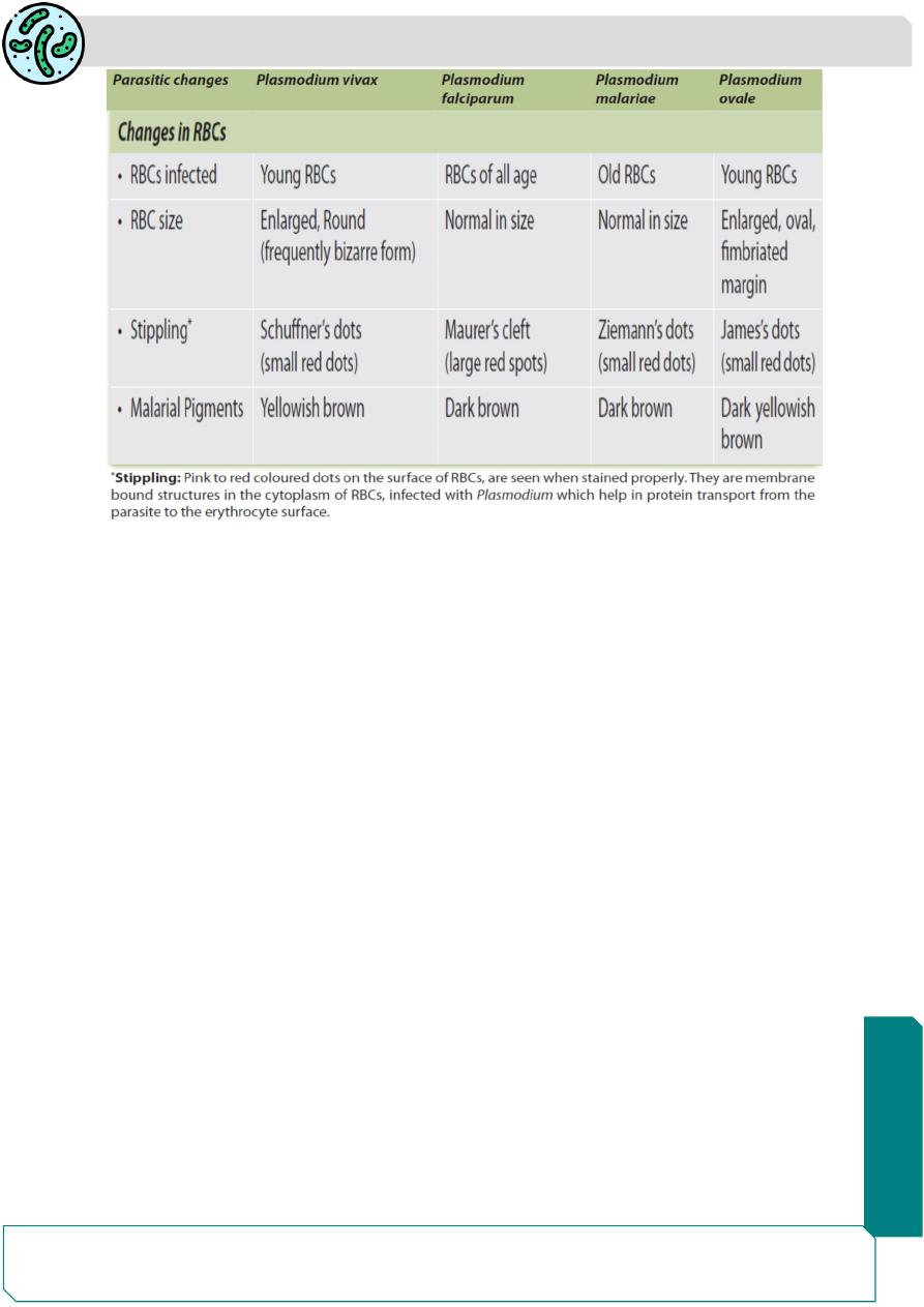
Merozoites of P. vivax preferentially infect reticulocytes and young erythrocytes,
the infected erythrocytes are enlarged and show red granules known as
Schuffner's dots on the surface -believed to be an allergic reaction from the host
to the presence of the parasite.
All stages of erythrocytic schizogony can be seen
in peripheral smears.
The degree of parasitization is not generally heavy;
not more than 2-5% of the red cells being affected.
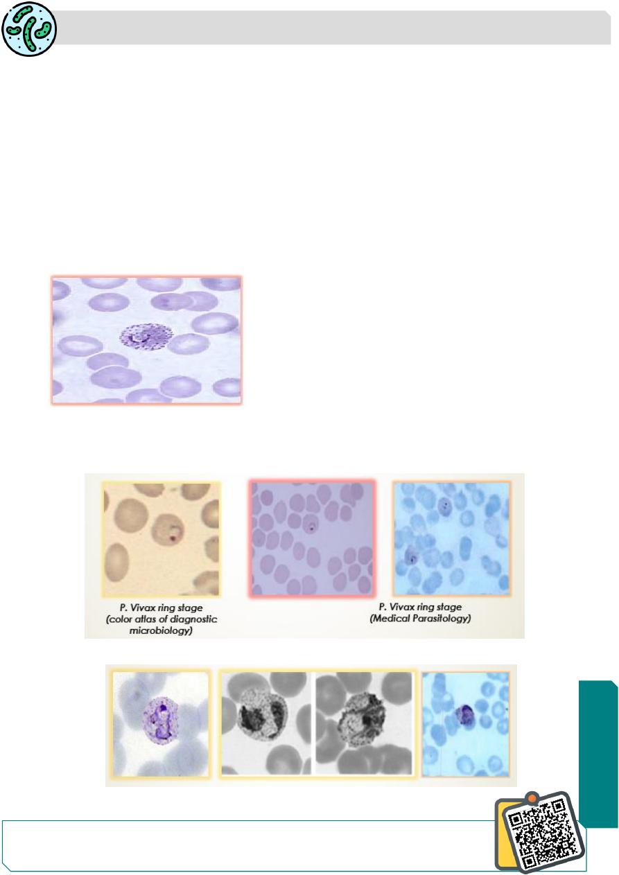
Morphology: The ring form has prominent central
vacuole. One side of the ring is thicker and the other
side thin. Nucleus is situated on the thin side of the ring (Signet ring appearance).
The ring is about 2.5-3 μm in diameter, about a third of the size of an erythrocyte.
The
cytoplasm
is
blue
and
the
nucleus
red
in
stained
films.
The ring develops rapidly to the ameboid form which is irregular in shape.
ameboid form of P.vivax
N
eed S
om
e H
el
p?
Parasitology
Notes…
2
The immature schizont form of P. vivax.
P. vivax immature schizont
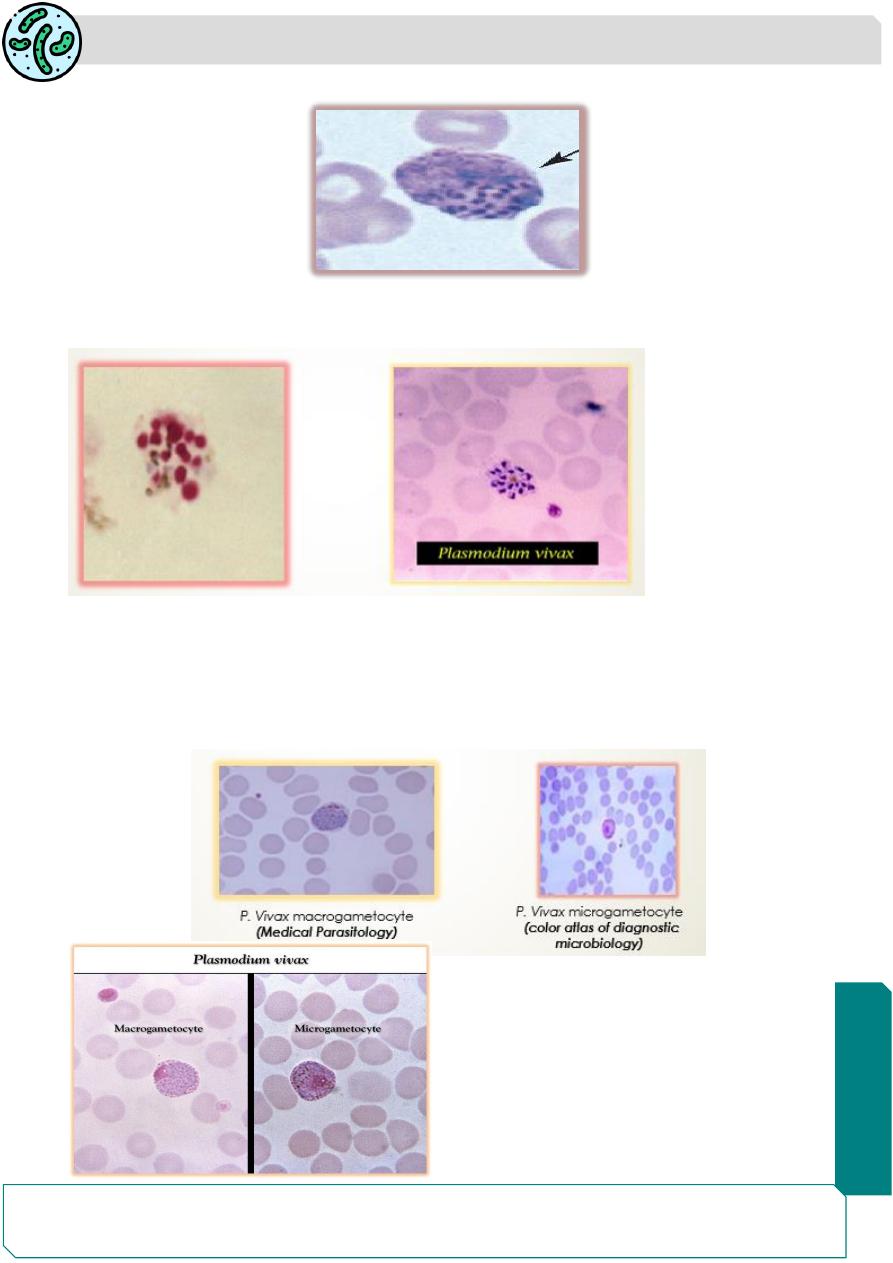
The mature schizont contains about 12-24 (usually 16) merozoites per schizont.
P. Vivax mature schizont (color atlas of diagnostic microbiology)
Both male and female gametocytes are large, nearly filling the enlarged red cell.
macrogametocyte has dense cytoplasm staining deep blue and a small compact
nucleus.
microgametocyte has pale-staining cytoplasm and a large diffuse nucleus.
Parasitology
Notes…
3
Plasmodium Falciparum
This is the highly pathogenic of all the plasmodia and hence, the name malignant
tertian or pernicious malaria for its infection.
The disease has a high rate of complications and unless treated, is often fatal. This
species is responsible for almost all deaths caused by malaria.
Recrudescence occur in this species. Hypnozoites do not occur.
They attack ALL types of erythrocytes and so the population of cells affected is
very large(25% of RBC infected).
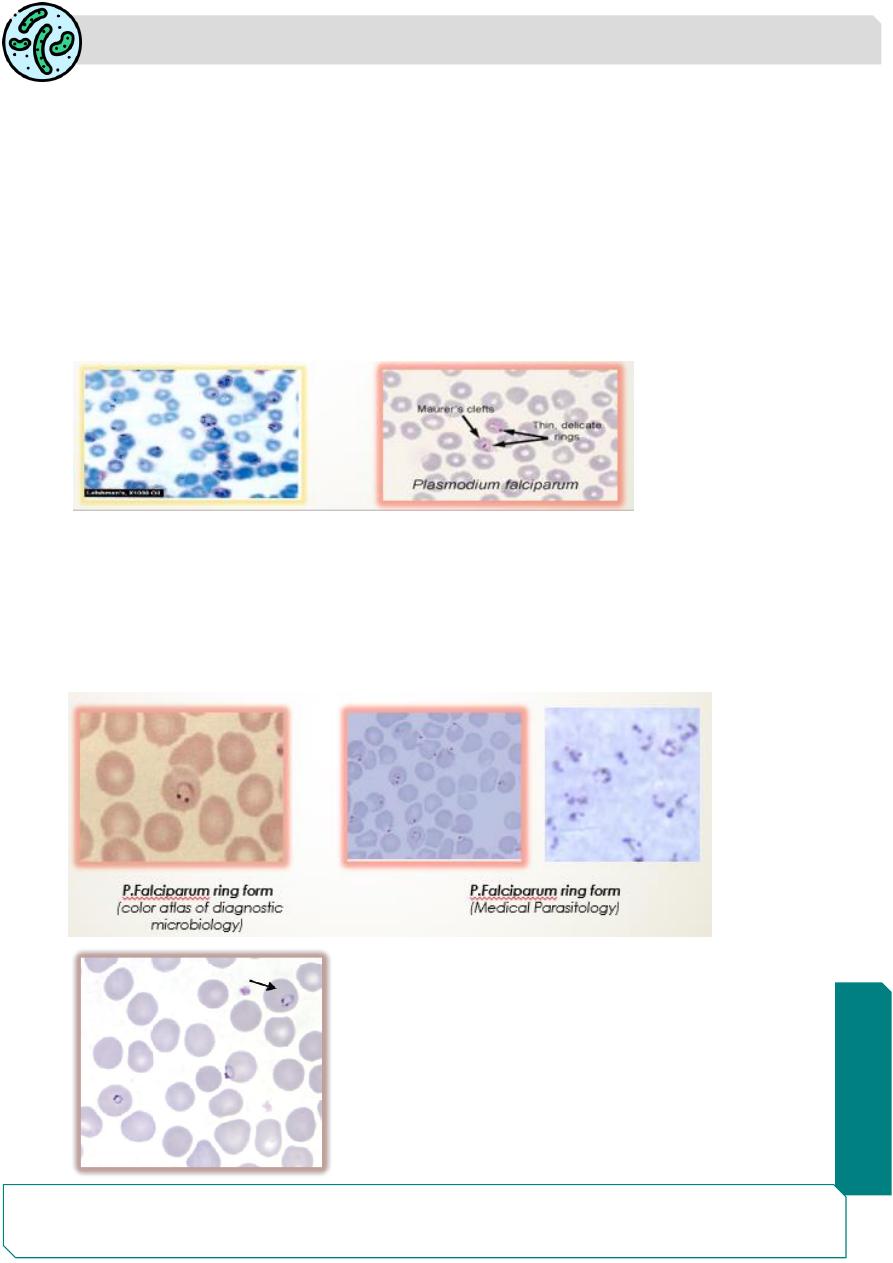
The infected erythrocytes are of normal size. They show a few (6- 12) coarse brick-
red dots which are called
Maurer ‘s clefts.
Morphology:
Ring form: The ring form in the erythrocyte is very delicate and tiny, measuring
only a one-sixth of the red cell diameter. Rings are often seen attached along the
margin of the red cell. Binucleate rings (double chromatin) are common resembling
stereo headphones in appearance. Several rings(multiple infection) may be seen
within a single erythrocyte.
double dot (headphone shaped) ring form of
Plasmodium falciparum
Parasitology
Notes…
4
The subsequent stages of the asexual cycle- ameboid early and mature schizonts-
are not seen in peripheral blood, except in very severe malaria, because these
stages of erythrocytic cycle occur in the capillaries of the brain and internal
organs.
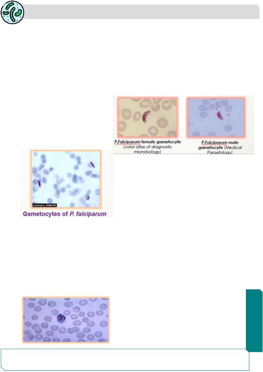
The mature gametocytes, which are seen in peripheral smears are curved oblong
structures, described as crescentic, kidney, sausage, or banana-shaped.
Male gametocytes: are broad and sausage-shaped or kidney-shaped, with blunt
rounded ends.
Female gametocytes: are thinner and more typically crescentic, with sharply
rounded or pointed ends.
The mature gametocyte is longer
than the diameter of the red cell
and so produces gross distortion
and sometimes disappearance of
the infected red cell.
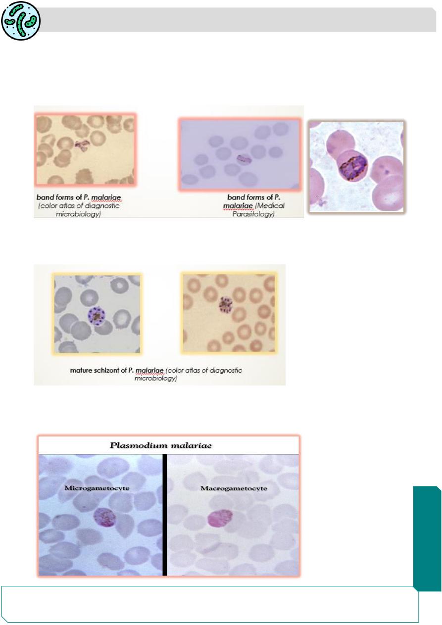
Plasmodium Malariae
It causes quartan malaria, in which febrile paroxysms occur every 4th day, with 72
hours interval between the bouts. Recrudescence occur in this species.
Hypnozoites do not occur.
P. malariae preferentially infects older erythrocytes and the degree of
parasitization is the lowest. The infected erythrocytes may be of the normal size
or slightly smaller.
Fine stippling, called Ziemann’s stippling, may be seen with special stains.
Parasitology
Notes…
5
Morphology:
The ring forms resemble those of P. vivax, although thicker and more intensely
stained.
The ameboid sometimes seen stretched across the erythrocyte as a broad band.
These band forms are a unique feature of P. malariae.
band forms of P. malariae
The mature schizont has (6-10 merozoites), which usually present a rosette appearance.
Hemozoin pigment ,brown in color, is also visible.
Gametocytes occupy nearly the entire red cell.
The male has pale blue cytoplasm with a large diffuse nucleus, while the female
has deep blue cytoplasm and a small compact nucleus.
Parasitology
Notes…
6
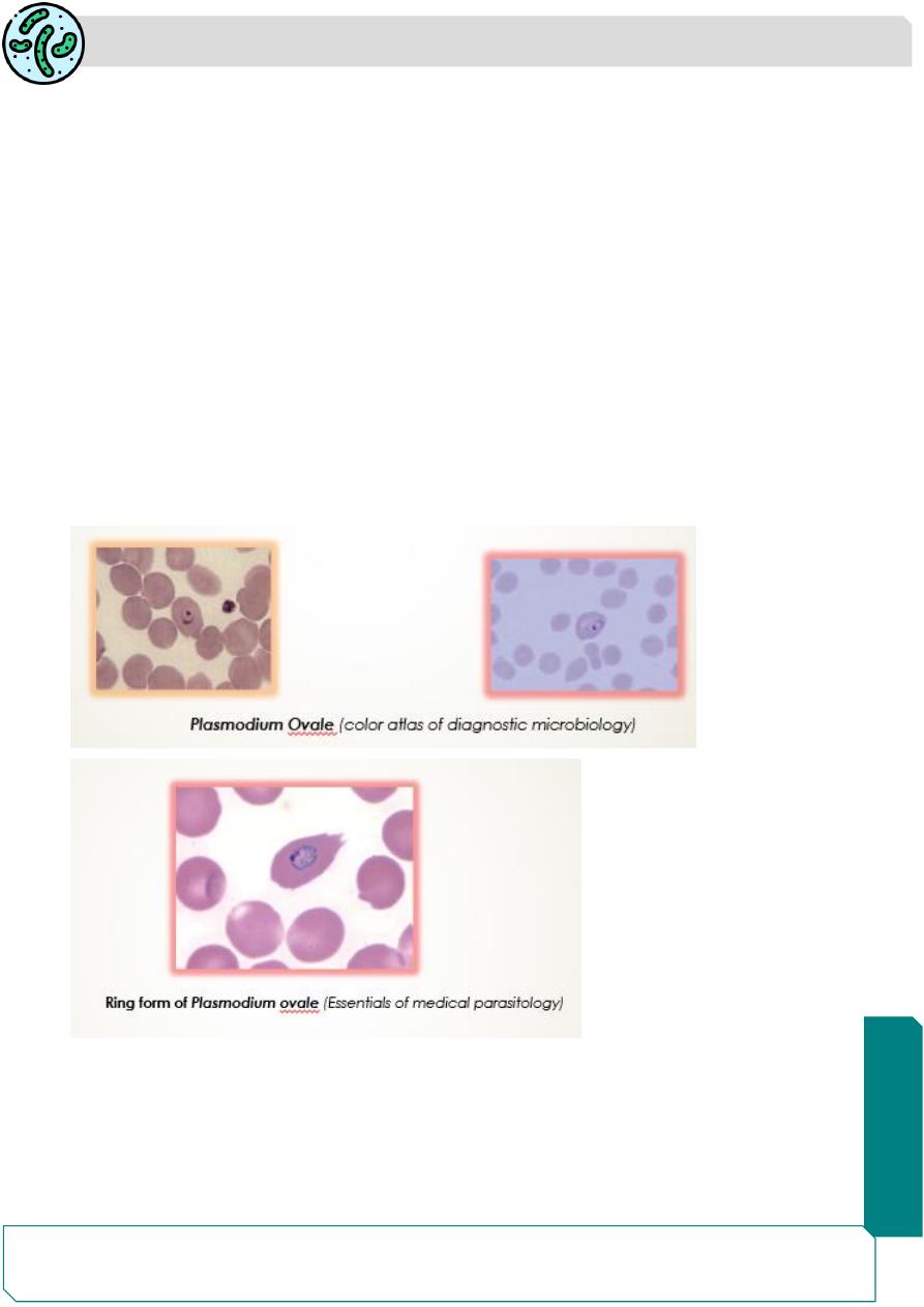
Plasmodium Ovale
This parasite cause tertian fever resembling vivax malaria, but with milder
symptoms.
It is the rarest of all plasmodia infecting humans
The infected erythrocytes are slightly enlarged and has irregular or fimbriated
appearance of the edges of the infected R.B.C. This oval appearance of the
infected erythrocyte is the reason for the name ovale given to this species.
Hypnozoites are present.
Morphology:
The trophozoites resemble those in vivax malaria. Schuffner's dots appear earlier
and are more abundant and prominent than in vivax infection.
The schizonts resemble those of P. malariae, except that the pigment is darker
and the erythrocyte is usually oval, with prominent Schuffner's dots.
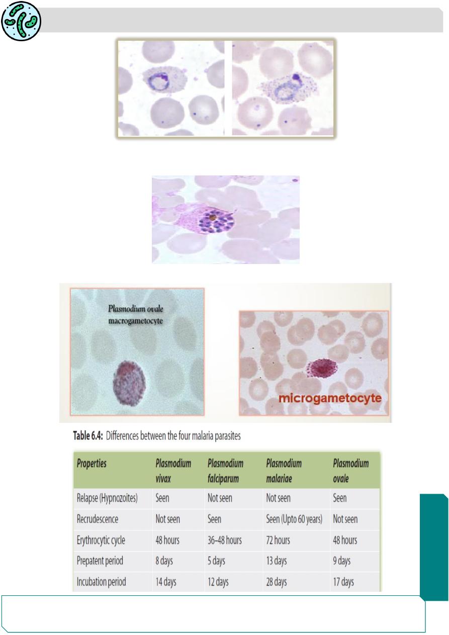
Trophozoites: P. ovale trophozoites have large chromatin dot, and can be compact to
slightly irregular and
showing Schüffner’s dots.
Parasitology
Notes…
7
Schizonts: P. aovale schizonts have 6 -14 merozoites with large nuclei, clustered
around a mass of dark-brown pigment.
Microgametocyte and Macrogametocyte as in P.vivax
Parasitology
Notes…
8
Summary
Schujfner's dots are seen in P .vivax and P.ovale.
Maurer ‘s clefts are seen in P. falciparum.
Ziemann’s stippling are seen in P.malariae.
In each spp. we havering, ameboid, immature and mature schizont.