College of Medicine University of Mosul /Department of: Microbiology
Subject: Parasitology Stage: 3
rd
2021-2022
Lecturer: Dr.Ahmed Alharbi Date: 17/11/2021 No.2
Page | 1
Blood and tissue protozoa
Objectives:
1. Introduction to kinetoplastids
2. Hemoflagellates :
General features
Leshmaniasis
Epidemiology, transmission and risk factors
Biology and life cycle
Clinical types of leishmaniasis :
Cutaneous leishmaniasis
Mucocutaneous leishmaniasis
Introduction
• Several pathogenic protozoa can infect human and localized in the blood and tissues:
A. Kinetoplastids
• Kinetoplastids are a large , diverse group of flagellated protozoa characterized by a
Geimsa-stained structure known as a kinetoplast.
• The kinetoplast is a specialized region of the mitochondria and the intense staining by
Geimsa stain is due to mitochondrial DNA. The kinetoplastids have more mitochondrial
DNA than other eukaryotes resulting in a more intense staining.
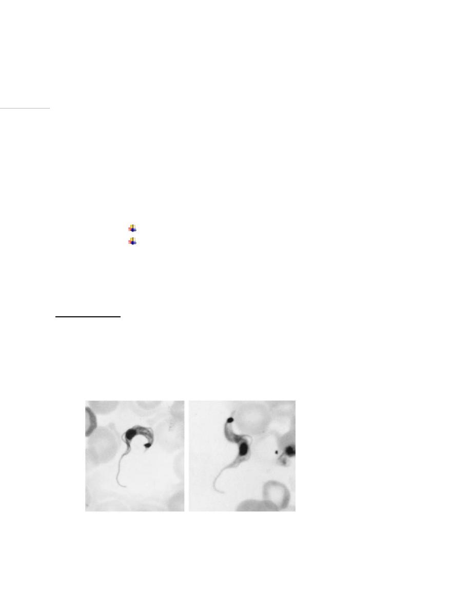
Kinetoplasstid( T.cruzi). (1)

College of Medicine University of Mosul /Department of: Microbiology
Subject: Parasitology Stage: 3
rd
2021-2022
Lecturer: Dr.Ahmed Alharbi Date: 17/11/2021 No.2
Page | 2
Kinetoplastids include:
Leishmania spp.: that cause leishmaniasis:
• Leishmania donovani complex
• L. infantum
• L. tropica complex
• L. mexicana
• L. braziliensis
• L. chagasi
Trypanosoma
• Trypanosoma brucei gambiense: sleeping sickness.
• Trypanosoma brucei rhodesiense: sleeping sickness.
• Trypanosoma brucei brucei, does not infect humans.
• Trypanosoma cruzi causes Chagas ’ disease
• Trypanosoma rangeli
B. Apic(K)omplexa(Sporozoa, Apikomplexia)
• Unicellular obligate intracellular (endoparasites) spore-forming protozoa.
• Possess a complex apical structure containing plastid and organelles used for
penetration of a host cell.
They include:
Plasmodium : Causes malaria
• Plasmodium vivax
• Plasmodium falciparum
• Plasmodium ovale
• Plasmodium malariae
Babesia : babesiosis
Toxoplasma gondii : toxoplasmosis
College of Medicine University of Mosul /Department of: Microbiology
Subject: Parasitology Stage: 3
rd
2021-2022
Lecturer: Dr.Ahmed Alharbi Date: 17/11/2021 No.2
Page | 3
Hemoflagellates
Classification of flagellates: لالطالع
• Phylum: Sarcomastigophora
• Subphylum: Mastigophora
• Class: Kinetoplastidea
• Order: Trypanosomatida
• Family: Trypanosomatidae
• Genera: Leishmania and Trypanosoma
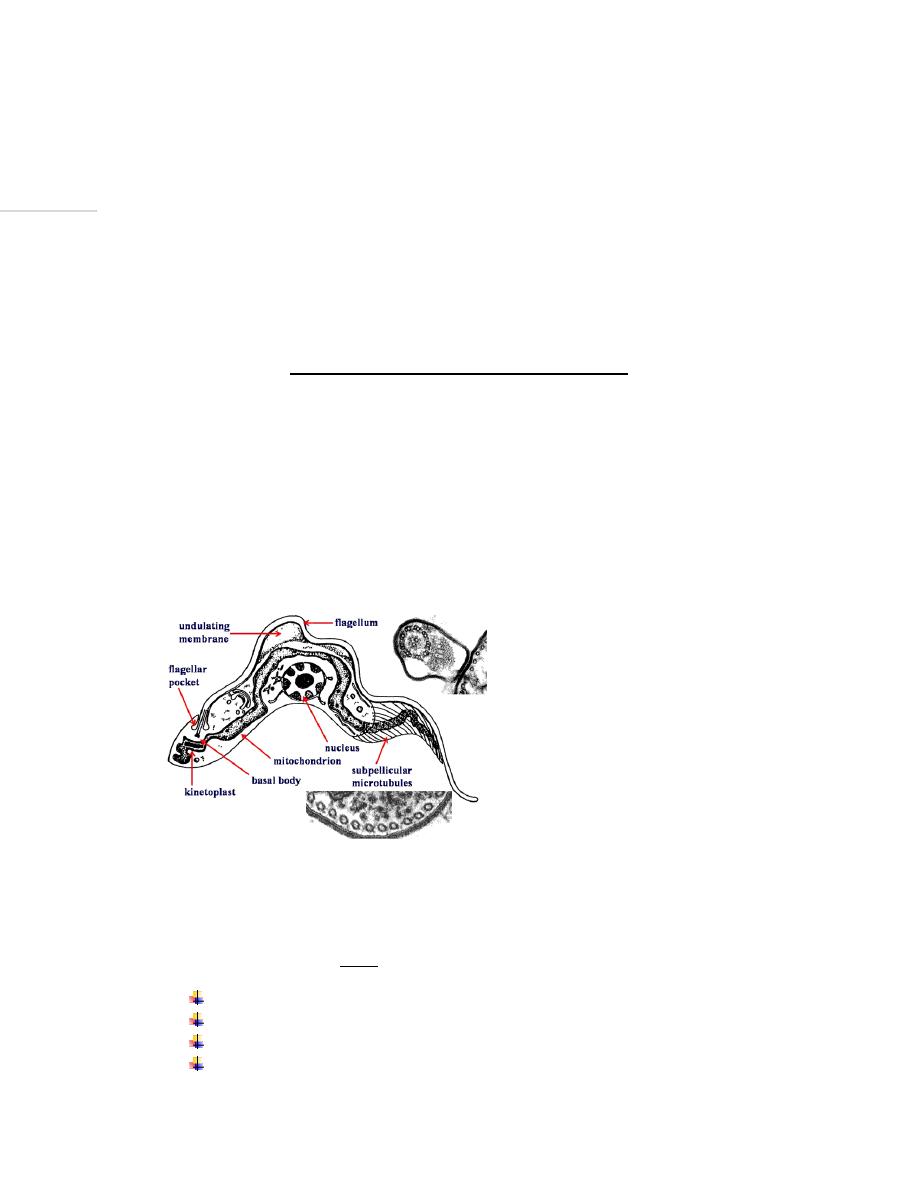
General features of flagellates
• They live in the blood & tissues of man & other vertebrates, and in the gut of the insect
vectors.
• They have a single nucleus, a kinetoplast and a single flagellum
• The flagellum is a thin, hair-like structure, which originates from the blepharoplast.
• A free flagellum at the anterior end traverses on the surface of the parasite as a narrow
undulating membrane .
Flagellates(2)
• Nucleus is round or oval, and is situated in the central part of the body.
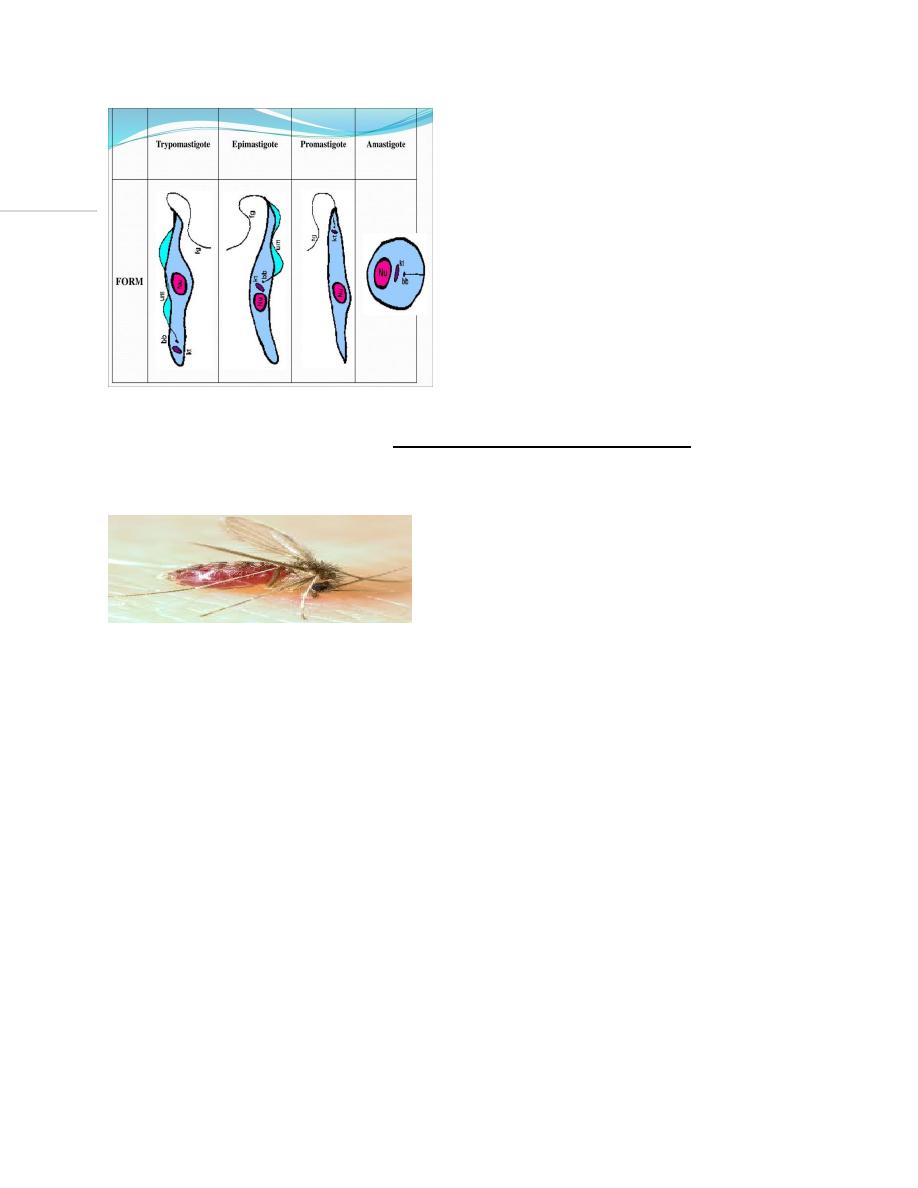
• Hemoflagellates exist in TWO or more of four morphological stages:
Amastigote
Promastigote,
Epimastigote
And Trypomastigote.
College of Medicine University of Mosul /Department of: Microbiology
Subject: Parasitology Stage: 3
rd
2021-2022
Lecturer: Dr.Ahmed Alharbi Date: 17/11/2021 No.2
Page | 4
Forms of kinetoplastids(3).
• Members of this family require an insect vector as an intermediate host.
• Multiplication in both the vertebrate and invertebrate hosts is by binary fission .
• Have no sexual cycle.
• Staining characteristics :
o For smears of body fluids: Romanowsky's Wrights stain, Giemsa stain and Leishman's
stain are suitable for identifying internal structures. The cytoplasm appears blue, the
nucleus and flagellum appear pink, and the kinetoplast appears deep red.
o For tissue section:Hematoxylin-eosin staining is done for demonstrating structures of
the parasite.
College of Medicine University of Mosul /Department of: Microbiology
Subject: Parasitology Stage: 3
rd
2021-2022
Lecturer: Dr.Ahmed Alharbi Date: 17/11/2021 No.2
Page | 5
Leishmaniasis
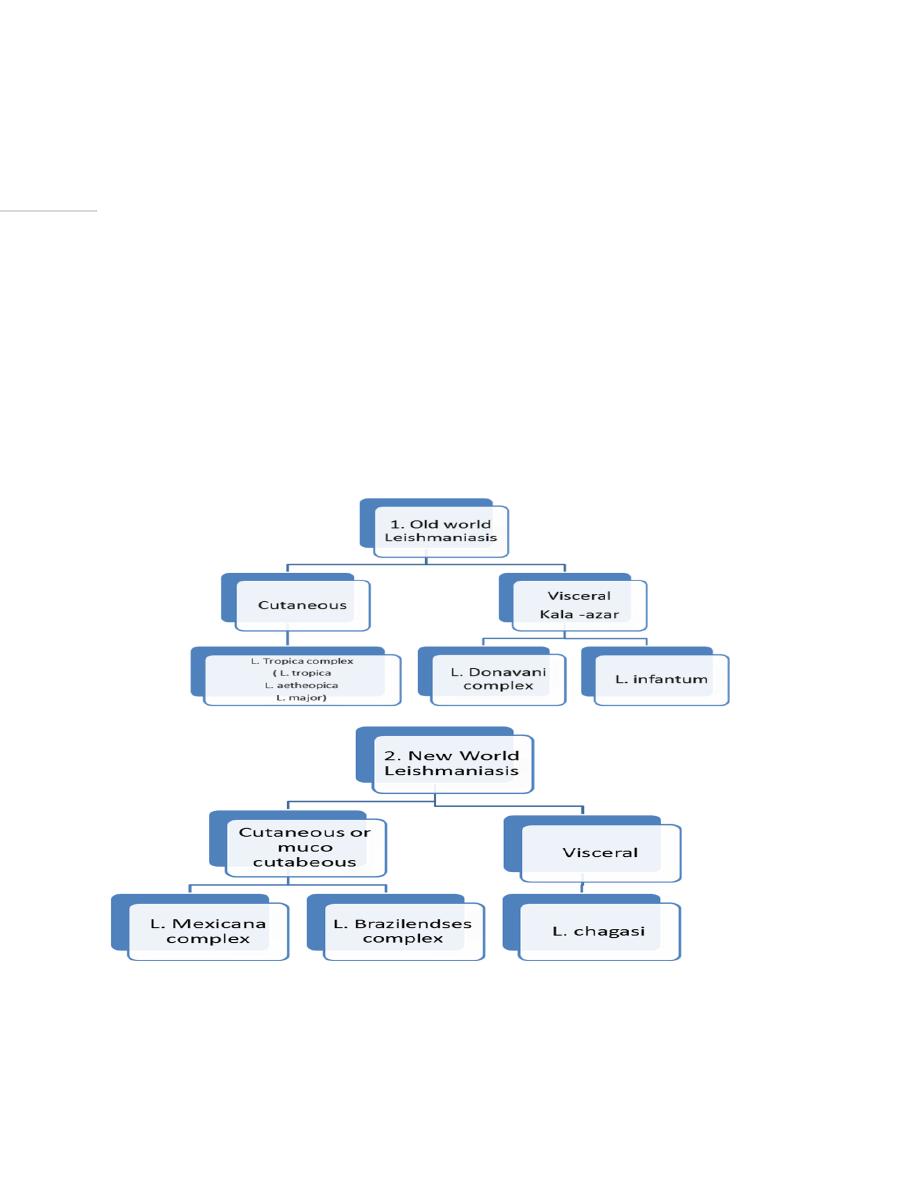
• Leishmaniasis is a tropical and subtropical disease caused by an intracellular parasite .
• The parasite is categorized in two main groups:
• The old world species: occur in Europe, Africa and Asia.
• The new world species : occur in America.
• More than 50 species of the parasite have been described from different regions of the
world.
• 31 species are known to be parasites of mammals, 20 species are pathogenic for human
beings.
Distribution and diseases caused by Leishmania spp

College of Medicine University of Mosul /Department of: Microbiology
Subject: Parasitology Stage: 3
rd
2021-2022
Lecturer: Dr.Ahmed Alharbi Date: 17/11/2021 No.2
Page | 6
Epidemiology
• Worldwide distribution.
• It is found in about 102 countries.
• It is endemic in Asia, Africa, the Americas, and the Mediterranean region.
• Between 12 and 15 million people in the world are infected.
• 350 million are at risk of acquiring the disease.
• An estimated 1.5 to 2 million new cases occur each year.
• It causes 70,000 deaths per year.
Transmission:
Leishmania spp. are spread by sandflies( vectors, intermediate host):
• a. Genus Phlebotomus in the Old World
• b. Genus Lutzomyiain the New World
Leishmania commonly infect canids, rodents( reservoir host) , and humans (final host).
Transmission via blood transfusion has been approved in animal experiments.
Transmission may occur through contaminated syringes and other paraenteral routes.
People at risk:
The main risk groups to get infection are:
• Farmers
• loggers
• Hunters
• Military personnel
• Biologists
• Ecological tourists
• Elderly and children
• Malnourished and immune deficient diseases.
• Those live under overcrowding, poor housing conditions and low socioeconomic status
can increase chance to get infection.
College of Medicine University of Mosul /Department of: Microbiology
Subject: Parasitology Stage: 3
rd
2021-2022
Lecturer: Dr.Ahmed Alharbi Date: 17/11/2021 No.2
Page | 7
Biology
Morphological forms(variants)
Depending on the stage of their lifecycle, they exist in two
structural variants:
A. Amastigote
B. Promastigote
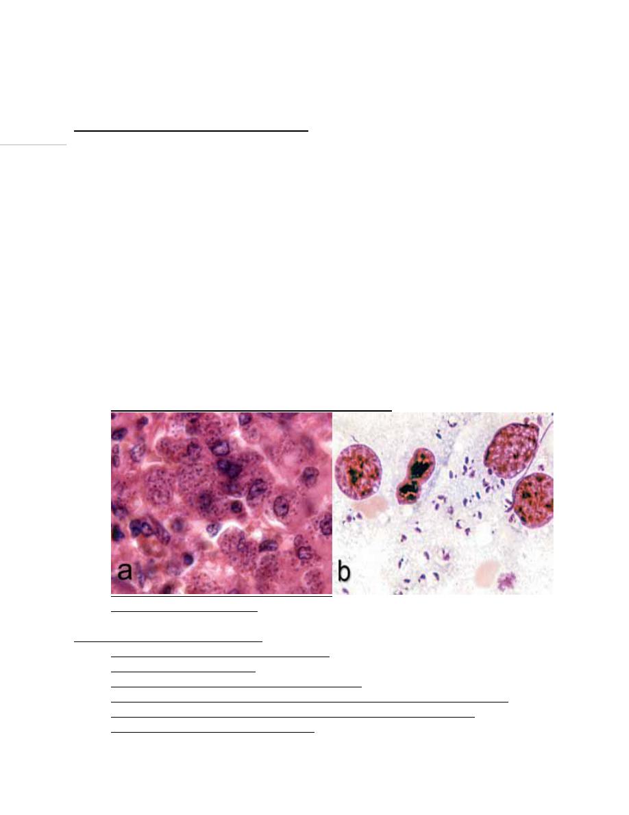
1. Amastigote form (Leishman-Donovan (LD) body):
• It is found in the mononuclear cells, neutrophils and reticulo endothelial system.
• Round or oval in shape.
• 3-6 micron X 1.5-3.0 micron.
• Have single nucleus with large central karyosome.
• The kinetoplast (which consists from blepharoplast and parabasal body beside
it) lies at
right angle to the nucleus.
• No visible flagellum (non motile).
• Amastigotes are usually grown inside tissue culture cells
Amastigote of Leishmania(4).
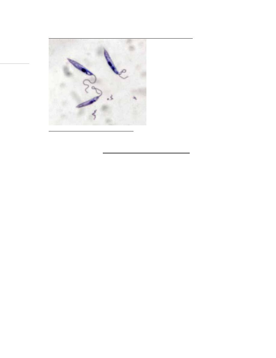
2. Promastigote (leptomonad) form:
• Is found in the alimentary tract of sandflies.
• Elongated (spindle in shape).
• 15-30 micron in body length and 5micron in width
• Have centrally located nucleus and the kinetoplast situated at the anterior end.
• From blepharoplast, single free flagellum projects from the anterior end.
• This form has no undulating membrane.
College of Medicine University of Mosul /Department of: Microbiology
Subject: Parasitology Stage: 3
rd
2021-2022
Lecturer: Dr.Ahmed Alharbi Date: 17/11/2021 No.2
Page | 8
• Promastigotes can be grown in vitro at 24-26
o
using NNN media.
Promastigote stage of Leishmania (4).
Life cycle of Leishmania spp.:
• Leishmaniasis is transmitted by the bite of infected female sandflies.
• The sandflies inject the infective stage (i.e., promastigotes) from their proboscis during
blood meals.
• Promastigotes that reach the puncture wound are phagocytized by macrophages and
other types of mononuclear phagocytic cells. Progmastigotes transform in these cells
into the tissue stage of the parasite (i.e., amastigotes).
The amastigotes multiply by simple division and proceed to infect other mononuclear
phagocytic cells
• Parasite, host, and other factors affect whether the infection becomes symptomatic and
whether cutaneous or visceral leishmaniasis result.
• Sandflies become infected by ingesting infected cells during blood meals.
• In the gut of sandflies, amastigotes transform into promastigotes.
• The promastigotes then migrate to the proboscis of the insect.
• The duration of the life cycle in the vector varies from 4 to 18 days, depending on the
species of Leishmania and climate.
• Laboratory studies suggest that a L. donovani parasite load of 20,000 per mL of blood in
the host is required to infect the sand fly insects.

College of Medicine University of Mosul /Department of: Microbiology
Subject: Parasitology Stage: 3
rd
2021-2022
Lecturer: Dr.Ahmed Alharbi Date: 17/11/2021 No.2
Page | 9
Clinical types
Cutaneous leishmaniasis:
Muco-cutaneous leishmaniasis, cutaneous-mucosal, American cutaneous, or “Espundia”
Visceral leishmaniasis (Black fever, Dumdum fever)
Cutaneous leishmaniasis:
• The most common pathogens causing this lesion are Leishmania tropica complex
• Cause skin sores (also known as oriental sore, Baghdad boil, tropical
sore, Aleppo boil,
or Delhi Boil).
• It usually produces ulcers on the exposed parts of the body, such as the face, arms and
legs.
• The incubation period is from 1 to 4 weeks.
Clinical forms of cutaneous leishmaniasis
• Leishmania major: Zoonotic cutaneous leishmaniasis: wet lesions with severe reaction
• Leishmania tropica: Anthroponotic cutaneous leishmaniasis: Dry lesions with minimal
ulceration
Cutaneous leishmaniasis lesion is characterized by:
• Local increase in temperature, and swelling.
• An erythematous asymptomatic papule (1 to 10 mm in diameter) appears at the site of
the bite, although pruritus may be present.
• After 2 days, it turns into a vesicle and later into a pustule.
• It breaks either spontaneously or by scratching to give volcano like ulcer, with a raised
edge and central crater.
• Such ulcers can last from 3 months to 20 years.
• The bottom of the ulcer shows granulation tissue that bleeds when rubbing and a pink
periphery and sometimes is covered by a whitish pseudomembrane.
• The lesion is not painful if it is not secondarily infected.
• Ulcers may be solitary or multiple.
• The clinical picture is usually afebrile with regional adenopathy.
Muco-cutaneous leishmaniasis, cutaneous-mucosal, American
cutaneous, or “Espundia”
• New world leishmaniasis in Central and South America. Caused by L. braziliensis
complex and L. mexicana complex
• The amastigote form is seen inside the macrophages of skin and mucous membrane of
the nose and buccal cavity. The promastigote form occurs in vector species Lutzomyia.
• It causes invasion and destruction of the nasopharyngeal mucosa.
• Usually, lesions start in the nasal mucosa and spread to the oral and pharyngeal mucosa,
the larynx, and the skin of the nose and lips.

College of Medicine University of Mosul /Department of: Microbiology
Subject: Parasitology Stage: 3
rd
2021-2022
Lecturer: Dr.Ahmed Alharbi Date: 17/11/2021 No.2
Page | 10
• Lesions of the oral mucosa usually produce symptoms that range from simple to severe
in extreme cases.
• Early in the disease, there is inflammation of the mucosa with superficial ulcerations;
later on, when the ulcers are well developed, their borders have a necrotic appearance
and are torn and detached.
• The uvula, pillars of the palate roof, and tonsils can be destroyed. If the larynx is
involved, the voice changes as well.
• Secondary infections usually occur.
• Destruction of the cartilaginous septum of the nose with foul smell can occur
• In extreme cases, it ends to death.
Chiclero ulcer
• also called as self healing sore of Mexico
Is a kind of American leishmaniasis caused by Leishmania Mexicana. The disease is
characterized by cutaneous ulcers on the head and may last 6 months but if occurs on
the pinna of the ear, it may last up to years leaving scarring and deformities.
Images for cutaneous leishmaniasis:
Muco cutaneous leishmaniasis :
--------------------------To be continued next lecture-------------
References:
1. Garcia Lynne.Diagnostic medical parasitology.Washington DC.ASM Press.5
th
edition.2007
2. Tulane.com( flagellates)
3.
www.slideserve.com/Gabriel/haemoflagellates-leishmaniasis-trypanosomiasis
4. Dickson D. Despommier, Daniel O. Griffin, Robert W. Gwadz, Peter J. Hotez,
and Charles A. Knirsch. Parasitic diseases 6
th
edition.New yourk Parasites
Without Borders, Inc. NY. 2017
5.
BURTON J. Bogitsh,Clint E. Carter, and Thomas N. Oeltmann, 2013, Human parasitology
4
th
edition, USA and UK, Elsevier.2013.
6.
CDC.gov
7.
Encyclopedia of Microbiology (Third Edition)
, 2009
8.
Paniker J CK , 2018, Paniker
,
s textbook of medical parasitology, 8
th
edition, Newdelhi,
London , Panama, JAYPEE.
9.
World health organisation. Photos on leishmaniasis
10.
Zeibig A. Elizabeth, 2013, Clinical parasitology, USA, Elsevier