
Trematodes lec. 4
د.اسماء زكي شيتاوي
Intestinal flukes
Several genera of intestinal flukes cause human infection, particularly in
the Far East.
Fasciolopsis buski,
Heterophyes heterophyes,
Metagonimus yokogawai
Infection is usually asymptomatic unless the worm burden is large.
Fasciolopsis buski
Distribution
It is a common parasite of humans and pigs in China, in Southeast Asian
countries and Indian subcontinent, especially in areas where humans raise
pigs and consume freshwater plants
.
Disease : fasciolopsiasis
Morphology
Adult warm: It is the largest of the intestinal trematodes. The adult is a
large fleshy worm, 20–75 mm long, 8–20 mm broad and 0.5–3 mm in
thickness.
It is elongated, ovoid in shape, with a small oral sucker and a large
acetabulum. It has no cephalic cone as in F. hepatica. The adult worm
has a lifespan of about 6 months.
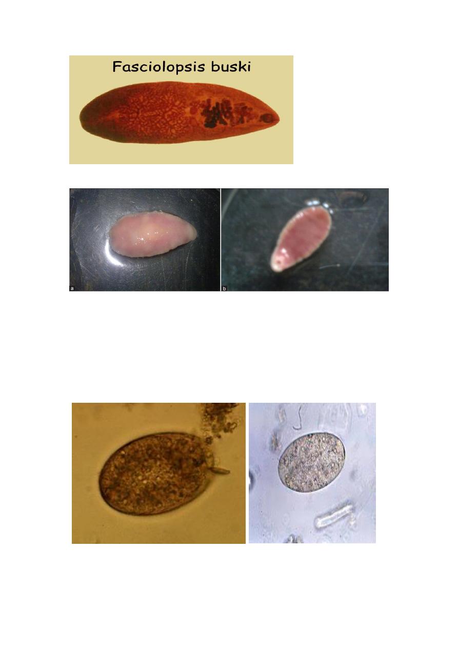
Egg :The operculated eggs are similar to those of
Fasciola hepatica.
Eggs are laid in the lumen of the intestine in large numbers, about 25,000
per day
. Pass
Unembryonated.

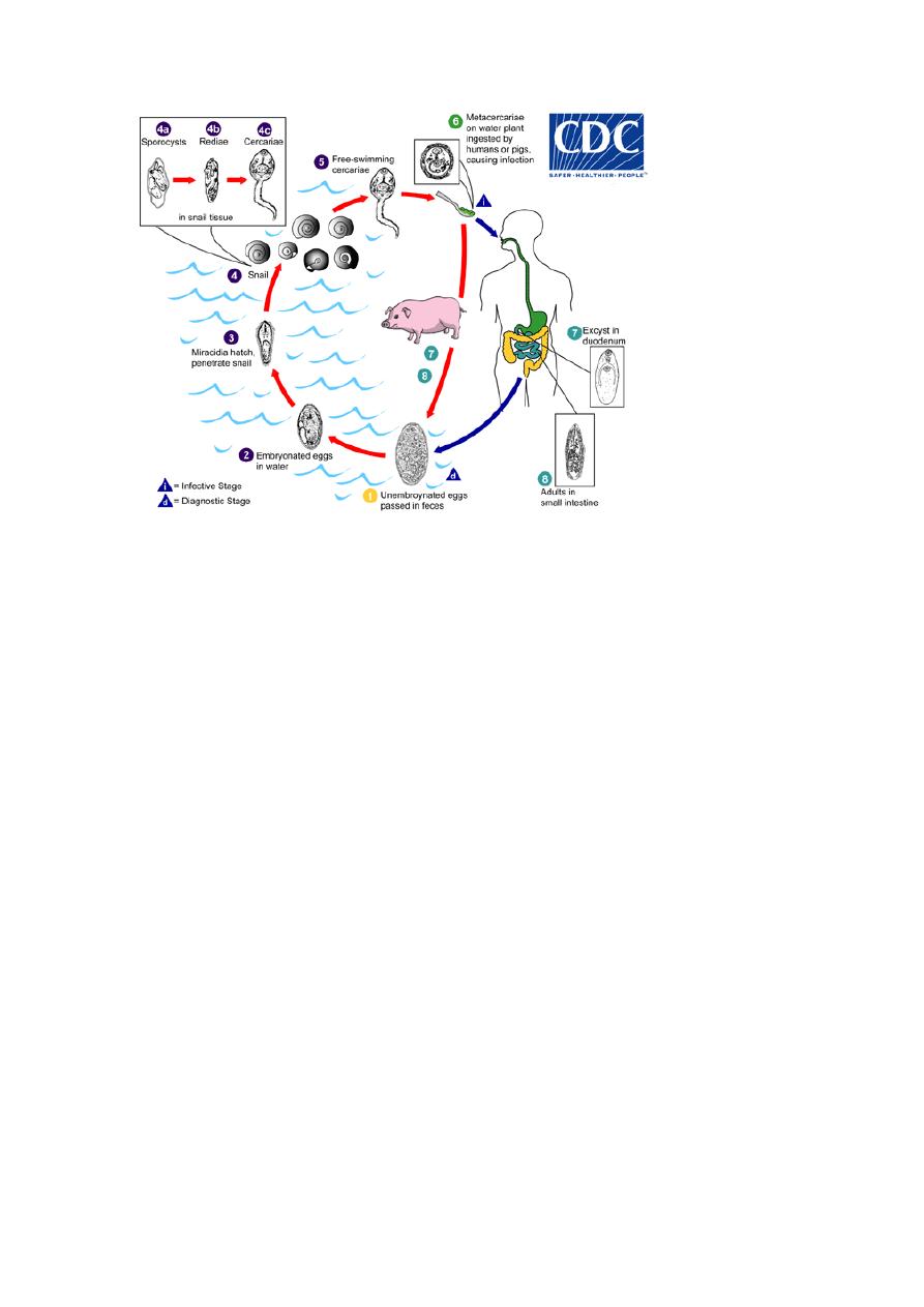
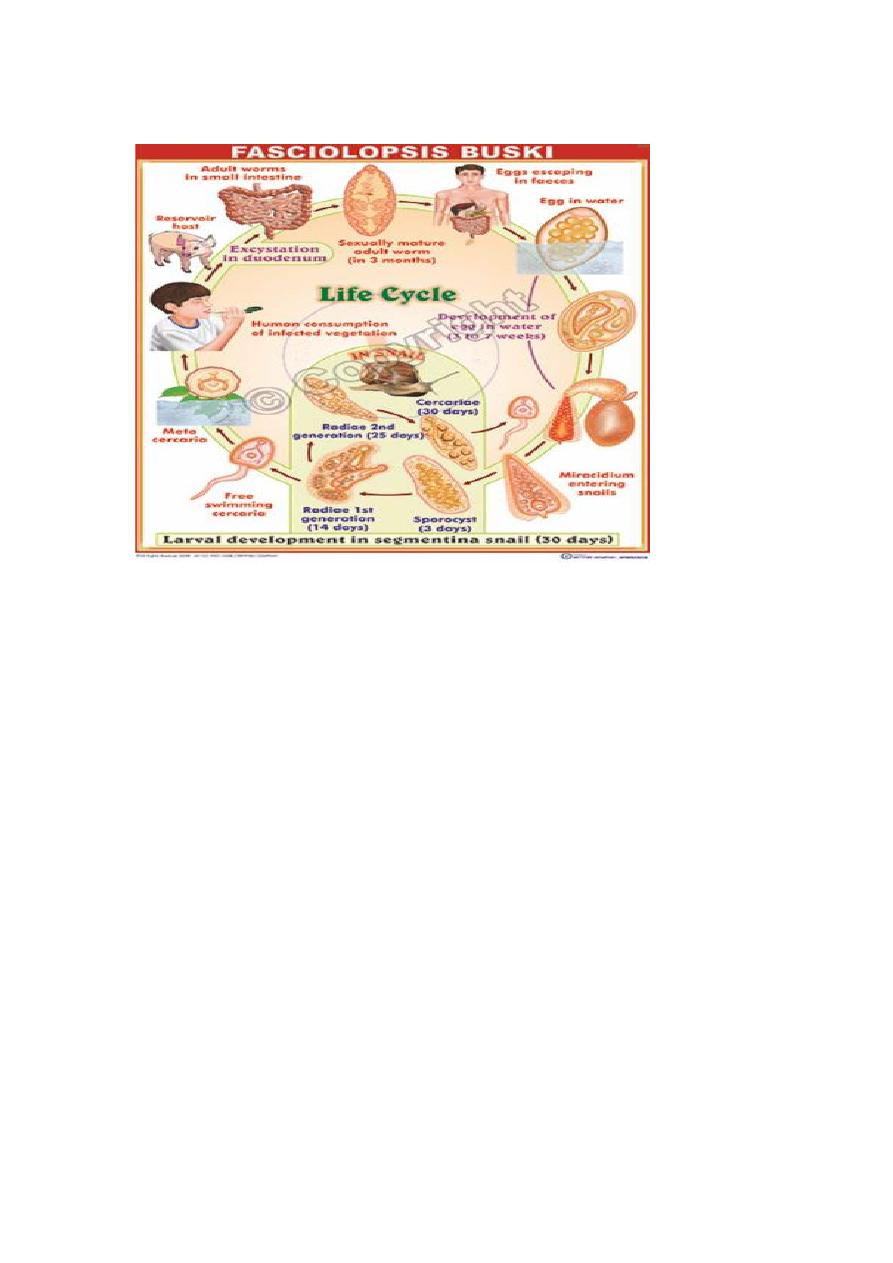
Life cycle :
F. buski passes its life cycle in definitive host and two intermediate
host.
Habitat The adult worm lives in the duodenum or jejunum of pigs and
humans.
Infective stage encysted metacercaria
Infection is acquired by ingestion of raw water chestnuts or caltrop
1
st
Intermediate host fresh water snail
2
nd
intermediate host
water plants water chestnut
Infective form: Encysted metacercariae on aquatic vegetation
.
• The eggs passed in feces of definitive host hatch in water in about 6
weeks, releasing the miracidia which swim about.
• On coming in contact with a suitable snails of the, miracidia penetrates
its tissues to undergo development in the next few weeks as sporocyst,
first and second generation rediae and cercariae .
• The cercariae, which escape from the snail, encyst on the roots of the
lotus, bulb of the water chestnut or on other aquatic vegetation.
• When they are eaten by man, the metacercariae excysts in the
duodenum, become attached to the mucosa and develop into adults in
about 3 months.
Pathogenesis and Clinical Features
The pathogenesis of fasciolopsiasis is due to traumatic, mechanical and
toxic effects.
• Larvae that attach to the duodenal and jejunal mucosa cause
inflammation and local ulceration. Intoxication and sensitization also
account for clinical illness.
• In heavy infections, the adult worms cause partial obstruction of the
bowel, malabsorption, protein-losing enteropathy and impaired vitamin
B12 absorption.
• The initial symptoms are diarrhea and abdominal pain.
• Toxic and allergic symptoms appear usually as edema, ascites, anemia,
prostration and persistent diarrhea.
• Paralytic ileus is a rare complication.
Prevention and Control
1. Treatment of infected cases
2. Wash water vegetation, preferably in hot water or cook well before
consumption
3. Prevent contamination of ponds and other water sources with pig or
human excreta
4. Control of snails
Heterophyes heterophyes and Metagonimus yokogawai
Heterophyes
This is the smallest intestinal trematode of human
(smallest
trematode)
Distribution
H. heterophyes is found in China, Egypt, India, Iran, Israel,
Japan, Korea, Sudan, the Philippines, Tunisia, and Turkey
M. yokogawai is found in the Balkans (a cultural region of southeastern
Europe), China, Indonesia, Israel, Japan is considered the most common
intestinal fluke infection in the Far East.
Disease : heterophyiasis
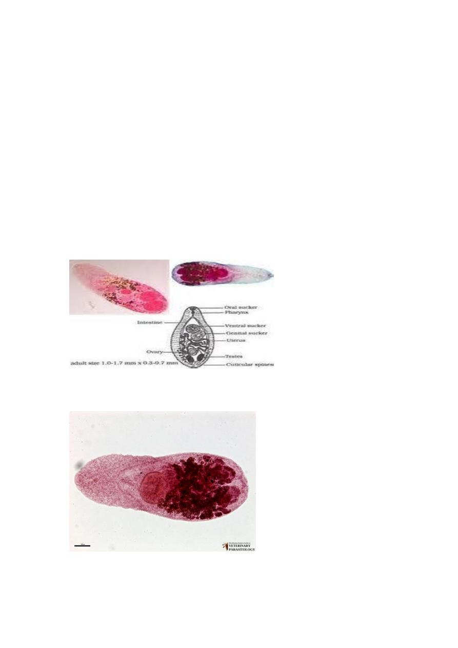
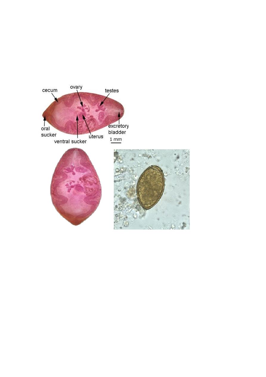
Morphology
Adult H. heterophyes worms range in size from 1.0 to 1.7 mm in length
and 0.3 to 0.4 mm in width. The worm is also pyriform in shape, with
tapering at the anterior end and rounding at the posterior end. It has an
extra powerful muscular sucker around the genital pore called genital
sucker. Metagonimus has the same morphology but slightly larger
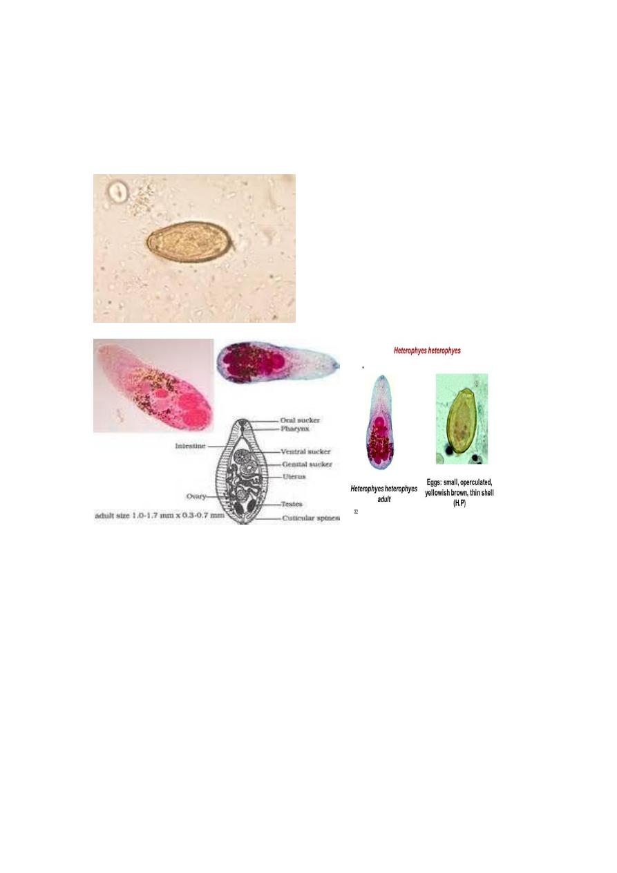
The eggs are small, yellow-brown, embryonated, and operculated and
may have minimal opercular shoulders. Eggs range in size from 26 to 30
µm long and 15 to 17 µm wide and may be indistinguishable between the
two species.
Life cycle
Habitat:
adult worm in duodenum and jejunum
Definitive hosts: humans, pigs, dogs and cats.
Intermediate hosts:
The first intermediate host is the freshwater snail
The second intermediate host is the freshwater fish.
Infective stage encysted metacercaria
Adults release embryonated eggs each with a fully-developed
miracidium,
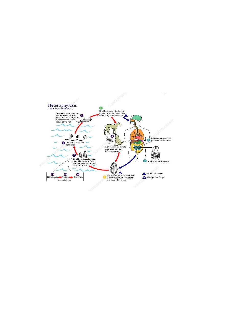
After ingestion by a suitable snail (first intermediate host), the released
miracidia develop into cercariae. The cercariae are released from the
snail and encyst as metacercariae in the tissues of a suitable fresh water
fish (second intermediate host). The definitive host becomes infected by
ingesting undercooked or salted fish containing metacercariae. After
ingestion, the metacercariae excyst, attach to the mucosa of the small
intestine and mature into adults.
Pathogenicity and clinical presentation
Infections with a small number of worms may be asymptomatic.
Symptoms in heavy infections may include abdominal pain, diarrhea with
a large amount of mucus, and ulceration of the intestinal wall. Eggs may
gain entry into the intestinal capillaries and the lymphatic system, where
they can be carried to the heart, brain, spinal cord, or other tissues,
causing emboli or granuloma formation.
Prevention

Infection can be prevented by avoiding ingestion of raw, inadequately
cooked, and pickled or salted fish. The risk of infection could be greatly
reduced by improved sanitary conditions and health education programs.
Laboratory Diagnosis of intestinal flukes
Identification of the intestinal trematodes is made by recovery of eggs, or
in rare cases adults, from stool specimens using a sedimentation method
such as formalin-ethyl acetate. The sediment may be examined in a wet
mount with or without iodine.
Because the eggs of F. buski are identical to those of F. hepatica, and
those of H. heterophyes and M. yokogawai are very similar, diagnosis
may also require assessment of symptoms, obtaining a travel history,
and/or recovery of adult worms.
Treatment
The drug of choice for treatment of intestinal trematode infection is
praziquantel, an isoquinoline derivative administered orally in three doses
for 1 day.
Lung Fluke
Paragonimus westermani
Common name Oriental lung fluke
Distribution
The parasite is endemic in Japan, Korea, Taiwan, China and Southeast
Asian countries.
Disease paragonimiasis
Morphology
The adult worm is coffee bean shaped about 10 mm long, 5 mm broad
and 4 mm thick. It is reddish brown in colour. It has an oral sucker and a
ventral sucker. It has a lifespan of up to 20 years in humans.
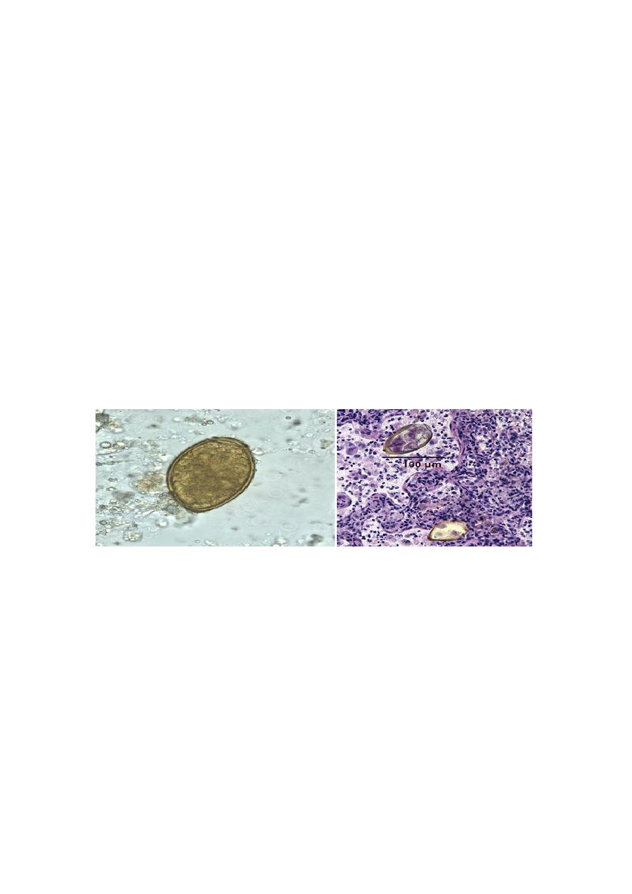
The eggs are operculated with opercular shoulders, golden brown in
colour and about 100 μm by 50 μm in size thick shelled. Eggs are
unembryonated when freshly laid.
.
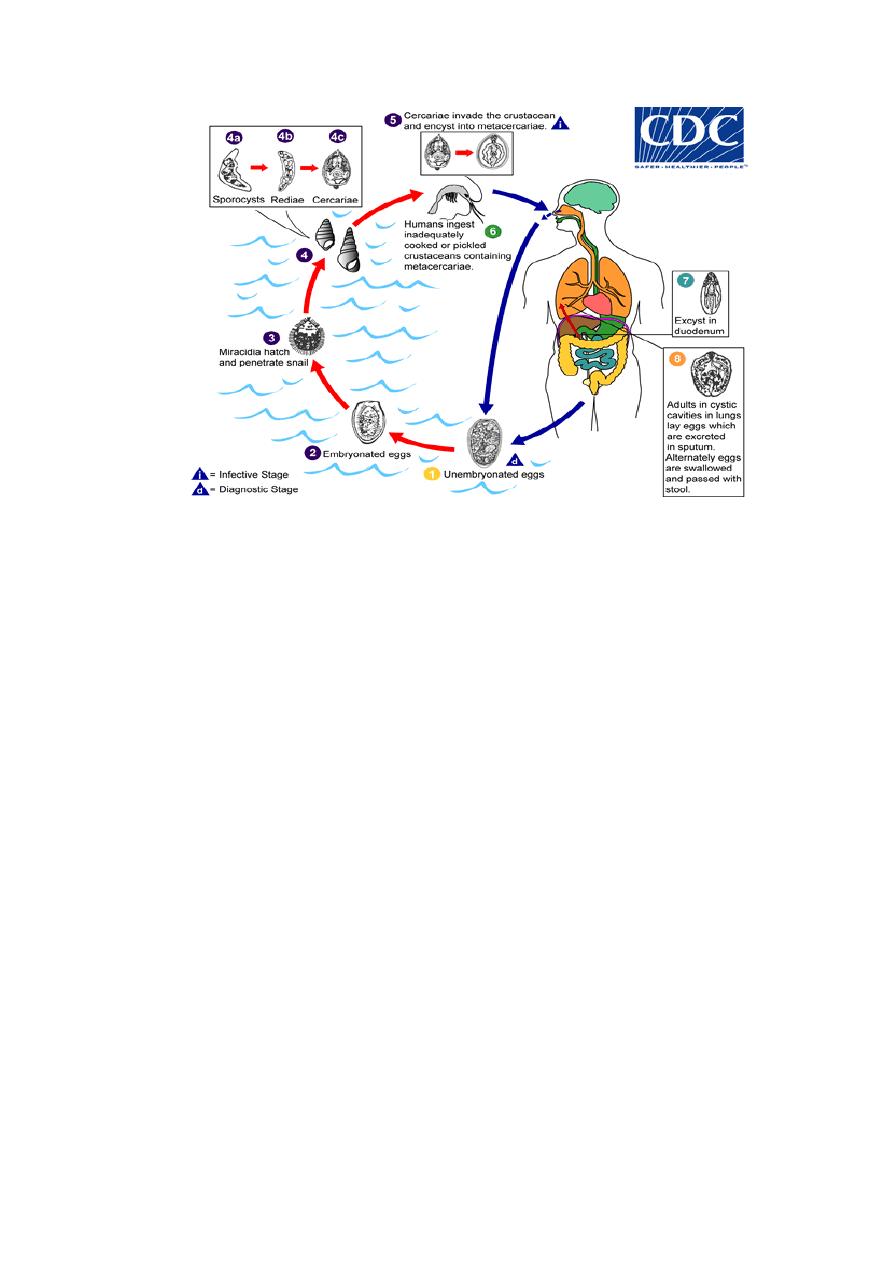
Life Cycle
Habitat Adult worms live in the lungs in cystic lesion
Definitive host man, dogs, pigs
1
st
Intermediate host fresh water snail
2
nd
intermediate host crustacean (crayfish, crab)
Infective stage encysted metacercaria
Mode of infection: Man acquires infection by eating undercooked crab or
crayfish containing metacercariae.

• The adult worms live in the respiratory tract of the definitive host.
• Unembryonated eggs escape into the bronchi and are coughed up and
voided in sputum or swallowed and passed in feces.
• The eggs mature in about 2 weeks and hatch to release free-swimming
miracidia.. These infect the first intermediate host (snail)
• Cercariae that are released from the snails swim about in streams are
drawn into the gill chambers of the second intermediate crustacean host,
crabs or crayfish
• They encyst in the gills or muscles as metacercariae.
• Definitive hosts are infected when they eat such crabs or crayfish raw or
inadequately cooked.
• The metacercariae excyst in the duodenum and penetrate the gut wall,
reaching the abdominal cavity in a few hours.
They then migrate up through the diaphragm into the pleural cavity and
lungs finally reaching in the vicinity of the bronchi, where they develop
into adult worms in 2-3 months.
The worm is hermaphroditic but usually it takes 2 for fertilization.
Sometimes, the migrating larvae lose their way and reach ectopic sites
such as the mesentery, groin and brain.
Pathogenicity and Clinical Features
Pulmonary features: In the lungs, the worms lie in cystic spaces
surrounded by a fibrous capsule formed by the host tissues.
Inflammatory reaction to the worms and their eggs lead to peribronchial
granulomatous lesions, cystic dilatation of the bronchi, abscesses,
pneumonitis and eosinophilia.
• Patients present with cough, chest pain and hemoptysis. The viscous
sputum is speckled with the golden-brown eggs. Occasionally, the
hemoptysis may be profuse.
• Chronic cases may resemble pulmonary tuberculosis.
Extrapulmonary features: The clinical features depend on the site of
involvement.
• Abdominal paragonimiasis: Occasionally the fluke migrates to liver
and intestinal wall resulting in enlarge liver, abdominal tenderness and
bloody diarrhea.
• Cerebral paragonimiasis: Encapsulated cyst of Paragonimus is found
in brain and spinal cord. Symptoms include headache, fever, paralysis,
visual disturbances and convulse seizures
.
Diagnosis
1. Microscopic examination
Detection of the eggs in sputum,
The egg may be observed in the feaces (if the patient swallows the eggs).
Or in the tissue of other tissue sample
2. Serodiagnosis detection of IgE useful in cerebral infection
3. Molecular diagnosis PCR on clinical specimens.
4. Biopsy Identification of species is made when adult or egg is recovered
from the lesion in the lungs.
5. imaging CXR and CT scan
Treatment
Praziquantel (25 mg/kg orally 3 times per day for 2 days) is the drug of
choice. Bithinol and niclofolan are also effective.
Prevention and Control
1. Adequate cooking of crabs and crayfish
2. Treatment of infected cases
3. Control of snails