Parasitology
Notes…
1
Helminths Lec.1
The term 'helminth' (Greek helmins-
’worm’) originally referred to intestinal worms, but now
found other worms, including tissue parasites and free-living species
Helminthes are multicellular (metazoa) bilalerally symmetrical animals , kingdom
Metazoa.
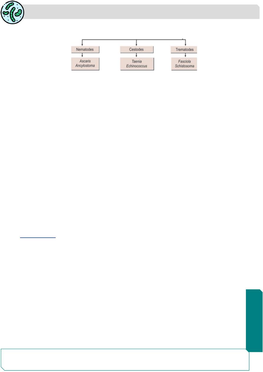
Helminths, which occur as parasite in humans belong to 2 phyla:
Phylum Platyhelminthes (flatworms)
It includes 2 classes:
1- Class
– Cestoda (tapeworms)
2- Class
– Trematoda (fl ukes )
Phylum Nemathelminthes
– It includes class
3-Nematoda
It includes 2 subclasses:
Subclass
– Adenophoraea (Aphasmidia)
Subclass
– Secernentea (Phasmidia).
General Features of Helminths
Adult Worms
Helminths have an outer protective covering, the cuticle or integument, which may be
tough and armed with spines orhooks. The cuticle of live helminths is resistant to intestinal
digestion.
- The mouth may be provided with teeth or cutting plates. Many helminths possess
suckers or hooks for attachment to host tissues.
-They do not possess organs of locomotion, but in some species the suckers assist in
movement
- Locomotion is generally by muscular contraction and relaxation.
-Many helminths have a primitive nervous system.
-The reproductive system and the excretory system is better developed

Parasitology
Notes…
2
The adult Female
1- monoecious (with functioning male and female sex organs in the same individual) the
hermaphroditic helminths, both male and female reproductive systems are present in the
same worm and self-fertilization as well as cross-fertilization take place. (e.g. Taenia
solium)
2-diecious (the two sexes, male and female, separate).
In the diecious species, males and females are separate, the male being smaller than the
female. (e.g. Ascaris lumbricoides)
Rarely the femal is parthenogenic, being able to produce fertile eggs or larvae without
mating with males (e.g. Strongyloides).
Eggs
The eggs or larvae are produced in large numbers
— as many as 200,000 or more per
female per day.
Various helminths have distinct morphology of eggs, which can be used to diff erentiate
the helminths
Larval Forms
There are various larval forms of helminths found in man and other hosts. These forms
are as follows:
1-Cestodes: The various larval forms are cysticercus, coenurus, coracidium,
cystecercoid, procercoid, hydatid cyst, and plerocercoid forms.
2-Trematodes: The various larval forms are miracidium, cercaria, redia, metacercaria, and
sporocyst.
3-Nematodes: The various larval forms are microfi laria, filariform larva, and rhabditiform
larva
Helminths differ from protozoans in their inability to multiply in the body of the host.
Protozoans multiply in the infected person, so that disease could result from a single
infection.
But in the helminths a single infection does not generally leads to disease Heavy worm
infection lead to disease .
Parasitology
Notes…
3
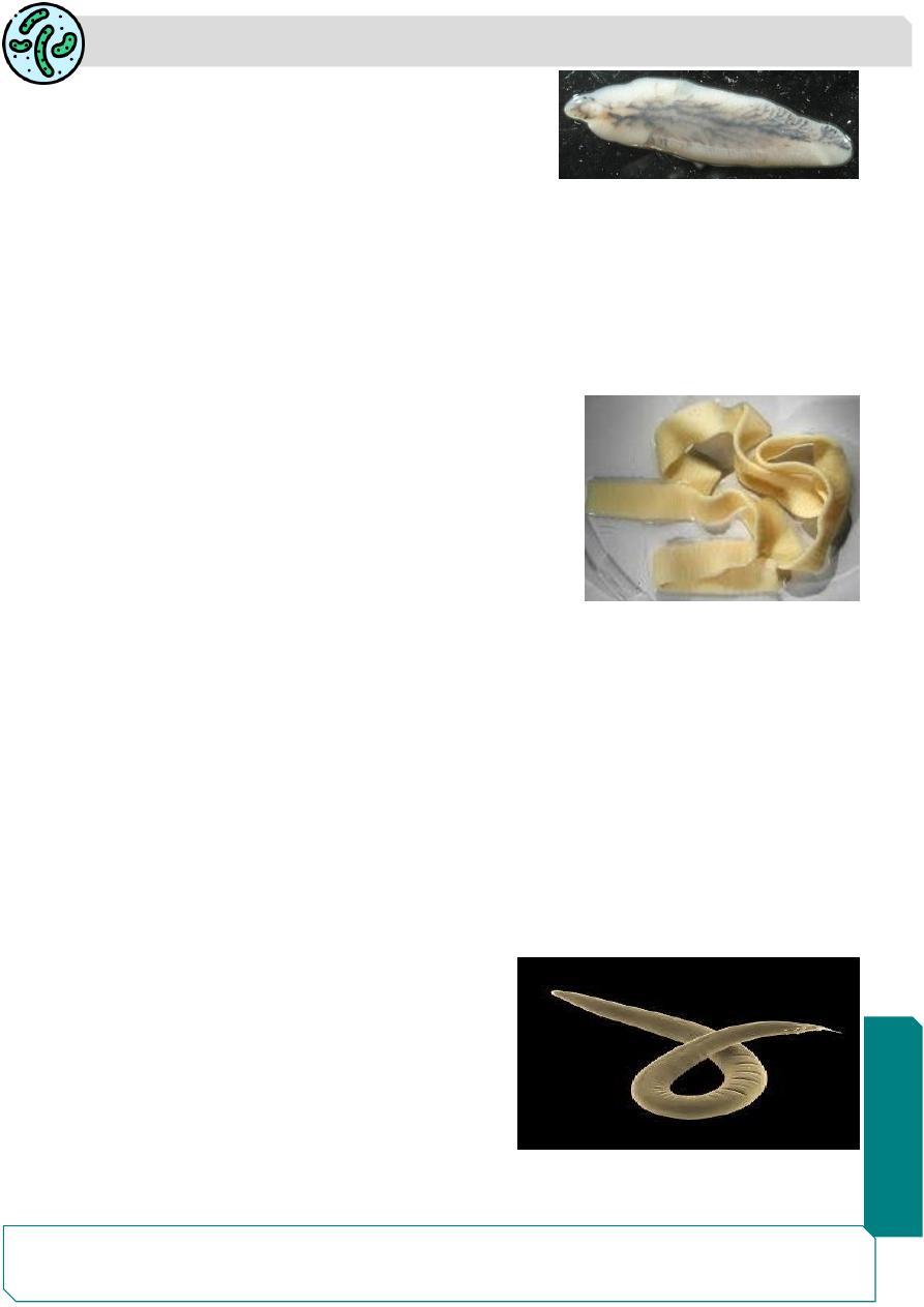
Class Trematoda
Trematodes have flat or fleshy, leaf-like unsegmented
bodies.
-The alimentary canal is present but is incomplete i.e., without an anus.
-They possess suckers but no hooks.
- The sexes are separate in the schistosomes, while the other flukes are hermaphroditic.
-They are oviparous.
Class Cestoda
Cestodes have tape-like, dorsoventrally flattened segmented bodies.
-They do not possess an alimentary system.
-The head carries suckers and some also have hooks.
- They possess scolex, neck, and proglottids.
- They are monoecious and body cavity is absent.
-They are oviparous
(Nematoda)
Nematodes are elongated, cylindrical worms with an unsegmented body
-They possess a relatively well-developed complete alimentary canal, with an anus.
- Body cavity is present.
- The head does not have suckers or hooks, but may have a buccal capsule with teeth or
cutting plates.
-The sexes are separate (diecious).
-They are either oviparous or Viviparous
Nematodes are generally resemble the common earth worm in appearance,However,
taxonomically earthworms are not nematodes .
The name nematode means thread-like (nema
meaning thread)
Nematodes
are
elongated,
cylindrical,
unsegmented worms with taper
ing ends.’.
Unlike trematodes and cestodes the most
nematodes are free-living forms found in soil and
water.

Parasitology
Notes…
4
Several species are parasites of plants, many nematodes parasitize invertebrate and
vertebrate animals. ( economic importance).
Their body is covered with a tough outer cuticle, which may be smooth, striated, or spiny.
The middle layer is hypodermis and the inner layer is the somatic muscular layer.
The body cavity is a pseudocele, in which all the viscera are suspended.
The digestive system is complete, consisting of a anteriorly placed mouth leading to the
esophagus, which characteristically varies in shape and structure in different groups.
The intestine is lined with a single layer of columnar cells and leads to the rectum, opening
through the anus.
In the male the rectum and the ejaculatory duct open into the cloaca.
Nematodes have simple excretory and nervous systems
The nematodes are diecious i.e. the sexes are separate.
The male reproductive system consists of a single delicate tubule differentiated into testis,
vas deferens, seminal vesicle, and ejaculatory duct, which opens into the cloaca.
It also includes copulatory structures such as spicules or bursa or both.
The female reproductive system consists of the ovary, oviduct, uterus, and vagina.
Female nematodes may produce eggs (oviparous) or larvae (viviparous).
Some lay eggs containing larvae which immediately hatch out
((ovoviviparous
Life Cycle
The life cycle of nematodes consists typically of larval stages and the adult form.
Man is the optimum host for all the nematodes.
The life cycle need only one host, except the superfamilies
Filarioidea and Dracunculoidea, where two hosts are required. Insect vectors and Cyclops
constitute the second hosts in these superfamilies, respectively.
Nematodes localize in the intestinal tract and their eggs pass out with the feces of the
host.
They undergo few developmental changes before they enter new host.
Modes of Infection
1-By ingestion of:
- Eggs: Ascaris, Enterobius. Trichuris
- Larvae within intermediate host: Dracunculus
-Encysted larvae in muscle: Trichinella
Parasitology
Notes…
5
2-By penetration of skin: Ancylostoma, Necator, Strongyloides
3- By blood-sucking insects: Filariae
4- By inhalation of dust containing eggs: Ascaris, Enterobius
The female nematodes may be divided as follows:
1- Oviparous (laying eggs):
A- Unsegmented eggs: Ascaris, Trichuris
B-Segmented eggs: Ancylostoma, Necator
C-Eggs containing larvae: Enterobius
2- Viviparous (producing larvae): Trichinella, Wuchereria, Brugia, Dracunculus.
3-Ovoviviparous (laying eggs containing fully formed larvae, which hatch out
immediately): Strongyloides
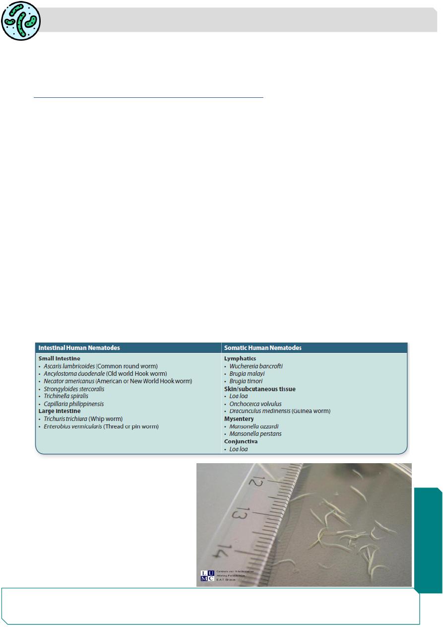
The largest number of helminthic parasites of humans belong to the class of nematodes
species
About one-half of the nematodes parasitic for man are intestinal, the others are found in
various tissues.
Pathogenicity of intestinal nematodes may be due to larval migration through tissue,
piercing of intestinal wall, blood loss, or allergic reactions to secretions of adults or larvae
Nematodes on the Basis of the Habitat of Adult Worms
Enterobius vermicularis
Enterobius vermicularis ,human
pinworm , threadworm or seat
worm,previously
called
Oxyuris
vermicularis)
Human is the only host of
E.vermicularis.

Parasitology
Notes…
6
The name Enterobius vermicularis means a tiny worm living in the intestine (Greek
enteron
—intestine, bios— life, and vermiculus—small worm).
The term Oxyuris means ‘sharp tail’, a feature of the female worm, from which the name
‘pinworm’ is also derived.
Leuckart (1865) first described the complete life cycle of the parasite.
Habitat
Adult worms are found in the caecum, appendix, and adjacent portion of ascending colon.
It has worldwide distribution
E. vermicularis is more common in temperate countries than in the tropics.
In the United States 40 million persons are infected with pinworms,
Pinworm infection is prevalent in large family groups and in schools and mental
institution,infections are more prevalent in the poor people
The infection is more common in children than adults
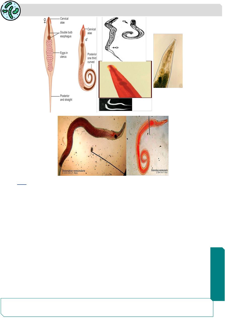
Morphology:
The adult male and female have an anterior expansion from both side (ventral and
dorsal) called (cervical alae).
the mouth is surrounded by 3wing-like cuticular expansions ,the oesophagus has a
double-bulb structure (oesophagial bulb)
The male
measures 2-5 mm long and width of 0.1- 0.2mm .
The posterior end is strongly curved with copulatory spicule
Male live for about 7
–8 weeks
Female
length 8- 13 mm ,0.3-0.5 mm width. They are light yellowish to white thread
like .
The posterior end is sharply pointed
The vulva is located just in front of the middle third of the body and opens into the single
vagina, which leads to the paired uteri, oviducts, and ovaries.
In the gravid female the whole body is filled by the distended uteri carrying thousands of
eggs.
The gravid female is oviparous.
Females survive for 5
–12 weeks
Parasitology
Notes…
7
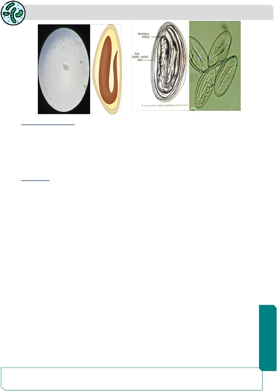
Egg
The egg is colorless (with out bile-stained), floats in saturated salt solution.
It has a characteristic shape, being elongated ovoid, fl attened on one side, and convex
on the other (D shape ), measuring 50
–60 μm by 20–30 μm
The egg shell is double layered ,colorless transparent inner layer and The outer
albuminous layer makes the eggs stick to each other and to clothing and other objects.
The egg contains a tadpole-shaped coiled embryo, which is fully formed, but becomes
infectious only 6 hours after being deposited on the skin.
Under cool moist conditions, the egg remains viable for about 2 Weeks.
A single female worm lays 5,000
–17,000 eggs.
Parasitology
Notes…
8
Mode of infection:
1) By swallowing fully developed eggs with food or water.
2) Inhalation of eggs (light infection).
3) Autoinfection
4) Retroinfection
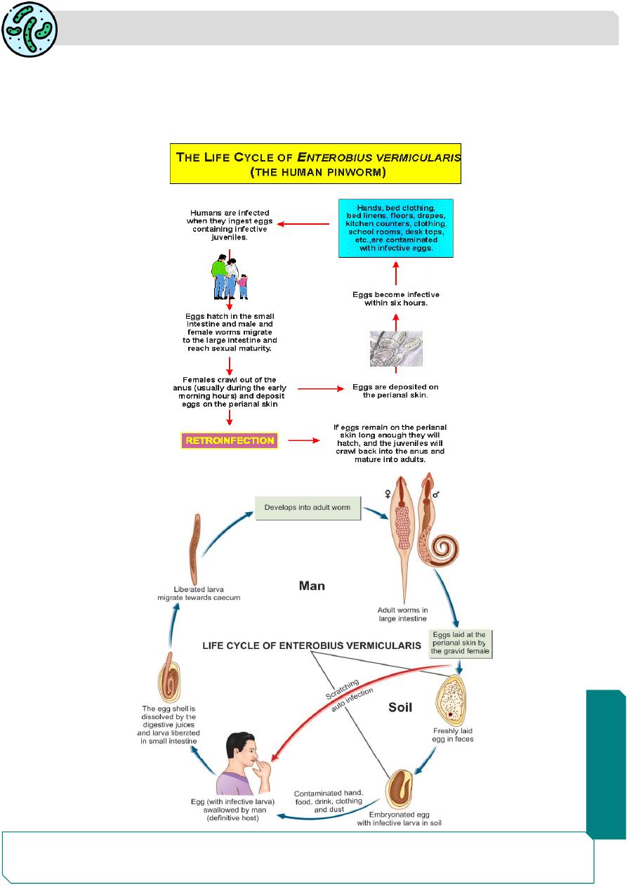
Life Cycle
life cycle only in one host.
No intermediate host and does not undergo any systemic migration
Natural host: Man
Infective form: Embryonated eggs
Mode of infection: Man acquires infection by ingesting embryonated eggs containing
larva by means of
1-Contaminated fingers
2-Autoinfection
Eggs laid on perianal skin containing infective larvae are swallowed and hatch out in the
intestine.
They moult in the ileum and enter the caecum, where they mature into adults.
It takes from 2 weeks - 2 months from the time the eggs are ingested, to the development
of the gravid female, ready to lay eggs
The gravid female migrates down the colon to the rectum.
At night, when the host is in bed, the worm comes out through the anus and crawls about
on the perianal skin to lay its sticky eggs.
Parasitology
Notes…
9
The female worm may wander into the vulva, vagina and even into the uterus and fallopian
tubes, sometimes reaching the peritoneum.
The male is seldom seen as it does not migrate,It usually dies after mating and is passed
in the feces

Parasitology
Notes…
10
When all the eggs are laid, the female worm dies or gets crushed by the host during
scratching.
The worm may often be seen on the feces, having been passively carried from the rectum.
The eggs rarly found in feces, as the female worm lays eggs in the perianal area .
Crawling of the gravid female worm leads to pruritis and the patient scratches the perianal
area.
These patients have eggs of E. vermicularis on fingers and under nails leading to
autoinfection
Autoinfection:
Ingestion of eggs due to scratching of perianal area with fingers leading
to deposition of eggs under the nails. This type of infection is mostly commo in children.
This mode of infection occurs from anus to mouth.
Retroinfection:
In this process, the eggs laid on the perianl skin immediately hatch into
the infective stage larva and migrate through the anus to develop into worms in the colon.
This mode of infection occurs from anus to colon
Some time pinworm eggs which inhaled and then it reach the pharynx and pass down the
oesophagus and migrate to the large intestine.