Parasitology
Notes…
1
Helminths Lec.2
Clinical features
pinworm-infected persons are asymptomatic.
The adult worms may cause slight irritation of the intestinal mucosa
Infection common in the children, more in females than male
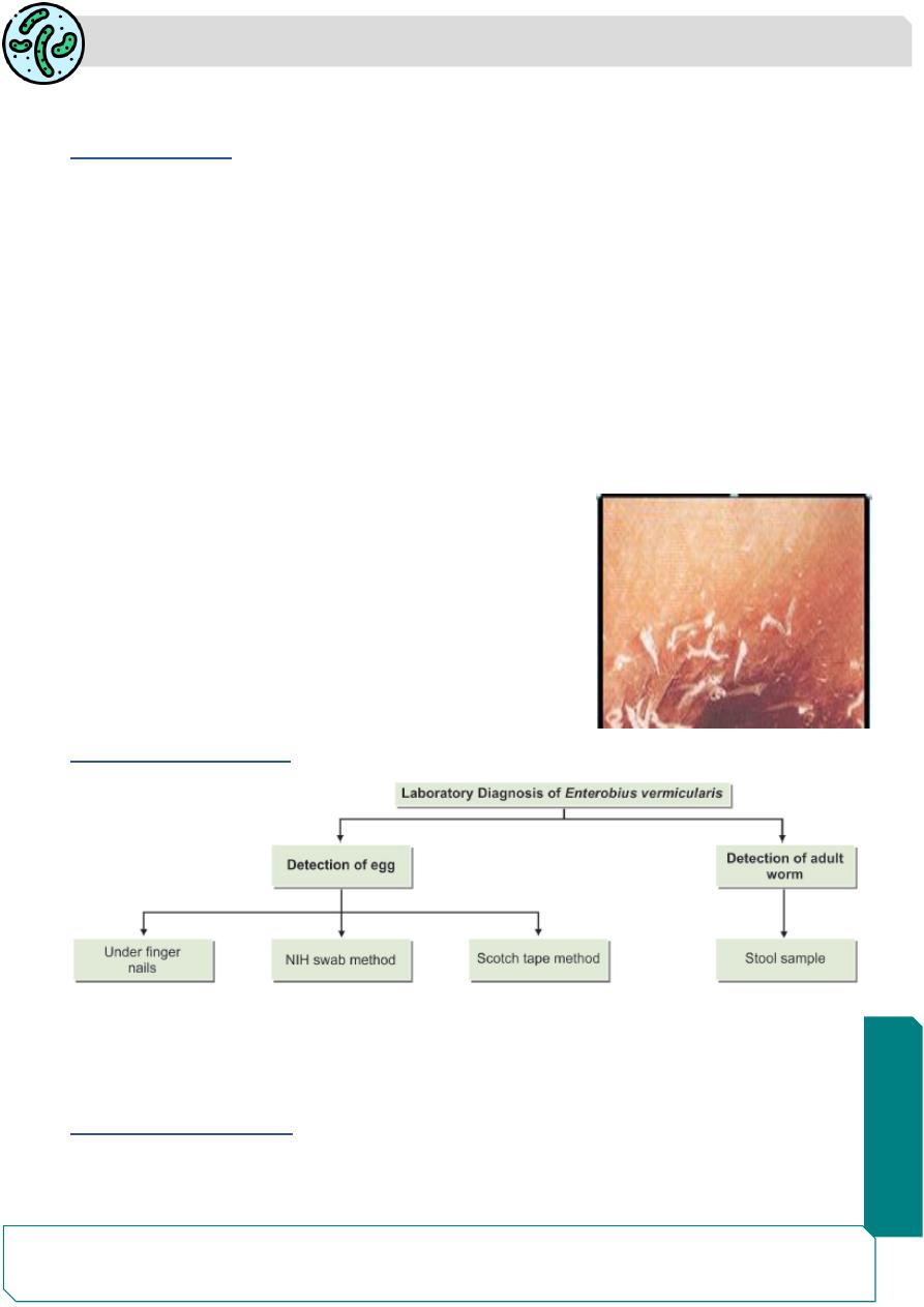
the adult worm rare can be seen in the stool , but in the skin of perianal area
About one-third of pinworm infections are asymptomatic. Perianal pruritus , irrtation is the
cardinal symptom ,the itching which is often worse at night as a result of the nocturnal
migration of the female worms to laying the eggs into perianal area which may lead to
excoriation and bacterial superinfection
worm migrates at night induce sleep disturbance ,nocturnal enuresis
Heavy infections have been alleged to cause abdominal
pain and weight loss.
On rare occasion pinworms invade the female genital
tract, causing vulvovaginitis ,salpingitis or granuloma of
the pelvic or peritoneal cavity .
appendicitis especially in children.
Eosinophilia is uncommon.
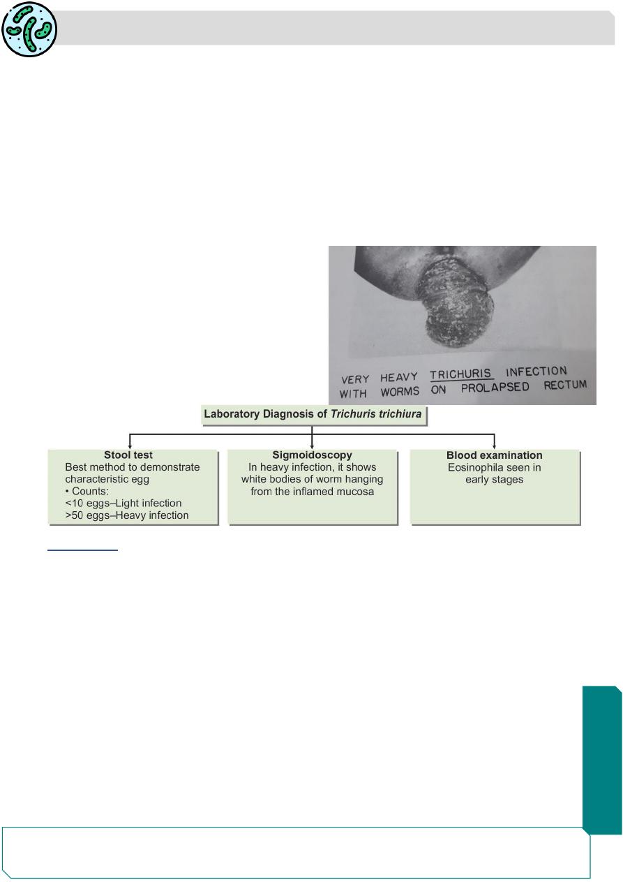
Laboratory Diagnosis :
Pinworm infestation can be suspected from the history of
perianal pruritus.
Diagnosis depends on the demonstration of the eggs or adult worms
Demonstration of Eggs
Eggs are present in the feces only in a small proportion of patients and so feces
examination is not useful in diagnosis.
Parasitology
Notes…
2
Egg can be demonstrated in swabs collected from the sites perianal area in early
morning, before going to the toilet or bathing.
Swabs from perianal folds are most often positive.
The eggs may sometimes be demonstrated from beneath the finger nails in infected
children
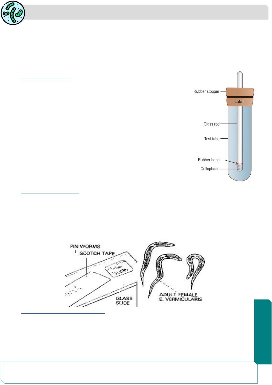
NIH Swab Method
The NIH swab (named after National Institutes of Health,USA)
NIH swab. A piece of transparent cellophane
is attached with rubber band to one end of a glass rod, which is
fi xed on a rubber
stopper and kept in a wide test tube
The cellophane part is used for swabbing by rolling over the
perianal area .
It is returned to the test tube and sent to the laboratory, where the
cellophane piece is detached, spread over a glass side and
examined microscopically
Scotch Tape Method
Another method for collection of specimens is with scotch tape (adhesive transparent
cellophane tape)
The tape is transferred to a glass slide, sticky side down, with a drop of toluene for clearing
and examined under the microscope
Demonstration of Adult Worm
The adult worms may sometimes be noticed on the surface of stools
They may occasionally be found crawling out of the anus while the children are asleep.
They may be detected in stools collected after an enema and may be in the appendix
during appendectomy.

Parasitology
Notes…
3
Note: Unlike the other intestinal nematodes, Enterobius infection is not associated with
eosinophilia or with elevated IgE
Treatment
Infected children and adults should be treated: Albendazole (400 mg once) or
mebendazole (100 mg once) can be used for single dose therapy. while piperazine has
to be given daily for one week.
It is necessary to repeat the treatment after 2 weeks
As pinworm infection usually a ffects a group, it is advisable to treat the whole family or
group of children in school
Prevention and control
1-All the patients sould be treated.
2-Health education for community and personal hygiene such as frequent hand washing,
finger nail cleaning and cut, and regular bathing .
3-Frequent washing of night clothes and bed linen..
Trichuris trichiura
Trichuris trichiura or whipworm.
Disease called Trichuriasis,whipworm infection.
first described by Linnaeus in 1771.
Trichuris means a ‘hair-like tail’ (Greek trichos—hair, oura—tail).
This name is not quite correct because it is the anterior end of the worm that is hair-like
and not the tail.
The name whipworm is more apt as the thick posterior part resembles the stock and the
thin anterior end resembles the lash of a whip.
It is worldwide in distribution, but is much more common in the tropics.
The infection is widespread in tropical Africa, South America, and South-east Asia.
Some 800 million people are estimated to be infected with this worm
Habitat
T. trichiura lives in the large intestine.
The adult worms are found attached to the wall of the caecum and less commonly in
appendix, colon, and anal canal
Parasitology
Notes…
4
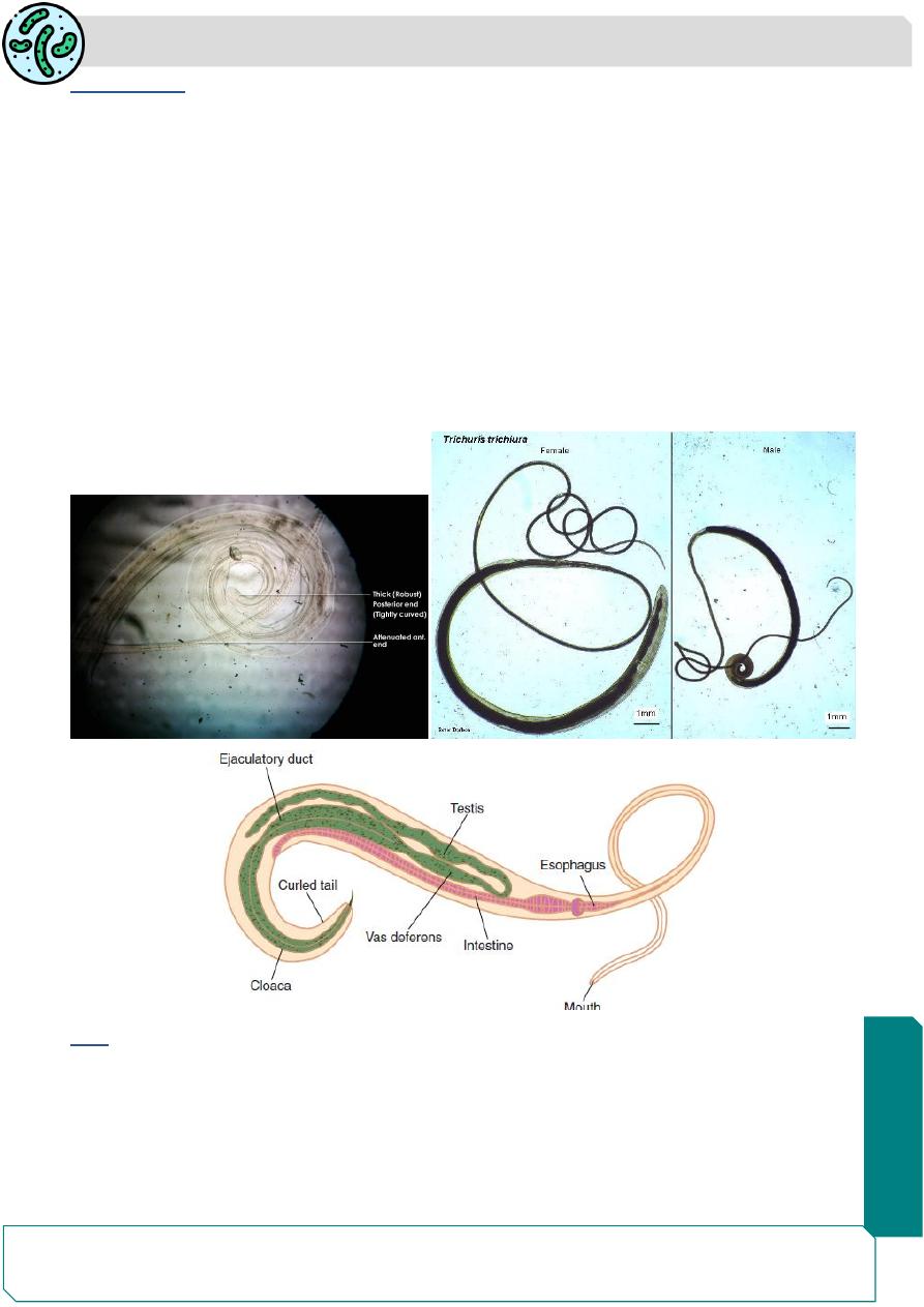
Morphology
The adult worm is flesh colour ,the shape it resembles a whip, with the anterior 3/5 is
thin and thread-like .
the posterior 2/5 is thick and fleshy appearing like the handle of a whip .
the anterior portion which contains the capillary oesophagus embedded in the mucosa .
posterior part contains the intestines and reproductive organs
The male 30-45mm long , posterior end coiled ventrally .
The female 40-50mm long ,the posterior end straight ,blunt and rounded
Humans are the only natural host for Trichuris trichiura But similar worms are found in
pigs and monkeys
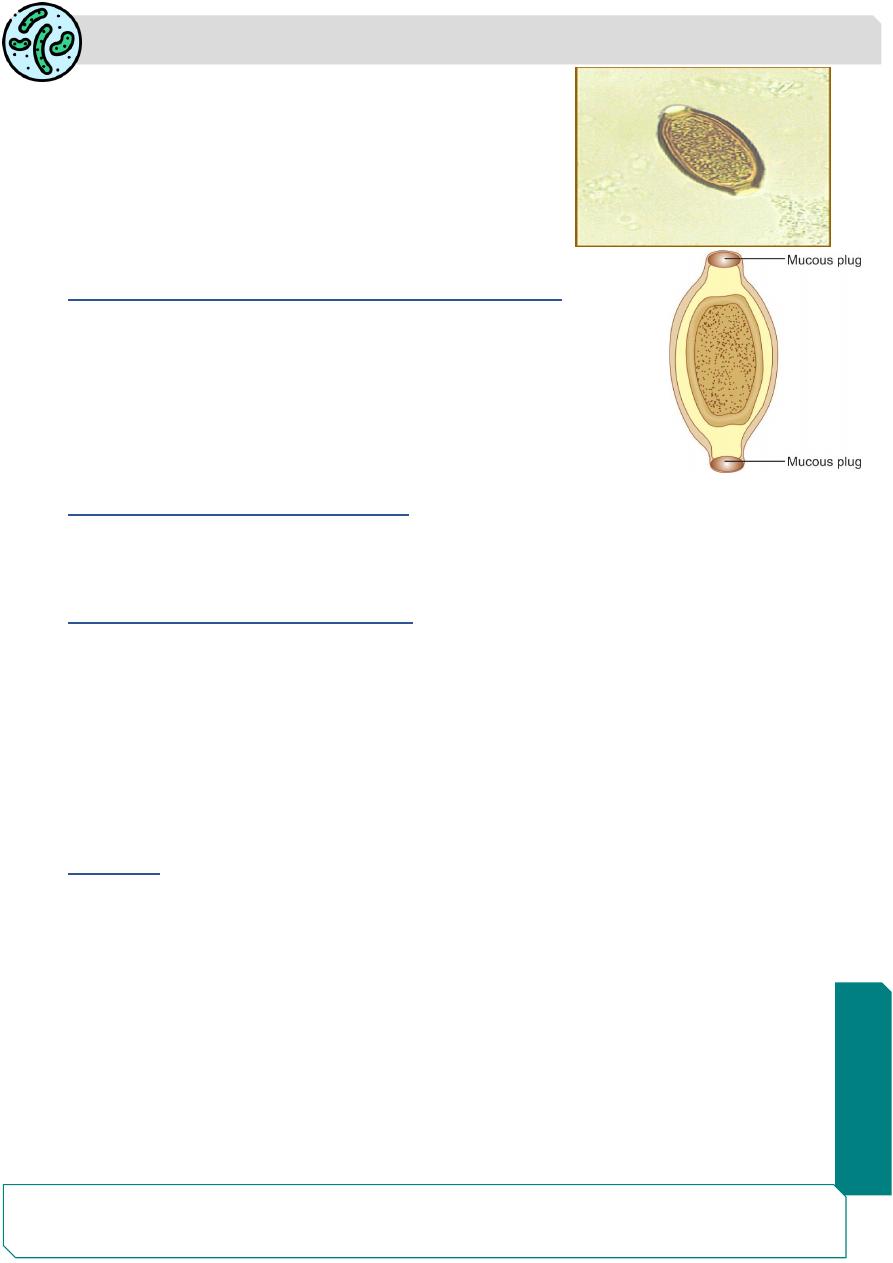
Egg
The egg brown in color being bile-stained.
It has a triple shell, the outer most layer of which is stained brown.
It is barrel-
shaped and about 50 μm long , 25 μm wide in the middle, with a projecting
mucus plug at each pole containing an unsegmented ovum .
male
Adults
Parasitology
Notes…
5
The plugs are colorless.
the egg foats in saturated salt solution.
When freshly passed, the egg contains an unsegmented
ovum,it is not infective for humans.
The fertilized female lays about 5,000 eggs per day
Helminths whose eggs float in saturated salt solution
• Enterobius vermicularis
• Ancylostoma duodenale
• Necator americanus
• Ascaris lumbricoides
• Trichuris trichiura
Nematodes present in large intestine
• Enterobius vermicularis
• Trichuris trichiura
Nematodes present in small intestine
•Strongyloides stercoralis
•Ascaris lumbricoides
•Ancylostoma duodenale
•Necator americanus
•Trichinella spiralis
•Trichostrongylus spp.
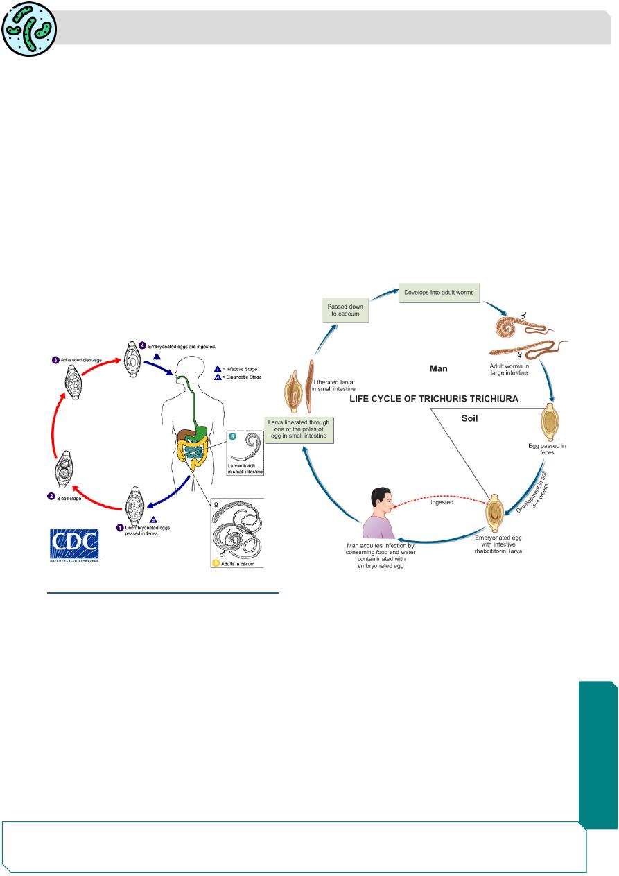
Life Cycle
Natural host:
Man.
No intermediate host is required.
Infective form:
Embryonated eggs contaning Rhabditiform larva.
Adult female worm lives in large intestine worm lays eggs which are discharged in feces.
The egg undergoes development in soil, optimally under warm, moist, shady conditions,
when the infective rhabditiform larva develops within the egg in 3
–4 weeks.
At lower temperatures, this may be delayed for 3 months or more ,these embryonated
eggs are infective to man.
Parasitology
Notes…
6
Mode of Transmission
: Infection occurs in humans when the mature embryonated eggs
containing the infective larvae are swallowed in contaminated food or water.
The eggs hatch in the small intestine and the larva penetrate and develop in the intestinal
villi with in 3-7days, and then return to lumen and migrate to the cecum.
In about 2
–3 months, they become mature adults and lie embedded in the cecal wall, with
the thread-like anterior portion piercing the mucosa and the thick posterior end projecting
out.
The gravid adult female lays eggs, which are discharged in feces and the cycle is repeated
Eggs start appearing in feces usually about 3 months after infection
The worm has a lifespan of 5
–10 years
Pathogenecity and Clinical Features
The disease called (trichuriasis, whipworm infection, or trichocephaliasis) is
asymptomatic, except when the worm load is heavy.
Disease may result either due to mechanical effects or
allergic reaction.
The worms lie threaded into the cecal mucosa and even though it is not a blood feeder,
oozing of blood may occur at the sites of attachment.
The blood loss is about 0.005 mL per worm per day. Over a period of time, this may lead
to anemia and malnutrition.
Parasitology
Notes…
7
mechanical blockage of the appendiceal lumen by masses of whipworms may cause
acute appendicitis
In heavy infection, the worm may be abundant on the colonic mucosa, even upto the
rectum.
Mucus diarrhea, chronic dysentery and abdominal pain, and weight loss .
Some patients particularly young children, may develop rectal prolapse. with whipworm
dysentery.
In heavy infection, sigmoidoscopy may show white bodies of worm hanging from the
inflamed mucosa called coconut cake rectum
Treatment:
Mebendazole (100 mg 12 hourly for 3
–5 days) or
Albendazole (single dose of 400 mg) are effective with cure rates of 80%.
Prophylaxis
1-Proper disposal of feces.
2-Avoiding consumption of unwashed fruits and vegetables.
3-Treatment of infected persons.