Red blood cells specialisations
2) no nucleus
extra space inside
3) contain haemoglobin
the oxygen carrying
molecule
250million molecules
/ cell
1) biconcave shape
increases the
surface area so
more oxygen can be
carried
Haemoglobin
•
gives red blood cells their colour
•
can carry up to 4 molecules of O
2
•
associates and dissociates with O
2
•
contains iron

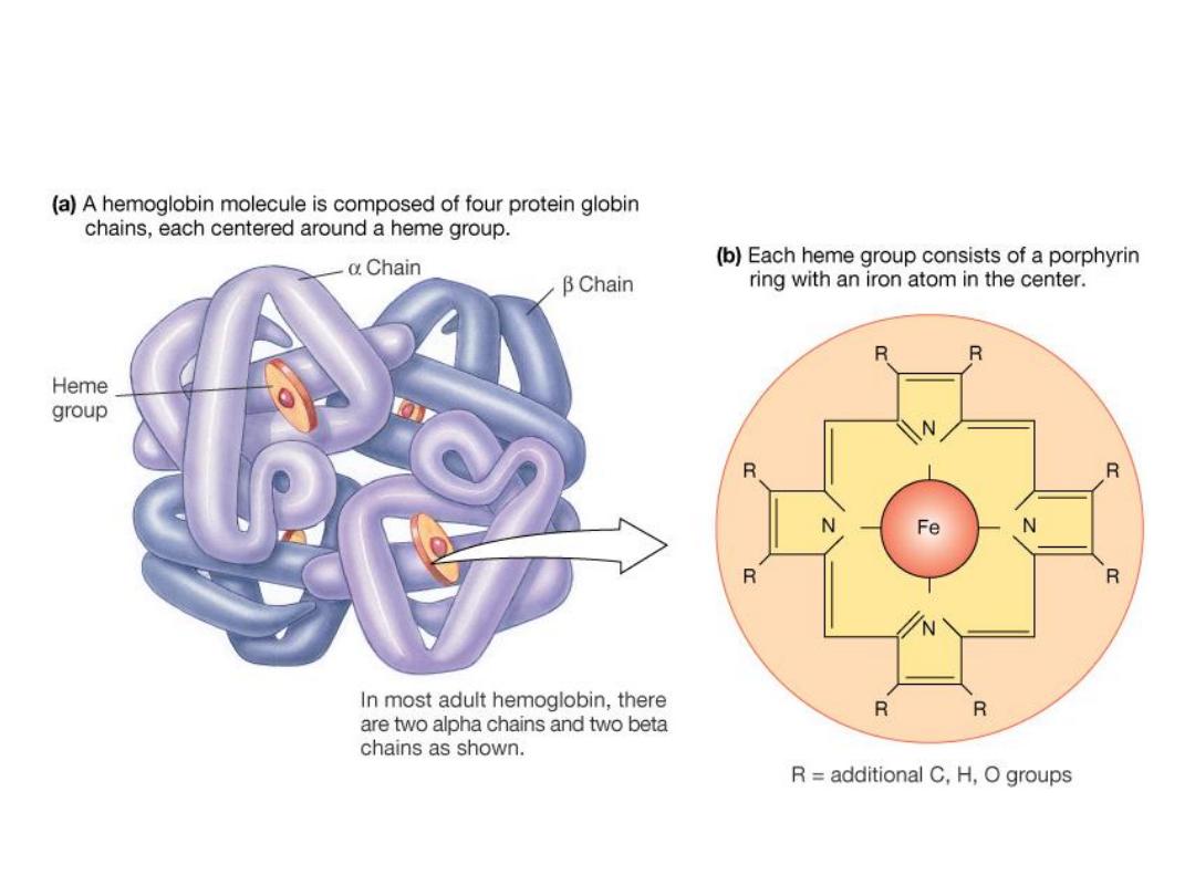
Description.
Hemoglobin
is
the
oxygen-carrying
pigment of the RBCs. It is composed of
amino acids that form a single protein
called
“globin” and a compound called
“heme.” Heme contains iron atoms and
the
red
pigment
porphyrin.
Each
erythrocyte contains approximately 250-
300 million molecules of hemoglobin.

SI Units
Females
12–16 g/dL
7.45–9.90 mmol/L
Pregnant
10–15 g/dL
6.3–9.9 mmol/L
Males
13.6–18.0 g/dL
8.44–11.17 mmol/L
Children
Neonates
14–27 g/dL
8.69–16.76 mmol/L
3 months
10–17 g/dL
6.21–10.55 mmol/L
1–2 years
9–15 g/dL
5.58–9.31 mmol/L
6–10 years
11–16 g/dL
6.82–9.92 mmol/L
Panic Levels
<5 g/dL
<3.10 mmol/L
>20 g/dL
>12.41 mmol/L
Focus on RBCs:
Figure 16-7a, b: Bone marrow
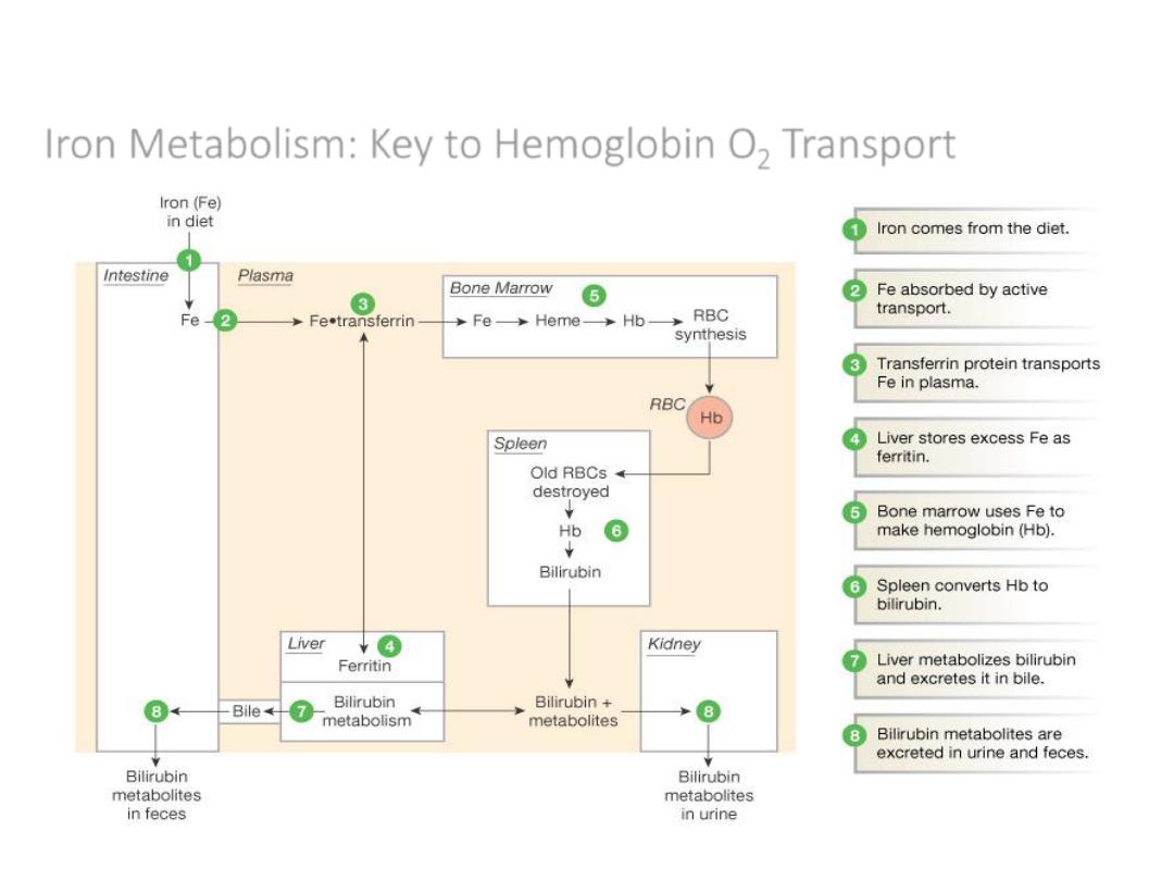
Iron Metabolism: Key to Hemoglobin O
2
Transport
Figure 16-8: Iron metabolism
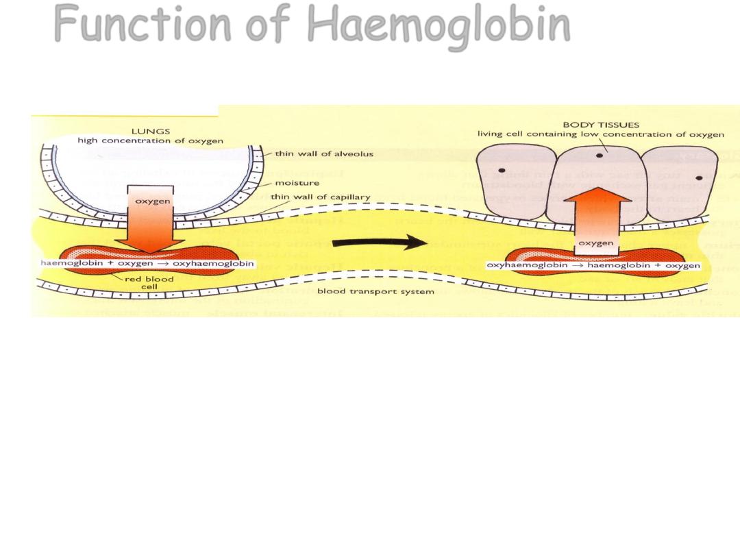
When there is a high concentration of oxygen e.g in
the alveoli haemoglobin combines with oxygen to
form oxyhaemoglobin. When the blood reaches the
tissue which have a low concentration of oxygen the
haemoglobin dissociates with the oxygen and the
oxygen is released into body tissues
Function of Haemoglobin

Increased.
Burns (severe), congestive heart failure, chronic
obstructive pulmonary disease (COPD),
dehydration, diarrhea, erythrocytosis,
hemorrhage, hemoconcentration, high altitudes,
intestinal obstruction (late), polycythemia vera,
and thrombotic thrombocytopenic purpura. Also
conditions that increase red blood cells (RBCs).
Drugs include gentamicin, methyldopa, and
pentoxifylline.

Decreased.
anemia
(iron
deficiency),
carcinomatosis,
cystic
fibrosis,
fat
emboli,
fatty
liver,
fluid
retention,
hemorrhage,
hemolysis,
hemolytic
reaction
to
chemicals
or
drugs
or
prosthetics,,hydremia
of
pregnancy, hyper thyroidism, hyper vitaminosis A,
hypothyroidism,
intravenous
overload,
leukemia,
lymphoma, platelet apheresis, pregnancy, renal cortical
necrosis, sarcoidosis, severe hemorrhage, systemic
lupus erythematosusof ,and transfusion of incompatible
blood. Also, conditions that decrease RBCs. Drugs
include antibiotics, antineoplastic agents, Apresoline
(hydralazine HCl with hydrochlorothiazide), aspirin,
hydantoin
derivatives,
indomethacin,
monoamine
oxidase inhibitors,, rifampin, sulfonamides, tridione,
and zidovudine (AZT); vegetarian diet.
1-Sahli Method
A-Material :
1- Graduated hemometer tube
2- Hb. Pipette
3-HCl (0.1 Normality)
4- Standard fluid
•The graduated Hemometer tube is filled up to
mark 10 with 0.1 Normality of HCl .
• add 0.02 ml of blood and mix then for about
10 minutes .
•When acid hematin is formed, then diluted with
D.W. until the color matches with the standard .
By using drabken solution
Method
1- add 0.02ml of blood by Hb Pipette to 5 ml of drabken
solution in test tube
left for about 10 minutes.
2- the reading is taken in colorimeter by using green filter
or 450um and
standard curve or by using hemoglobin meter directly
with out standard