Amyloidosis
A medical condition resulting from aggregation of extracellularly deposited abnormal proteins called amyloid fibrils that cause damage to organs and tissues.
Proteins aggregate and form fibrils called amyloid fibrils.●

● These fibrils are insoluble, linear, rigid and measures approximately 7.5 to 10 nm in width & indefinite length.
Classification of Amyloidosis
Chemical subtypes95% of amyloid material consists of fibril protein and 5% is the P-component which is a glycoprotein similar to C-reactive protein.
A- Chemically according to amyloid precursor protein amyloid is classified into:
1- AL (amyloid light chain) which is derived from the Ig light chain and deposited in the tissues of patients with plasma cell dyscrasias (like multiple myeloma).
2- AA (amyloid associate) which is derived from serum amyloid associated protein (SAA) synthesized by the liver in cases of chronic inflammations like bronchiectasis, Rheumatoid A, TB, osteomyelitis, abscesses.
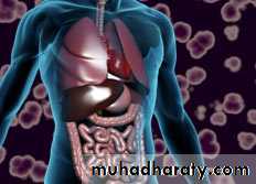
B- According To the Distribution of Amyloid deposition into: Systemic & Localized,
localized; close to cells producing it.
-Systemic; distant sites from the cells producing these abnormal proteins.
C- According to the etiology
● acquired (secondary) & Inherited (primary)
Clinical Manifestations
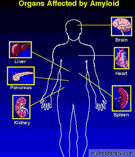
Clinical Manifestations depend on which organ is affected:
E.g. Heart:
-Cardiomyopathy
-Heart failure
-Ischemic heart disease
● Renal: Proteinuria, nephrotic syndrome, renal failure leading to kidney transplant or dialysis.
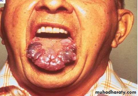
Muscle: Hypertrophy of muscles, macroglossia●
● Skin: Nodules, plaques, easy bruising
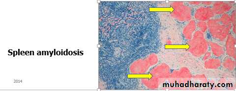
● GI: Organomegaly (Hepatomegaly, splenomegaly), abnormal bowel movement usually constipation, malabsorption
CNS/Neuro: Neuropathy, dementia.
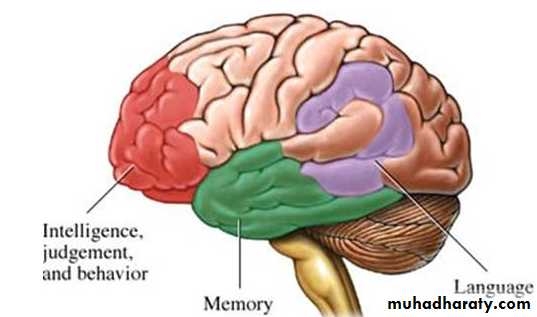
Senile cerebral amyloidosis in patients with Alzheimer’s Disease
Alzheimer's disease (AD) is a progressive degenerative disease of the brain from which there is no recovery. The disease slowly attacks nerve cells in all parts of the cortex of the brain and some surrounding structures, thereby impairing a person's movement and memory. Ult imately, patient loses memory and many other mental functions.

Diagnosis
● Clinical: Unexplained medical disorder and you suspect amyloidosis: e.g heart failure, proteinuria, hepatic dysfunction● Ultimately, you need tissue biopsy from: Abdominal fat, kidney, oral mucosa, rectal mucosa.
● Bone marrow biopsy
Characteristics common to all amyloid subtypes
● Hematoxylin and Eosin (HE) staining results in an extracellular deposition of amorphous eosinophilic appearance when viewed on light microscopy.
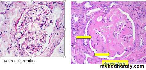
Kidney: Histologically amyloid is deposited primarily in the glomeruli (widening of the mesangial matrix with homogenous, structureless material + thickening of basement membrane).
Special tests for diagnosis of amyloidosis
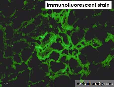
Diagnosis of amyloidosis is by a special stain for amyloidosis (like Congo red, Lugol’s iodine, Immunofluorescent stain), electron microscopy.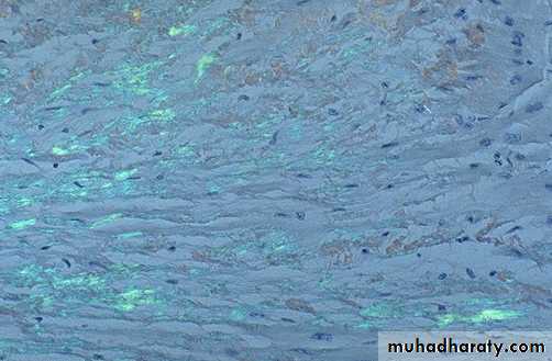
● Congo red staining results in bright green fluorescence/birefringe apple green color when viewed under special microscope (containing polarized light).
Congo red stain (apple green color)
Definite diagnosis is by electron microscope; non-branching fibrils of indefinite length and a diameter of approximately 7.5 - 10 nm.
End