Anorectal Anomalies
A perinium without an anal opening is traditionally described as
imperforated anus .This is inappropriate terminology because most of
anorectal malformations communicate by afistulous track with either the
urinary or genital system or actually open to the skin of the perineum .
Classification:
Development of the distal bowel is arrested at one of two levels ,each
with it’s subtypes .The principle of distinction is in the relation of the end
of the bowel to the chief muscle of continence the puborectalis component
of levator ani.
Arrested development at or above this sling (supralevator lesion)
produces rectal deformities .
Arrested development below the sling (translevator lesion) produce anal
deformities .
In each group the bowel may end blindly or communicate by a fistula with
neighboring viscus or the perineal skin.
Generally :Anorectal anomalies classified into 2 types :
1. Low type (Anal deformities),this is subdivided into :
• Stenosed anus
•
-Covered anus

•
-Ectopic anus
•
-Cutanous fistula ,extending to the perineum or to the scrotum or
ventral skin of penis in male.
• -Ano vestibular fistula in females There are some variations between
male and females .
2. High type (Rectal deformities),in this group the colon or rectum
terminates above the levator ani muscles ,there may be a fistulous
communication to the urinary tract in males(Recto-urethral fistula) or to
the genital tract in females(recto-vaginal fistula) ,some times there is no
connections .
There is a higher rate of associated anomalies in the urinary tract ,vertebral
column ,alimentary canal and heart ,for this reason the mortality is higher
than low type lesions in addition to technical difficulties (i.e. pull through
surgery).
Clinical features :
Any newborn baby should be examined for the presence of congenital
abnormalities .
Examination of the perineum is part of this routine examination, which
may reveal the absence or abnormal anal opening .
Some times the parents are unaware of the baby problem and they bring
him with abdominal distention , vomiting and the passage of meconium
through the urethra or vagina .
Infants with anal stenosis may present weeks or months later with
excessive screaming on defecation with the passage of toothpaste like
stool due to narrow anus.
Examination:
we may be able to differentiate low from high lesions :
Low type there is anal dimple with:
• Stenosed anus :you can’t admit you little finger .
• Covered anus with skin covering the anus with meconium under
the skin .
• Anterior ectopic anus .
Cutanous fistula filled with meconium.or vestibular fistula.
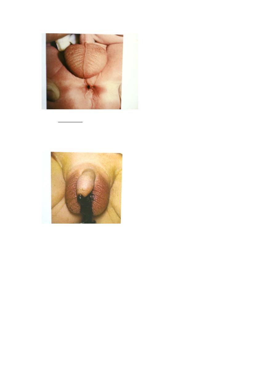
8B
93BU
High type:
9B
flat perineum(no anal dimple)
10B
Meconium may be passed from the urethra or vagina indicating the
presence of fistula .
45B
112B
TYPES OF DEFECTS ( classification )
113B
MALE DEFECTS
114B
1. Low Defects : Perineal fistula
138B
Median raphe fistula
139B
Bucket handle malformation
140B
Anal stenosis
141B
Anal membrane
115B
2. Rectourethral bulbar fistula
116B
3. Rectourethral prostatic fistula
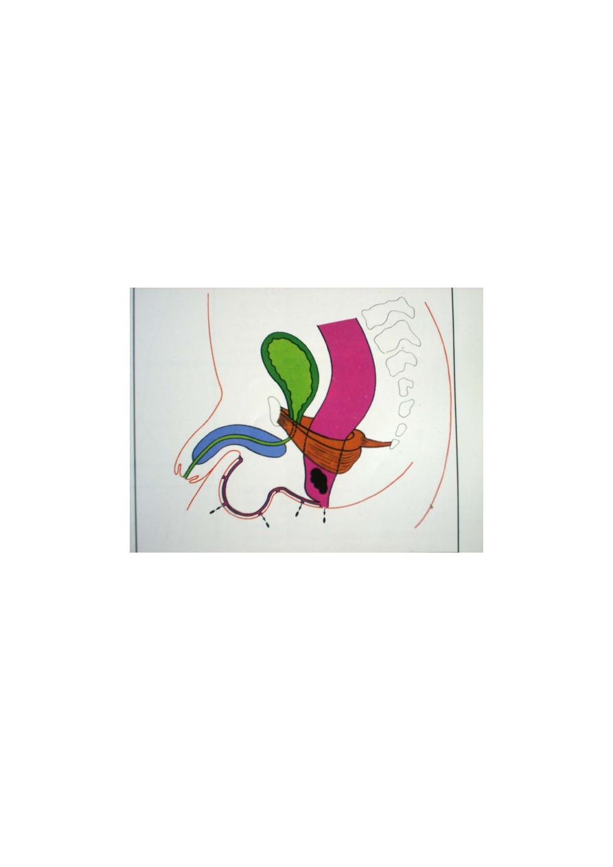
117B
4. Rectovesical ( bladder neck ) fistula
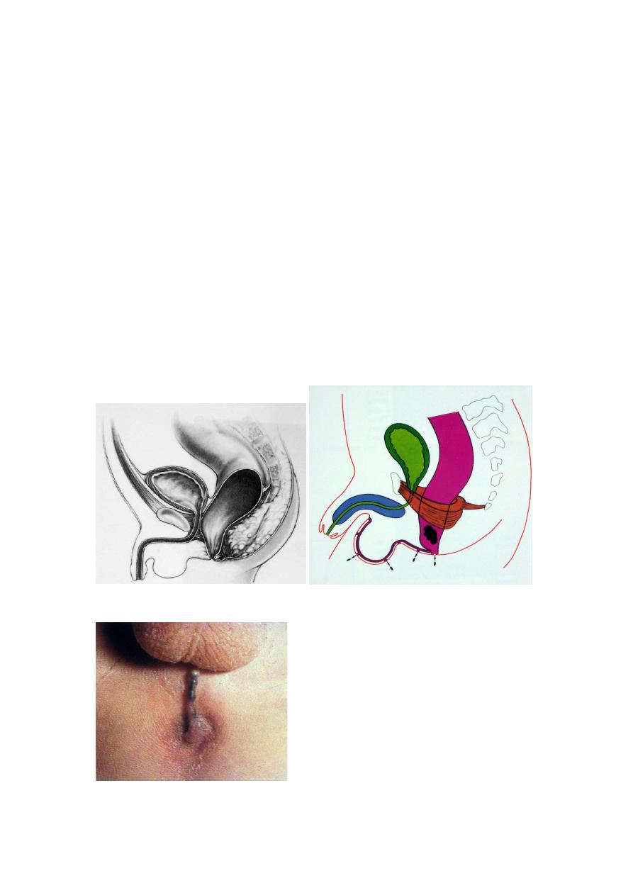
118B
5. Imperforate anus without fistula
119B
6. Rectal atresia and stenosis
46B
FEMALE DEFECTS
120B
1. Low defect : Perineal fistula
121B
2. Vestibular fistula
122B
3. Vaginal fistula
123B
4. Imperforate anus without fistula
124B
5. Rectal atresia and stenosis
125B
6. Persistent cloaca
47B
MALE DEFECTS
48B
1. Low Defects : Perineal fistula
49B
50B
Median raphe fistula
51B
Bucket handle malformation
53B
54B
Anal stenosis
Anal membrane
11B
2.Rectourethral bulbar fistula
12B
13B
3.Rectourethral prostatic fistula
14B
15B
4. Rectovesical (bladder neck ) fistula
16B
5. Imperforate anus without fistula
6. Rectal atresia and stenosis
57B
( as in female )
58B
Female Defects
59B
1. Low Defect : Perineal Fistula
60B
61B
2. Vestibular fistula : 3 orifices
62B
3.
63B
Vaginal Fistula : 2 orifices
64B
65B
4. Imperforate anus without fistula
( as in male )
66B
5. Rectal atresia and stenosis
67B
68B
6. Persistent cloaca : 1 orifice
69B
70B
N.B. Anteriorly displaced anus
is a normal anus
situated anteriorly
71B
ASSOCIATED DEFECTS
72B
Urogenital
73B
- Most common
74B
- 20 – 45 %

17B
- The higher the malformation the higher the incidence
75B
Sacrum and Spine
76B
- Sacrum frequently abnormal
•
77B
deformed
•
142B
reduced in number
•
143B
hemisacrum
144B
- Spine frequently shows hemivertebrae
78B
Investigations:
•
18B
Examination(GUE).the presence of meconium indicate the
presence of fistula to the bowel.
19B
Invertogram
•
20B
X-ray taken 12-18 hours after birth ,inverted position ,lateral view,
after putting a mark to see the distal bowel end and it’s relation to
pubococcygeal line and the distance to the mark.
•
21B
X-ray of the spine for vertebral anomaly (e.x.sacral agenesis).
•
22B
Very rarely :we may need to do micturating cysto-
urethrogram(MCU)for fistulas or other urological anomalies e.x.
neurogenic bladder or vesico-ureteric reflux .
79B
Management of Anorectal Malformations:
94B
Low type lesions have good prognosis .Surgical reconstruction is much simpler
and continence is usually normal .Colostomy is not required and the fistulas
don’t involve other viscera and the coexisting anomalies are less common and
less severe.
•
23B
Anal stenosis :repeated anal dilatation with dilators.
•
24B
Imperforated anal membrane (covered anus)need incision and dilatation .
•
25B
Anterior ectopic anus :mild form need nothing or only dilatation .Severe
form need cut back surgery or transposition .
•
26B
Anovestibular fistula(low type):transposition surgery with or without
colostomy.

80B
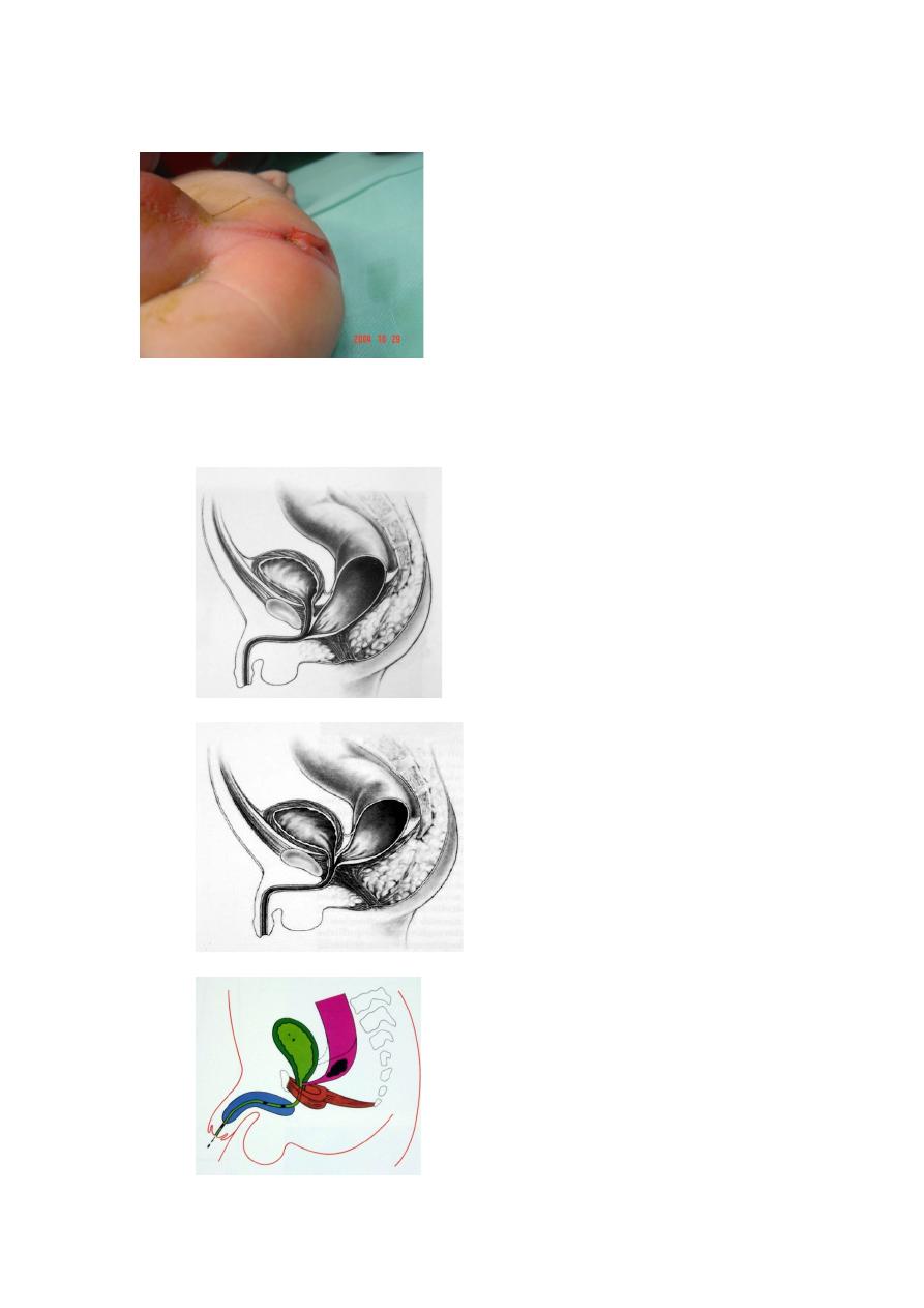
COLOSTOMY
95B
Type :
127B
sigmoid defunctioning with separate stomas
96B
Site :
128B
junction of descending with sigmoid
97B
Incision :
129B
left iliac , muscle cutting
130B
N.B. Distal loop should be cleared of meconium during operation
81B
DISTAL COLOSTOGRAM
-
98B
Most valuable test
-
99B
Water soluble contrast into distal stoma
-
100B
Significant pressure needed
-
101B
Under fluoroscopic control
-
102B
Contrast usually fills proximal urethra &bladder
-
103B
Injection must continue till a voiding episode
-
104B
Pictures taken during micturition
105B
It shows in a single picture :
-
131B
sacrum
-
132B
height of rectum
-
133B
perineum
-
134B
fistula location
-
135B
bladder
-
136B
vesicoureteral reflux
-
137B
urethra
82B
No need for voiding cystourethrography or cystoscopy
83B
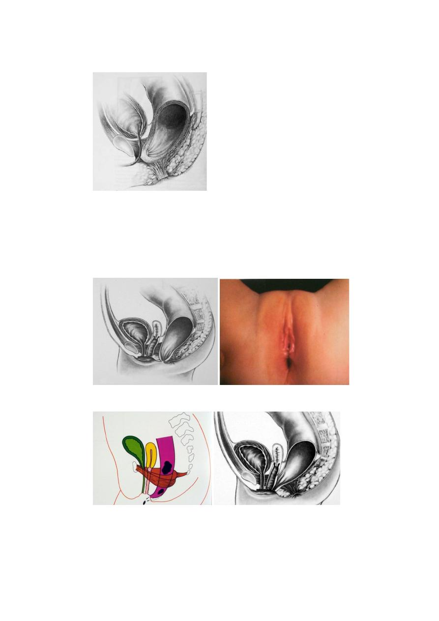
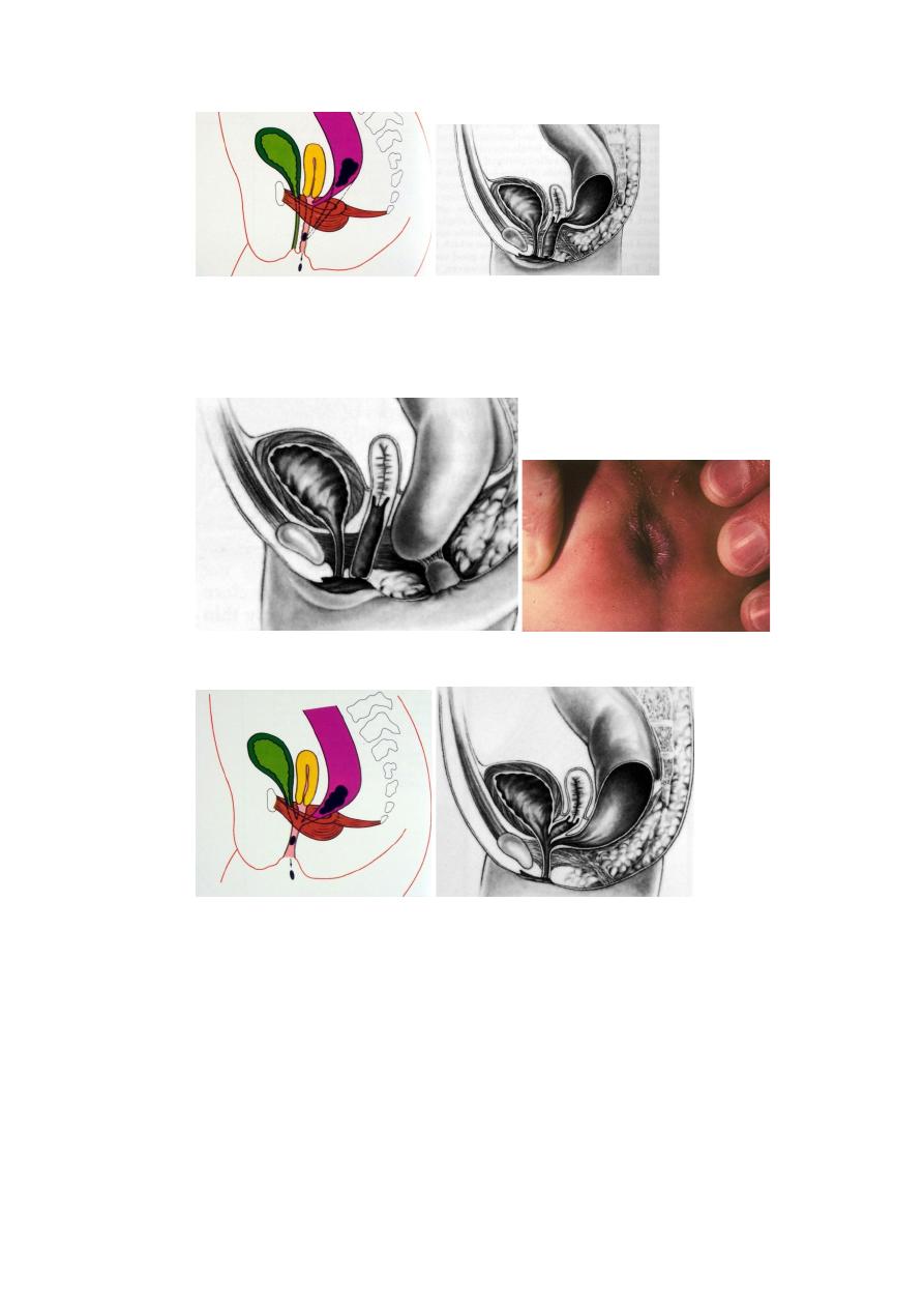
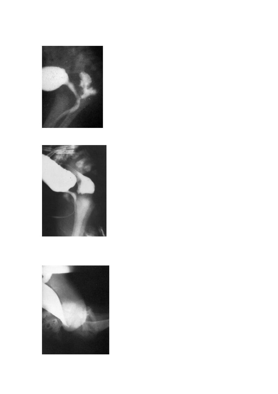
Rectourethral bulbar fistula
84B
85B
Rectourethral prostatic fistula
86B
87B
Rectovesical (bladder neck ) fistula
88B

High types:
The prognosis is not good especially in those with multiple congenital
anomalies .Continence is usually affected .
Surgery include:
Colostomy done at the neonatal period .
The definitive surgery done at 9-12months by penna technique or posterior
sagital anorectoplasty (PSARP),through posterior midline incision exploring the
end of the rectum & separating the fistula connected to the urethra in males or
the vagina in females .Then the rectum is pulled through the puborectalis muscle
,closing the muscle around the rectum &fixing the new opening in the perineum
.
Colostomy closure 4-6 weeks later .