EMBYRIOLOGY
الاثنين 25/11/2013أ.د.عبد الجبار الحبيطي
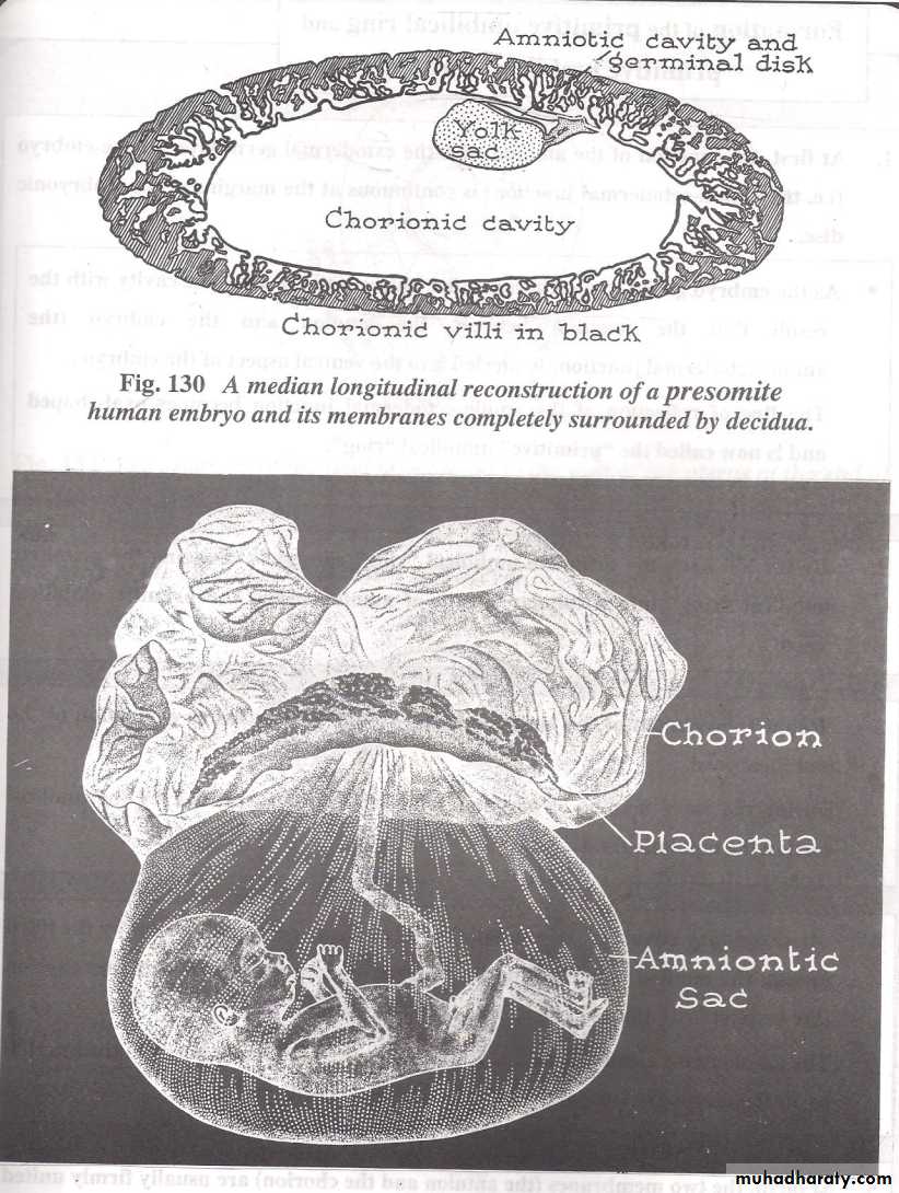
The amnion is a fetal membrane enclosing the amniotic cavity.The amniotic cavity is formed dorsal to the Embryonic Disc ,it's floor is formed by the Ectoderm while it's roof and walls sare formed by the amniogenic cells .The cavity starts to appear during 2nd week of development and increases in size gradually parallel to the increase in the size of the fetus.At first it lies on the dorsum of the embryonic disc ,but after folding of the embryo it encloses the fetus inside it where the fetus is suspended inside the fluid by the umbilical cord.The volume of the fluid just before delivery is around 1500 ml (As maximum)THE AMNIOTIC CAVITY
If it increases above this figure we call the condition as Hydramnious or Polyhydramnious which could be due :
1-Congenital atresia of the Esophagus.
2-Anencephally of the Brain as there is inability
of the fetus to swallow some fluids.
If the volume is less than 500 ml we call the condition as Oligohydramnious which is due tio Renal agenesis.
1-Serves as a protector shock absorber.
2-Prevents adhesion of the Fetus to the amniotic membrane.3-AS it is a bad conductor of heat it keeps the temperature of the
fetus nearly constant specially when the mother is feverish in
cases of illness or infection of the mother.
4-Allows the fetus to move & swim freely in side it which results in
proper development oh his musculo-skeletal system and nervous
system
5-Learn how to suck before his birth.
6-Provides a media for the fetus to pass urine & meconium before
birth.
7-At the end of pregnancy,it bulges through the cervix as a bag of
water to dilate Cervix.
8-After it's rupture ,the discharged fluid sterilizes the Vagina&
Birth Canal.
Functions of the amniotic fluids are the followings
It lies ventral to the Embryonic Disc.There are 3 stages of yolk sac formations as follows:
1-Primitive (Primary ) yolk sac ,which is the earliest stage .It's
roof is formed by the Endoderm while it's floor and sides
by the Heusers membrane(from Cytotrophoblast).
2-Secondary yolk sac which is complewtely lined by the
Endoderm and has the allantois as a diverticulum
extending into the connecting stalk & primitive umbilical
ring.
3-Definitive(Permanent) yolk sac which is the last stage and
is formed after folding of the embryonic disc,thus it is of
small size & communicates with the gut by Vitello-
intestinal duct.
THE YOLK SAC
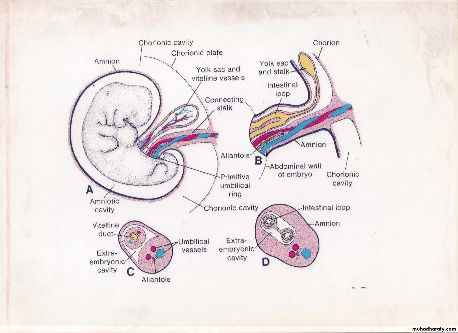
1-The first step is by the appearance of the connecting stalk.2-As the embryo grows ,the embryonic disc bulges into the
amniotic cavity& thus the line of reflection of the amnio-
ectodermal junction( oval ring) known as primitive umbilica
ring.
3-At 5th week of intrauterine life the ring constricts ,thus
converting the primitive umbilical ring into tubular sheath
of amnion called the primitive umbilical cord.
4-Due to the rapid enlargement of the amniotic cavity,the
primitive umbilical cord becomes enlarged & is suspended
in the amniotic cavity & becomes the true umbilical cord.
Formation of the Umbilical Cord
Usually the umbilical cord measures 50-60 cm just before labor & contains the followings:
a-Single umbilical vein.
b-Two umbilical arteries.
c-Wharton's jelly surrounds the umbilical
vessels.
If the umbilical cord is less than 25 cm is short & may leads to premature separation of the placenta with its troubles,but if the cord is very long(above 70cm) it may leads to:
1-Prolapsed cord with the beginning of labor& descends before the head of the fetus and may become compressed by the head & results in cut of the blood supply to the fetus & death of the fetus during labor.
2-Roll on itself & creating true knots which again endanger the life of the fetus before its birth
primitiveumbilical ring contains
a-The allantois.b-The umbilical Vessels.
c-Part of the extraembryonic coelom.
d-Vitelline duct.
The Primitive Umbilical Cord Contents
a-Allantois.
b-Umbilical Vessels.
c-Intestinal loop of the Physiological
hernia.
d-Yolk sac & Vitelline vessels.
e-Part of extraembryonic coelom.
Abnormalities of the Cord are
1-Vilamentous cord where it is attached to fetal
membranes and not to the Placenta.
2-Battledore Placenta where the cord is attached to
the edge of the Placenta.
3-Mid-Central attachment of the cord ,here it is
attached to the centre of the placenta while
normally it is attached near the centr.
4-Abnormally long or short cord.
5-True knots of the cord.
6-Double or Triple cord (this is very rare).
7-Umbilical Cord with a single umbilical artery
instead of two.