Dr:Muhannad / lec 6 written by:Noor Mohammad and Ban Hikmat
The function of the CNS :
1-sensory 2-motor 3-integrative function Cerebral cortex thin layer normally (3-5) mm thick of a wide surface area about one quarter square meter. There are about 100 billion neurons in the cerebral cortex (c.c.). There is a sub cortical large structure deep in brain work in co-operation with cerebral cortex called the thalamus.
The thalamus consist of many nuclei, each nucleus has bidirectional connection with the corresponding area in the cerebral cortex called according to the nucleus of the thalamus.
Q /What for is the bidirectional connection with the thalamus? To exchange excitatory signals between the c.c. and thalamus, so that there will be a sort of positive feed-back circuit between the c.c. and thalamus.
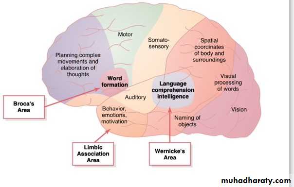
Sub- areas of the cerebral cortex and their functions:- The central fissure divides the cerebral cortex into anterior and posterior halves.
The gyrus immediately behind the central fissure is(post central gyrus) the primary somatosensory area, it's function is primary analysis of sensory signals. Immediately in front of central fissure is the motor cortex which consist of 3 areas: the primary motor area, premotor and supplementary motor areas .
The areas in between the main areas of the brain are called association areas.
1-between the parietal, occipital and temporal areas an area called (parietal-occipital-temporal association area).
The function of this area is a more detailed integrative function than the primary area, so it's higher degree integration sensory information that tell us many things about sensory information ex:- tell u about a special orientation of the body and relation of different parts of the body or it's relation to the surrounding for example :- Tell u are standing not sitting..
2-anterior to motor cortex is the prefrontal association area, people think that this area is the area of intelligence and thinking, it is differentiated in human from monkeys in that it is much more developed in human than monkey. But this is not true, because it is not the only area that participates in producing intelligence.
So it's not the only site of intelligence, but it's one of the areas that work together to produce intelligence.
Motor cortex in its anterior part responsible for make an image for motor movement and then send it to the posterior part to put a program then het program is sent deep in the brain to basal ganglion to check it then to the thalamus after that it will be sent back to the primary motor cortex, because the primary motor cortex is the only area that sends it's out puts as a long nerve fibers to the spinal cord and eventually to executers (the muscle).
This process can be described as motor thinking that occurs in the motor cortex. The prefrontal area can be described as the area of sensory thinking, because it receives many information through connection that pass subcortically from the somatic sensory area to convey the previously analyzed sensory signals to this area so that in this area there will be a sort of putting together many bits of information to make thoughts, therefore, this area may be called as the area of elaboration of thoughts, deep thoughts that characterizes human beings from animal .
3-another association area located in the under surface of the brain called as the limbic association area.
This area is a part of the limbic system which is concerned with emotions, motivation and behavior.
Auditory Area :-
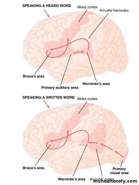
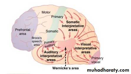
It is the hearing area, Posterior to auditory area is most important area of the entire brain which responsible understanding and comprehension of spoken and written language this area was discover by German neurologist and called in honor of his name Wernicke's area, he was interested in aphasic patients (difficulty in speech)and use to do post mortem to his patient, he have had found in all of his patients were having some damage in the same area that is called (Wernicke’s area). This area is located in the left hemisphere at the point of junction parietal ,occipital and temporal lobes so it receives information from all of these areas the sensory information after pre analyzed will be sent to the Wernicke’s area for the production of ideas and to choose the word to be used in expressing the idea.
Q / Which area in the motor cortex controls the muscles of speech?
A /the area that is laterally located in the motor cortex, this area has been discovered by French neurologist .who's name was (Broca 1886) who was interested in patients with aphasia and he also use to do postmortem to his patients and he have had noticed that all of them were having damage in the same area, (broca's area) again in the left hemisphere, which is the dominant hemisphere in right handed people..Q /if the patient has a problem in broca's area ,what will happen? A / Damage of this area will result in difficult speech what we call as aphasia.
Q /what could be the result of damaging Wernicke’s area? A /unable to produce idea he cannot understand and comprehend spoken and written languages he will be totally dull.
Q / How can we know the name and the material of the object.
A / We learn the names of objects through hearing but the material that the object is made of through vision.
Q / The area for face recognition ? A/ it is an area located under surface of the brain, this area is wide because of the large number of people that we meet every day. The damage of this area results in that the patient will no more be able to recognize the faces even of his close friends.
Q /The function of Wernicke's area (the comprehension and understanding area). This area is located at the point of junction of the parietal, occipital and temporal lobes. It receives pre analyzed sensory information from these areas (somatosensory, visual and auditory areas).The area develops an idea and choose the words to be used in expressing this idea, then it pass it to the Broca's area, through a connection between Wernicke’s and broca's area called (arcuate fasciculus) which is a neural connection joining the two areas, through which it receives information from Wernicke’s area.
Q /What does the word intelligence mean?
A / It means the ability to solve complex problems and to go through a long train of thought, to prognosticate and plan for the future. It requires the following:1-To have the areas of brain (Wernicke’s and broca’s) normally function.
2-have the ability to learn.
3-have the ability to store new information.
4-search out the stored information in the memory storage house.
Learning consist of two aspects the sensory aspect and the motor.
The sensory aspect, the inputs are through.
1-Audition is the first rout for learning before birth can start at the age of 26th week of gestation.
2-Visual rout starts after birth.
Q /when the auditory to start? A /it is start very early even before birth at the 26week the auditory system will be complete and function actually u can deliver many different information to the infant through applying the source of sound to his mom's abdominal wall.
After birth the new born baby starts using the other rout which is the visual.
2)THE MOTOR ASPECT of learning:
Is to produce words through the broca’s area that we talked about it previously.
The other function of the brain that is required for intelligence is (MEMORY):
Memory means the storage of information in the memory store areas.
How is information stored? Are they stored randomly?
Memories are not stored randomly, but they are stored after they are codified a process called codification.
Codification means giving the memory a code number. When a new information is to be stored an old memory will be withdrawn from the store houses to be compared with the new information, for similarities and differences, and according to these similarities and differences they will be stored i.e. if they are similar they will be stored in the same place but if they aren’t the new information will be stored in another area.
There are many types of memories:
1. Short term memory which lasts from few seconds to few minutes such as trying to save someone’s phone number .
2. If you keep thinking of the information and try to continue remembering it the memory will be converted from the short term to the intermediate long term which might stay for few minutes, hours, days or weeks.
3. After that if you keep rehearsing (repeating) the information, it will be converted from the intermediate long term to the long term which will be kept lifelong (with continuous many times rehearsal) This is the way how we keep information when we study.
That means if you don’t rehearse the information many times when you read or study you will lose them later and won’t be able to remember them .
Important areas of the brain that participate in producing intelligence are:
1) Intact association areas.
2) Intact inputs to learning, audition and vision and their areas.
3) Werincke's area that makes you capable to learn comprehend and understand.
4)store the information (memory) mainly in the cerebral cortex.
5) The thalamus which will search out the memory from the store houses and read it .
# the part which will search the memory is the (thalamus) ;it is a big structure in the brain that is required to find and get the information that is stored and read it out.
The other part of the brain that helps in producing intelligence is the (hippocampus) that helps in converting the information store from the short term to the long term memory.
# what will happen if the hippocampus is damaged?
This damage will be associated with forgetfulness which is called (amnesia) ,the patient with lesion of the hippocampus won’t be able to learn new information because of being unable to store the information in the long term memory. This is along with forgetfulness of the recent events ( anterograde amnesia ).
# In case of brain concussion as in the case of trauma to the head the hippocampus might be affected, so when the subject regains consciousness he might remember nothing in this case (anterograde amnesia) .
Damage of the hippocampus may not be only associated with loss of the new memories but the old memories will be lost too, but recent memories will be affected more than the old ones (because the old memories are rehearsed and repeated for many times) so the old memories will be partially affected.
The Brain waves and the different states of activities During our different daily activities.
The human beings passes through a different state of brain activities, sometimes we are awake fully alert, drowsy, disappointed ,happy or sad….. etc
These different states are associated with different brain activities, among these different states is sleep.
Sleep:- is defined as unconsciousness from which the subject can be aroused by sensory stimuli. Sleep is different from coma .
Coma:- is defined as unconsciousness from which the subject cannot be aroused by sensory stimuli unless removing the cause that induces the coma. During sleep one passes through two phases one of them different from the other in time and features .
Q /How many type of sleep are there? A /during the long hours of the night having 7-8hr of sleep we pass through 2 types of sleep ,they are :-
1-Slow wave sleep, its name is derived from the slow brain waves and high voltage in this type of sleep.
2-Rapid eye movement sleep, during this type of sleep there will rapid movements of the eyes.
Characteristic features of each type of sleep.
1-slow wav sleep:
a- This type is the restful type of sleep that we usually encounter during the first hour of sleep after being kept awake for many hours.
b- It is associated with decreased bodily activities such as respiratory rate ,blood pressure and metabolic rate the whole body will get rested. That is why when u get a wake u feel rested.
c-This type of sleep is not associated with dreams, dream free, but sometimes, dreams do occur and even night mares in this type.
d- The subject can easily be aroused from this type.
e- It takes a period from 1.5 hr (90 minutes).
2- Rapid eye movement sleep.
a- Associated with increased brain activity up to 20%.
b- Active dreaming, this type of sleep is associated with active dreaming.
c- The subject is more difficult to arouse during rapid eye movement than slow wave sleep.
for ex:- when u see a sleep person dreaming or in night mares u want to awaken him but it looks not easy.
d-the respiratory rate and heart rate will be irregular if u try to record the pulse of a sleep subject during rapid eye movement u will find it irregular also the metabolism of the brain will be increased about 20%.
e-the musculature system intensity of the body will be depressed the muscles of the body will be very lax except the muscle of eye that might show increased activity.
f- This type of sleep takes a period from 5-30 occurring every 90 minutes. when u go to sleep u will pass through a sequence of two type of sleep 1.5hr (slow wave sleep)→5-30 min(rapid eye movement sleep)→1.5 hr(slow wave sleep) and so forth.
Q /how u can pass to sleep from the state of wake fullness?
A / This is a matter of (debate) no document is provided to support the hypothesis to explain the mechanism of sleep. Among the most accepted hypotheses is the following .
Sleep is an active process, after long hours of wakefulness during the day the brain will get fatigue and its activity will slow down so the subject will pass from wakefulness to sleep but it prove that is absolutely wrong, sleep is an active process.
There are certain chemical substances released from certain areas of the brain to induce sleep. And to prove that through animal experiment. They keep an animal awake by forcing them not to sleep for 24-48 hrs, they withdraw blood sample or sometimes urine sample from these animals and inject it into the brain of awake animal they found that this blood or urine immediately induces sleep in the animal.
Q /This experiment indicates what? A /this blood sample or urine must contain a substance or substances that can induce sleep, so that we conclude that sleep is an active process and induced by chemical substances released from certain areas of the brain.
Q /where are these areas that are concerned with sleep?
A / 1-raphe Magnus nucleus :if u are able to do an animal experiment and stimulate into the raphe Magnus nucleus with a microelectrode, the animal immediately passes to sleep, that is why if u cut the brain above the level of the Raphe magnus nucleus, so that the brain is isolated from the Magnus nucleus and below structure, u will get a brain can never pass to sleep, because the higher centers will be freed from the effect of the chemical substances from raphe Magnus nucleus to inhibit the cerebral cortex and induced sleep.
2-nucleus of the tractus solitaries. It is the other area in the brain stem in the upper part of the medulla oblongata and lower pons, that when stimulated can induce sleep immediately in an experimental animal. These are the two main areas in the brain that can induce sleep.
Q /what are the chemical neurotransmitters in the brain that can induce the sleep?
A /1-serotonin : chemical neurotransmitters released from raphe Magnus nucleus . Note : the raphe Magnus nucleus have 2 neuronal projections. One of them that is responsible for pain relief, and the other neuronal projections to the cerebral cortex that is responsible for inducing the sleep.
2-muramel :- it is the other chemical neurotransmitters that can induced sleep.
Q /what is the importance of sleep? A /sure sleep is beneficial for:- 1-rest of brain 2-rest of the whole body.
During sleep the all system of the body and especially the brain get rested, as if restarting the brain as what we do with the personal computer when start work randomly .We will restart it or shutdown to make recover.
(( The brain waves))
There are 4 type of brain waves they are :
1/α-waves these waves can be recorded when u close your eyes and think of nothing with wondering mined in paradise looking at flowers and looking for butter flys, in this case if we record the brain wave, we will get α-waves that are characterized by:
a. Frequency ( 8-13 Hz)
b. Higher voltage than β-wave
2/β-waves: These waves are recorded from awake subjects with eyes opened thinking of something. They are characterized by :
a. Low voltage (irregular)
b. Frequency (more than 18 Hz)
3/θ-waves: Normally recorded in children, but in adults we cannot record this type of waves unless there is a functional abnormality in the brain such as disappointment and frustration. They are characterized by:
a)High voltage
b. Low frequency (3-7Hz)
4/ ˠ-waves this type of waves are record in young infants normally, but in adults they always indicate the presence of organic damage (brain lesion). They are characterized by.
a) slow wave low than 3.5 Hz.
b) High voltage.
Both of θ and ˠ-waves recording is abnormal indicating abnormal function and brain organic disease respectively. Recording θ-waves in the presence of functional abnormality of the brain for ex: epilepsy, while ˠ-waves recorded in case of the presence of organic damage (lesion in brain). This is the importance of studying the different brain waves.
Epilepsy:
It is an abnormal functional disease of the brain .This abnormality might be associated with generalized over activity of the brain (whole brain is affected and brain stem) .
we classified it into 3 types
Grand mal (seizure): is the first type of the epilepsy, it is associated with generalized tonic and clonic contractions of almost all muscle of the body. The subject will undergo convulsions (serve contraction of the muscle of the body) this convulsion might last up to 3-4min. after that the seizure will cease (end up). Some of the lay people try to stop the seizure by holding the subject, but this do nothing. The convulsion will continue then after 3-4min. the seizure will end up. The subject after convulsion will be tired, exhausted and fatigue and might sleep for many hours. The brain waves recorded are of,
Θ-waves will observed because there is functional abnormality in the brain
2. petit mal :very mild type to epilepsy characterized by abnormal movement in the head and eye usually blinking of the eyes .The patient don't lose consciousness as in grand mal epilepsy, the patient while sitting in his chair to do some task he will stop for a period that might last up to (3-30 sec)then he resumes his activity. The is isolated from the surrounding for 3-30 sec there for we called it sometime the absence syndrome. The brain waves recorded during this type of epilepsy is (spike and dome).
Note: petit mal epilepsy may end up in grand mal epilepsy.
3)focal epilepsy : The abnormality is localized over a certain area of the brain, this over activity may spread to affect another area of the brain to reach the motor area to induce abnormal movements of the head beginning around the mouth and spreading downward. This case is called (jacksonian)epilepsy.
4) Other type is called psychomotor epilepsy characterized by behavioral and emotional features .