Light or Compound Microscope
Lab-1-Sunday 6/12/2015
The optical microscope, often referred to as the "light microscope", is a type of microscope which uses visible light and a system of lenses to magnify images of small samples.
Optical microscopes are the oldest design of microscope and were possibly designed in their present compound form in the 17th century.
Basic optical microscopes can be very simple, although there are many complex designs which aim to improve resolution and sample contrast.
History of (L.M)
It is difficult to say who invented the compound microscope.Dutch spectacle-makers Hans Janssen and his son Zacharias Janssen 1590.
Galileo Galilei , He developed an occhiolino or compound microscope with a convex and a concave lens in 1609.
Giovanni Faber , Faber coined the name from the Greek words μικρόν (micron) meaning "small", and σκοπεῖν (skopein) meaning "to look at", a name meant to be analogous with "telescope", another word coined by the Linceans.
Christiaan Huygens, another Dutchman, developed a simple 2-lens ocular system in the late 17th century.
Anton van Leeuwenhoek , (1632–1723) is credited with bringing the microscope to the attention of biologists, even though simple magnifying lenses were already being produced in the 16th century. Van Leeuwenhoek's home-made microscopes were very small simple instruments, with a single, yet strong lens.
Single lens (simple) microscope
A simple microscope is a microscope that uses only one lens for magnification, and is the original design of light microscope.Van Leeuwenhoek's microscopes consisted of a small, single converging lens mounted on a brass plate, with a screw mechanism to hold the sample or specimen to be examined.
Compound microscope
Compound microscope is a microscope which uses multiple lenses to collect light from the sample and then a separate set of lenses to focus the light into the eye or camera.
Compound microscopes are heavier, larger and more expensive than simple microscopes due to the increased number of lenses used in construction.
Compound microscope also makes more advanced illumination setups, such as phase contrast.
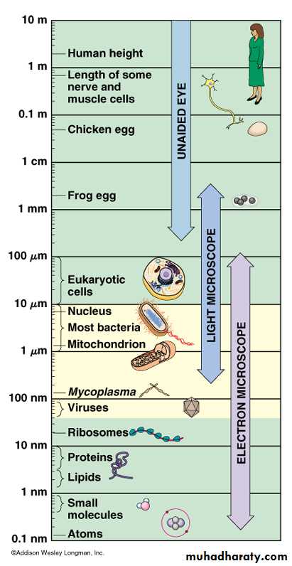
(Logarithmic Scale of Cell Measurement)
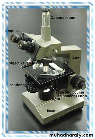
ComponentsAll modern optical microscopes designed have the same 'structural' components:-
Ocular lens (eyepiece)
Objective turret or Revolver or Revolving nose piece (to hold multiple objective lenses)
Objective lens
Focus wheel to move the stage ,coarse adjustment & fine adjustment
Frame
Light source, a light or a mirror
Diaphragm and condenser lens
Stage (to hold the sample)
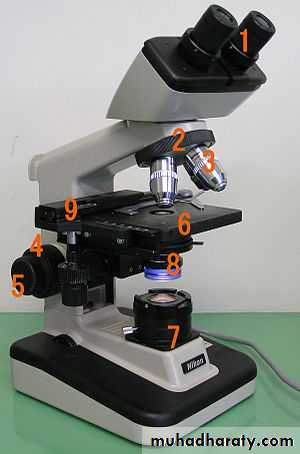
(Components of light microscope)
The eyepiece, or ocular lensIs a cylinder containing two or more lenses.
Its function is to bring the image into focus for the eye.
Objective or Revolving nose piece
Objective turret or Revolver is the part that holds the set of objective lenses.It allows the user to switch between objectives.
Objective lens
At the lower end of a typical compound optical microscope there are one or more objective lenses that collect light from the sample.The objective is usually in a cylinder housing containing a glass single or multi-element compound lens.
Oil immersion objective
Some microscopes make use of oil-immersion objectives for greater resolution at high magnification.These are used with index-matching material such as immersion oil and a matched cover slip between the objective lens and the sample.
The larger numerical aperture allows collection of more light making detailed observation of smaller details possible.
An oil immersion lens usually has a magnification of 40 to 100×.
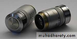
(Objective lenses)
Two Leica oil immersion microscope objective lenses; left 100x, right 40x.Focus wheels
Adjustment wheels move the stage up and down with separate adjustment for coarse and fine focussing.The same controls enable the microscope to adjust to specimens of different thickness.
In older designs of microscopes, the focus adjustment wheels move the microscope tube up or down relative to the stand and had a fixed stage.
Frame
The whole of the optical assembly is traditionally attached to a rigid arm, which in turn is attached to a robust U-shaped foot to provide the necessary rigidity.Light source
Many sources of light can be used. At its simplest, daylight is directed via a mirror. Most microscopes, however, have their own adjustable and controllable light source – often a halogen lamp.Condenser
The condenser is a lens designed to focus light from the illumination source onto the sample. The condenser may also include other features, such as a diaphragm and/or filters, to manage the quality and intensity of the illumination.Stage
The stage is a platform below the objective which supports the specimen being viewed. In the center of the stage is a hole through which light passes to illuminate the specimen. The stage usually has arms to hold slides.Magnification
The actual power or magnification of a compound optical microscope is the product of the powers of the ocular (eyepiece) and the (objective lens). The maximum normal magnifications of the ocular and objective are 10× and 100× respectively, giving a final magnification of 1,000×.
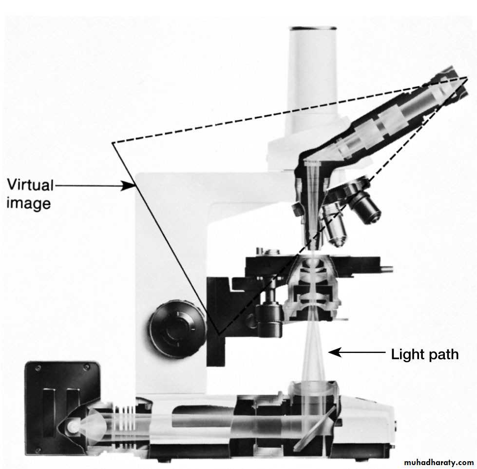
Lenses and the Bending of Light
Light is refracted (bent) when passing from one medium to another.Focus light rays at a specific place called the focal point.
Distance between center of lens and focal point is the focal length.
Short focal length more magnification.
Types of light microscope
There are many types of (L.M):-Bright-field microscope
Dark-field microscope
Phase-contrast microscope
Fluorescence microscopes
Are compound microscopes
Image formed by action of 2 lenses.
The Bright Field Microscope
Bright field microscopy is the simplest of all the optical microscopy illumination techniques.Sample illumination is transmitted (i.e., illuminated from below and observed from above) white light and contrast in the sample is caused by absorbance of some of the transmitted light in dense areas of the sample.
The typical appearance of a bright field microscopy image is a dark sample on a bright background, hence the name.
(The Bright Field Microscope)
ComponentsTransillumination light source, commonly a halogen lamp in the microscope stand.
Condenser lens which focuses light from the light source onto the sample.
Objective lens which collects light from the sample and magnifies the image.
Oculars and/or a camera to view the sample image.
Bright field microscopy may use critical or Köhler illumination to illuminate the sample.
(The Bright Field Microscope)
PerformanceBright field microscopy typically has low contrast with most biological samples as few absorb light to a great extent.
Staining is often required to increase contrast, which prevents use on live cells in many situations.
Bright field illumination is useful for samples which have an intrinsic colour, for example chloroplasts in plant cells.
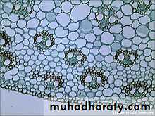
An example bright field micrograph this image shows a crossection of the vascular tissue in a plant stem

Bright field illumination, sample contrast comes from absorbance of light in the sample
The Dark-Field MicroscopeProduces a bright image of the object against a dark background.
Used to observe living, unstained preparations.
In optical microscopy, dark field describes an illumination technique used to enhance the contrast in unstained samples.
It works by illuminating the sample with light that will not be collected by the objective lens, and thus will not form part of the image.
This produces the classic appearance of a dark, almost black, background with bright objects on it.
(The Dark-Field Microscope)
(The Dark-Field Microscope)
(The Dark-Field Microscope)
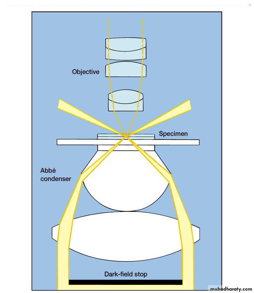
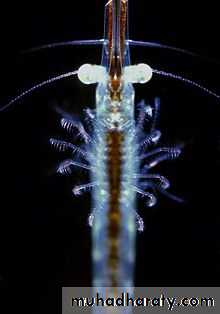
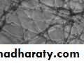
Dark field microscopy produces an image with a dark background

Dark field illumination, sample contrast comes from light scattered by the sample
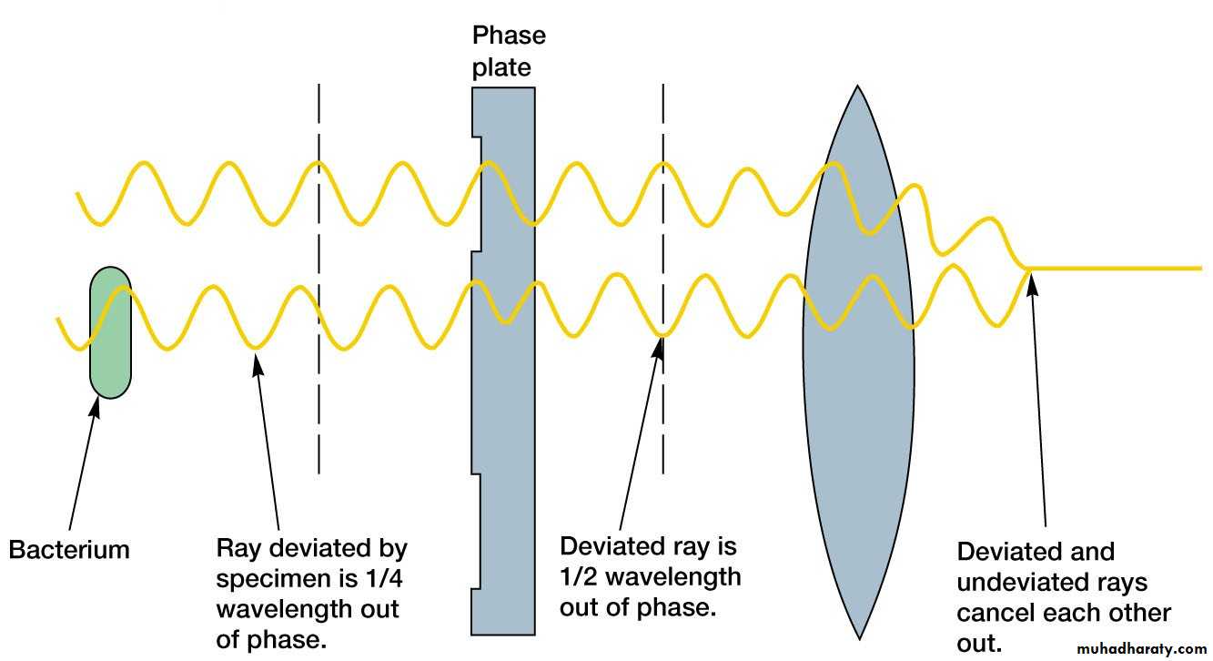
The Phase-Contrast Microscope
Enhances the contrast between intracellular structures having slight differences in refractive index.
Excellent way to observe living cells.
Phase contrast microscopy is an optical microscopy illumination technique that converts phase shifts in light passing through a transparent specimen to brightness changes in the image.
The phase shifts themselves are invisible to the human eye, but become visible when they are shown as brightness changes.
(The Phase-Contrast Microscope)
PerformancePhase contrast microscopy is particularly important in biology, as it reveals many biological structures that are not visible with a simpler bright field microscope.
These structures were often made visible to earlier microscopists by staining the slide.
This requires additional preparation and it also kills the cell.
Phase contrast microscopy of live cells without staining allowed for the in vivo study of the cell cycle.
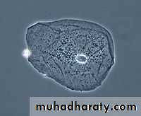
(Phase contrast image of a cheek epithelial cell)
Image appearancePhase contrast images have a characteristic grey background with light and dark features found across the sample.
Light and dark fringes appear around regions with a change in optical density, for example the boundary between water and a cell.
This normally manifests itself as a bright halo around a dark object.
Phase contrast illumination, sample contrast comes from interference of different path lengths of light through the sample
The Fluorescence Microscope
Exposes specimen to ultraviolet, violet, or blue light.Specimens usually stained with fluorochromes.
Shows a bright image of the object resulting from the fluorescent light emitted by the specimen.
A fluorescence microscope is an optical microscope that uses fluorescence and phosphorescence instead of, or in addition to, reflection and absorption to study properties of organic or inorganic substances.
The "fluorescence microscope" refers to any microscope that uses fluorescence to generate an image.
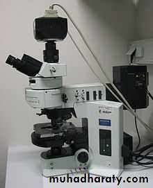
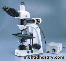
An upright fluorescence microscope (Olympus BX61) with the fluorescent filter cube turret above the objective lenses, coupled with a digital camera
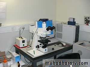
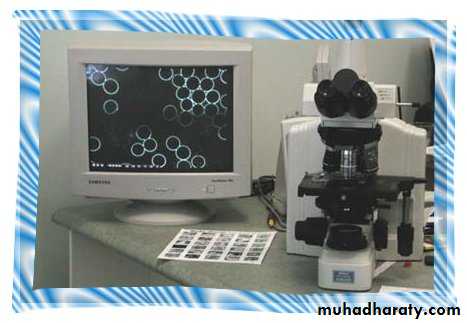
A modern microscope with a mercury bulb for fluorescence microscopy. The microscope has a digital camera, and is attached to a computer
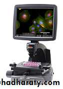
(An Inverted Fluorescence Microscope)
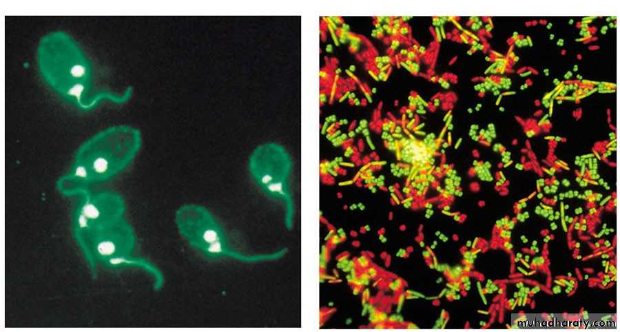
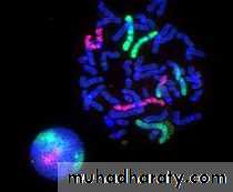
Fluorescence microscopy images
Fluorescence microscopy images of sun flares pathology in a blood cell showing the affected areas in red
Applications of (L.M)
Optical microscopy is used extensively in:-Microelectronics
Nanophysics
Biotechnology
Pharmaceutic research
Mineralogy
Microbiology
Medical diagnosis
Histopathology
Smear tests
In Industrial use