SMALL INTESTINE
Dr. ZAID MUWAFAQ AL-HAMID
MRCS England(UK), FJMC Jordan, HSM
SURGERY Jordan, MBChB Mosul
Specialist Laparoscopic Surgeon
Mechanical small bowel obstruction is the most
frequently encountered surgical disorder of the small
intestine.
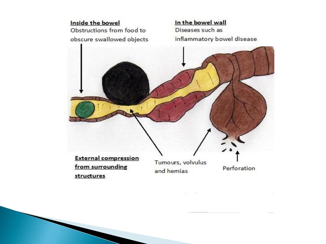
Aetiology:
1. Intraluminal
(e.g., foreign bodies, gallstones, or meconium)
2. Intramural
(e.g., tumors, Crohn’s disease–associated
inflammatory Strictures, Diverticulitis, Meckel’s diverticulum,
Hematoma), Congenital abnormalities (e.g., webs,
duplications, and malrotation)
3. Extrinsic
(e.g., adhesions, hernias(ext. or internal), or
carcinomatosis or local invasion by intraabdominal malig.)
Volvulus, Intussusception
Intra-abdominal adhesions
related to prior abdominal
surgery account for up to 75% of cases of small bowel
obstruction.
Less prevalent etiologies for small bowel obstruction include
hernias
,
malignant
bowel obstruction(extrinsic compression
or invasion by advanced malignancies arising in organs other
than the small bowel), and
Crohn’s disease
.
Pathophysiology
Obstruction
gas and fluid
accumulate proximal to the
site of obstruction
the intestinal activity increases
to overcome the obstruction colicky pain .
gas swallowed air, some is produced within the
intestine.
fluid
swallowed liquids and gastrointestinal secretions
(obstruction stimulates intestinal epithelial water secretion).
More gas and fluid accumulation the
bowel distends
i
ntraluminal and intramural pressures rise
The intestinal motility reduced , lumine small bowel (sterile)
organisms grows
Translocation of bacteria
intramural pressure high enough
intestinal ischemia
,
necrosis
strangulated bowel obstruction
.

partial small bowel obstruction
only a portion of the intestinal lumen is occluded.
Progression is slower .
strangulation is less likely
.
Continued passage of flatus and/or stool
beyond 6 to 12 hours
after onset of
symptoms is characteristic of partial
obstruction
closed-loop
obstruction
in which a
segment of intestine is
obstructed both proximally and distally
(e.g., with volvulus) leading to a rapid rise in luminal pressure
and a rapid progression to strangulation.

Clinical Presentation
The symptoms of small bowel obstruction are
1- colicky abdominal pain.
Is the first symptom ,sudden
and severe in umbilical region . Continuous sever pain suggestive of
strangulation.
2- nausea, vomiting
Vomiting is a more prominent symptom with proximal obstructions than distal.
The vomitus usually bile stained and when it is more feculent, suggesting a more
established obstruction.
3- obstipation(absolute constipation)
4-abdominal distention,
which is most pronounced if the site
of obstruction is in the distal ileum and may be absent if the site of obstruction is in
the proximal small
intestine.
5- Bowel sounds may be hyperactive initially
,
but in late stages , minimal bowel sounds may be heard.
The patient is dehydrated
Laboratory findings reflect intravascular volume depletion and consist of
hemoconcentration
and
electrolyte abnormalities
. Mild
leukocytosis
is common.

1- abdominal pain often disproportionate to the
degree of abdominal findings,
2- tachycardia
3- localized abdominal tenderness
4- fever
5- marked leukocytosis
6- acidosis
Any of these findings should alert the clinician to the
possibility of strangulation and need for early surgical
intervention.
1- History
: prior abdominal operations
abdominal disorders (e.g., intra-abdominal cancer or
inflammatory bowel disease)
2-Examination,
for hernias (particularly in the inguinal and femoral
regions)and the presence of abdominal scar.
Signs of dehydration, tachycardia , hypotension, may be fever(in strang.)
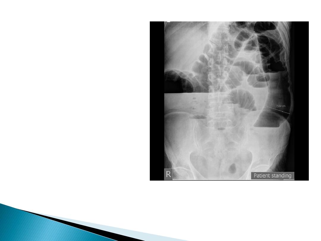
3- Radiological
:
- upright films: dilated small bowel loops (>3 cm in diameter), air-
fluid levels, and a paucity of air in the colon.
- Computed tomography (CT) scanning :
^ transition zone
^ proximal dilation of bowel
^ distal decompression of bowel
^ intraluminal contrast that does not pass beyond the
transition zone, and a colon containing little gas or fluid.
^ Strangulation, closed loop obstruction and the
etiology of obstruction can be suggested.

4- Complete blood count (hemoconcentration,leukocytosis)
serum electrolytes
(vomiting of intestinal contents result in
hypokalemia, ischemia and renal failure result in
hyperkalemia)
renal function test (bl.urea, serum creatinine)
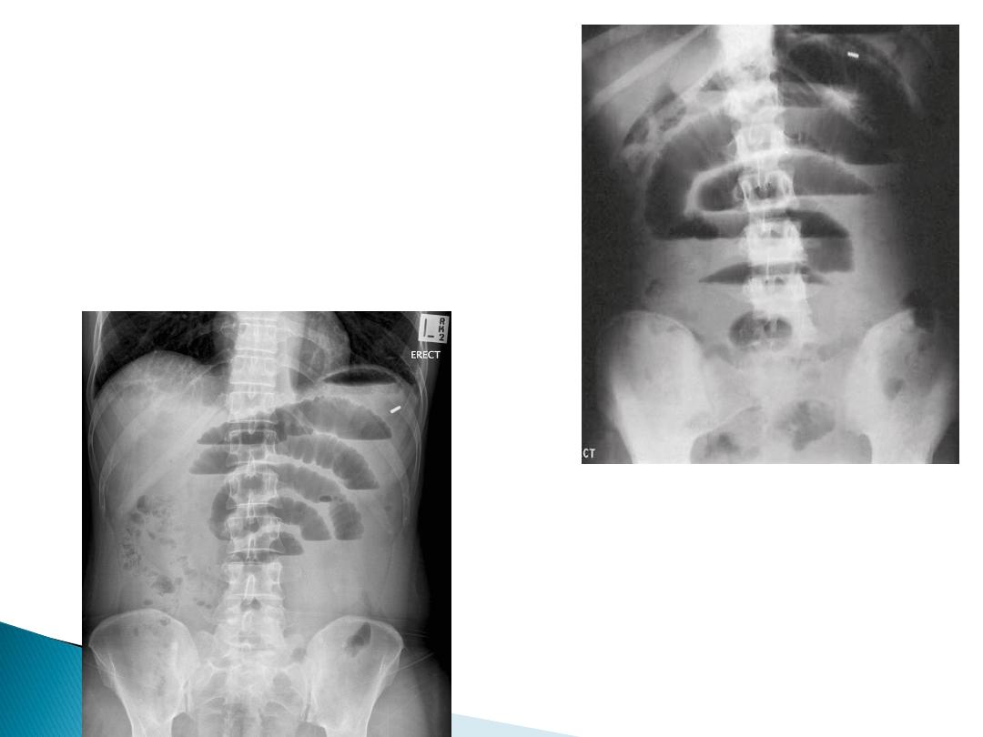
Small bowel obstruction
-The dilated bowel loops centrally
located and lie transversely.
-No/minimal gas is seen in the
colon.
-valvulae conniventes,which
completely pass across the width
of the bowel
-ladder pattern
Multiple air fluid level,
small and centrally
located.
-Dilated loops of bowel
-periphery located.
-Larger bowel diameter
-Huastration
(incomplete line)
-longer length airfuid
level , less in number
Large bowel
obstruction

1- NPO
2- fluid
resuscitation, Isotonic fluid should be given
intravenously
3- Nasogastric (NG) tube
: The stomach should be
continuously evacuated of air and fluid to decreases nausea,
distention, and the risk of vomiting and aspiration.
4-
an indwelling
bladder catheter
may be placed to monitor
urine output.
5- Central venous or pulmonary artery catheter
monitoring
may be necessary to assist with fluid management in
patients with underlying cardiac disease and severe
dehydration.
6- Broad-spectrum antibiotics
???

close observation and serial exams
.
“the sun should never rise and set on a
complete bowel obstruction.”
If there is any evidence of
closed-loop obstruction
or
intestinal
ischemia
, surgical exploration should be performed.
Conservative Therapy is commonly recommended for:
1. Partial small bowel obstruction(for 48 hours)
2. Obstruction occurring in the early
postoperative period (2-3 weeks)
3. Intestinal obstruction due to Crohn’s disease
4. Carcinomatosis
All those periods of conservative therapy should be coupled with
close
observation
and if signs of complete obstruction or intestinal ischemia
occurs , urgent surgical exploration should be performed.

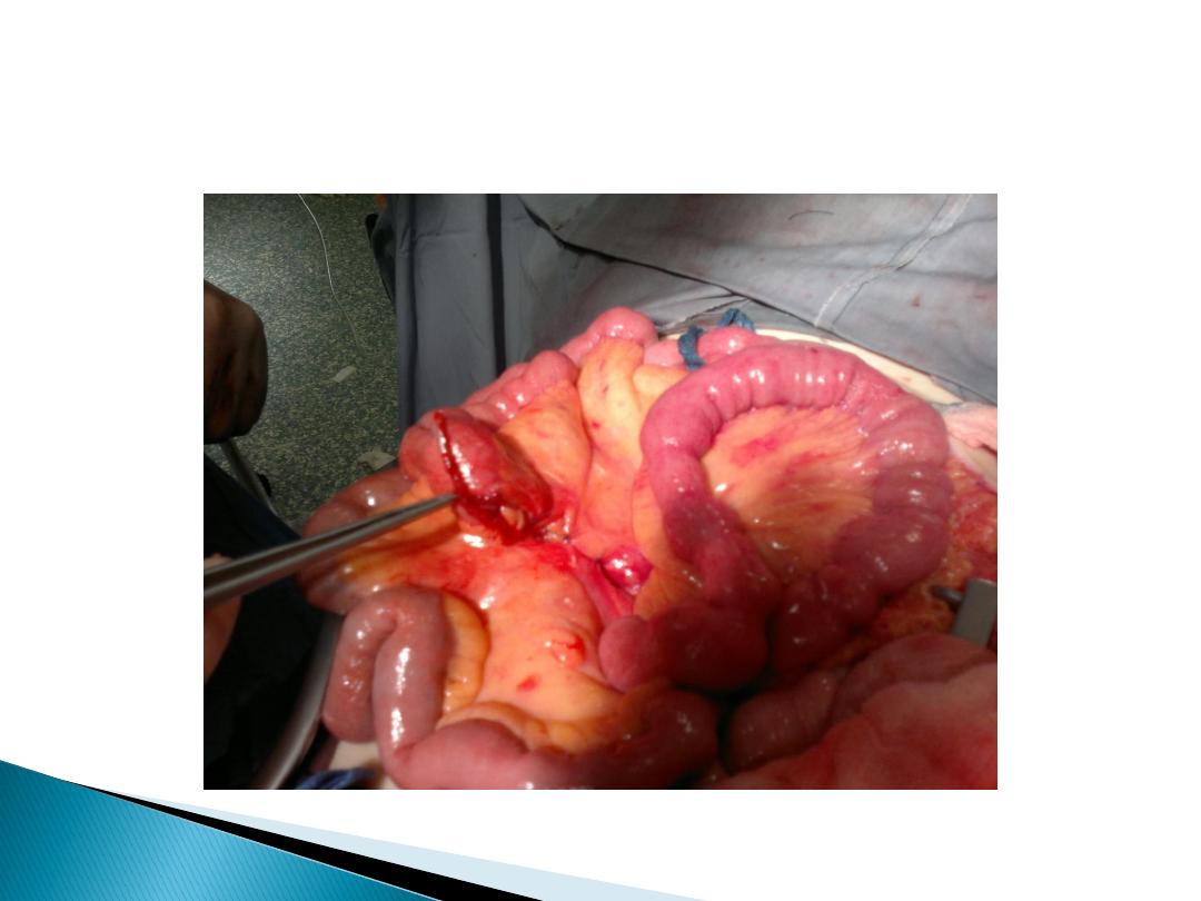
The operative procedure
performed for small bowel obstruction
varies according to the etiology of the obstruction.
Adhesions are lysed(adhesiolysis)
Tumors are resected,
Hernias are reduced and repaired.
Criteria suggesting viability of small intestine are
normal color, peristalsis
,
marginal arterial
pulsations
.
Regardless of the etiology, the affected intestine should be examined.
1- Nonviable bowel resected
.
2- Viable healthy bowel left intact.
3- Questionable viability
:
should be packed with gauze(socked with
warm saline) and rexamined for viability. If viability is questionable and the
patient is hemodynamically stable:
-
short lengths
of bowel of questionable viability should be resected and
primary anastomosis.
-
long length
of the intestine is in question, should be left intact and the patient
re-explored in 24 to 48 hours in a “secondlook” operation. At that time,
definitive resection of nonviable bowel is completed.

Prevention of postoperative adhesion:
1- good surgical technique.
2- careful handling of tissue
3- minimal use and exposure of peritoneum to foreign
bodies.
4- use of laparoscopy rather than open surgery.
Paralytic ileus
failure of transmission of peristaltic waves
secondary to neuromuscular failure with absence of a
lesion-causing mechanical obstruction.
The resultant stasis leads to accumulation of fluid
and gas within the bowel, with associated
distension,
vomiting
,
absence of bowel sounds
and
absolute
constipation
.

Ileus
is a temporary motility disorder that is reversed
with time as the inciting factor is corrected.
Chronic
intestinal pseudo-obstruction
comprises a spectrum of specific disorders associated
with irreversible intestinal dysmotility.
Following celiotomy
– small bowel- 24h, stomach- 48h, colon- 3-5d
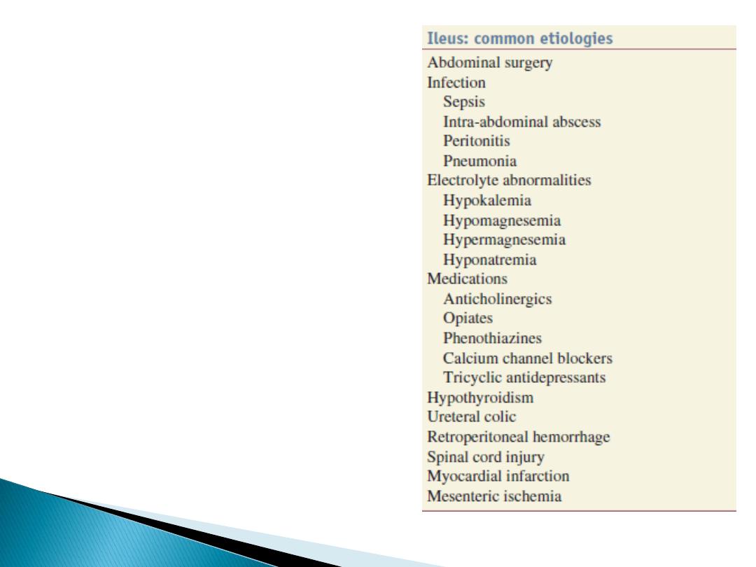
The most frequently
encountered factors are :
Abdominal operations
infection
inflammation
,
Electrolyte-abnormalities
drugs

Clinical Presentation
Paralytic ileus takes on a clinical significance if, 72 hours
after laparotomy:
• there has been
no return of bowel sounds
on auscultation;
• there has been
no passage of flatus
.
Abdominal distension
becomes more marked and
tympanitic.
Colicky pain is not a feature.
Distension
increases pain from the abdominal wound.
In the absence of gastric aspiration,
effortless vomiting
may occur.
Radiologically, the abdomen shows gas-filled loops of
intestine with multiple fluid levels (if an erect film is felt
necessary).

Management
Paralytic ileus is managed with :
1- Nasogastric suction
2- NPO
3- Electrolyte balance must be maintained.
4- If a primary cause is identified, this must be treated.
• There is
no place for
the routine use of
peristaltic stimulants.
• If paralytic ileus is prolonged, CT will
demonstrate any intraabdominal sepsis or mechanical
obstruction --laparotomy.

Small intestinal pseudo-obstruction
This condition may be
primary
(i.e. idiopathic or
associated with familial visceral myopathy)
or
secondary
.
The clinical picture consists of
recurrent subacute
obstruction
.
The diagnosis is made by the
exclusion
of a mechanical
cause.
Treatment consists of initial correction of any
underlying disorder
.
Metoclopramide and erythromycin
may be of use.
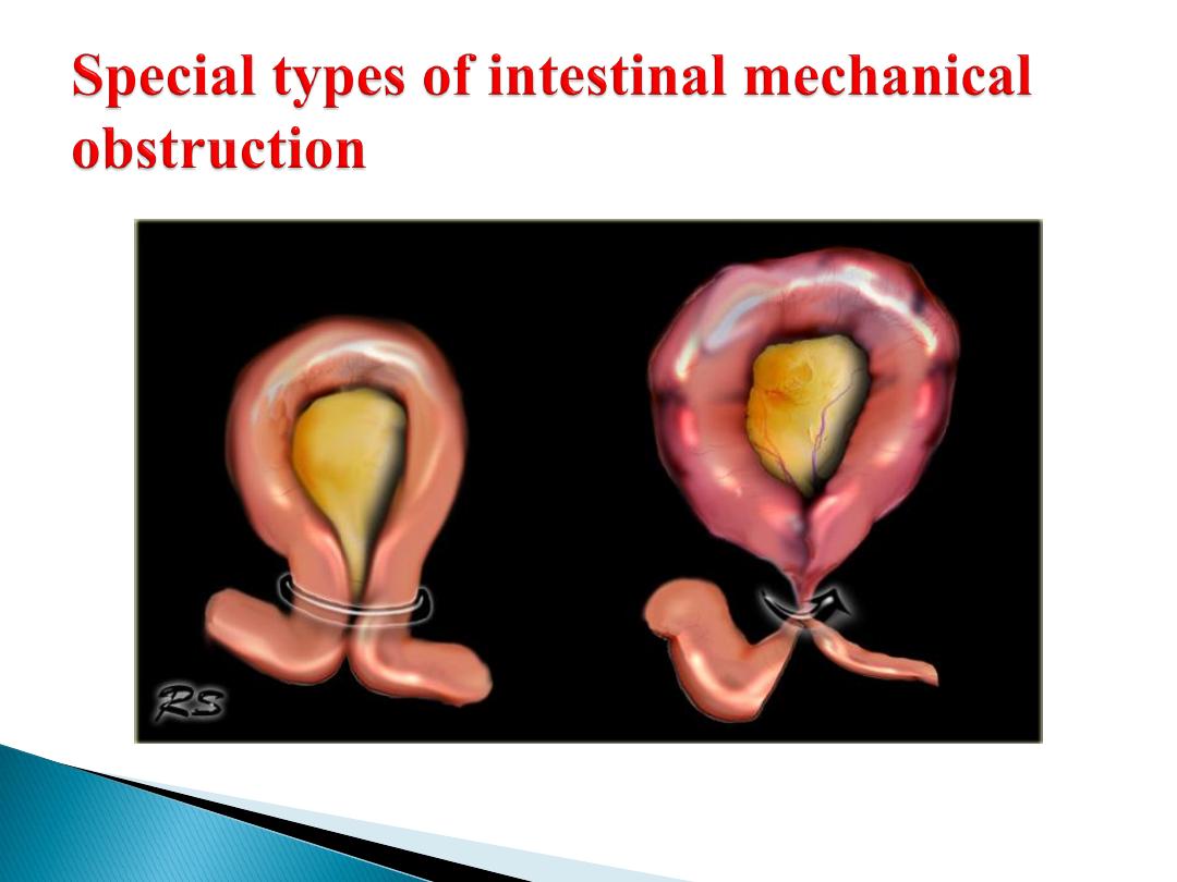

Volvulus
A volvulus is a
twisting or axial rotation of a portion of bowel about
its mesentery
. The rotation causes obstruction to the lumen (>180°
torsion) and if tight enough also causes vascular occlusion in the
mesentery (>360° torsion).
Bacterial fermentation adds to the distention and increasing intraluminal
pressure impairs capillary perfusion. Mesenteric veins become
obstructed as a result of the mechanical twisting and thrombosis results
and contributes to the ischaemia.
Volvuli may be primary or secondary.
The primary
occurs secondary to congenital malrotation of the gut,
abnormal mesenteric attachments or congenital bands. Examples
include volvulus neonatorum, caecal volvulus and sigmoid volvulus.
A secondary
volvulus, which is the more common variety, is due to
rotation of a segment of bowel around an acquired adhesion or stoma

Treatment :
-
Resuscitation(NPO, IV fluid, NG tube , antibiotic)
-
Surgery
untwist the bowel
resect non viable bowel and anastamose.
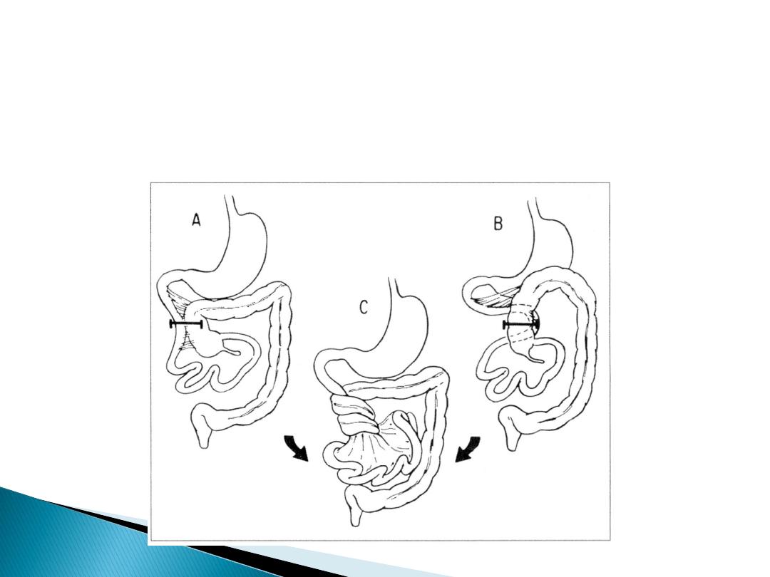
Volvulus neonatorum
This occurs secondary to intestinal malrotationand is potentially
catastrophic.less than one year old with bilious
vomitingurgent surgical exloration(ladd procedure)

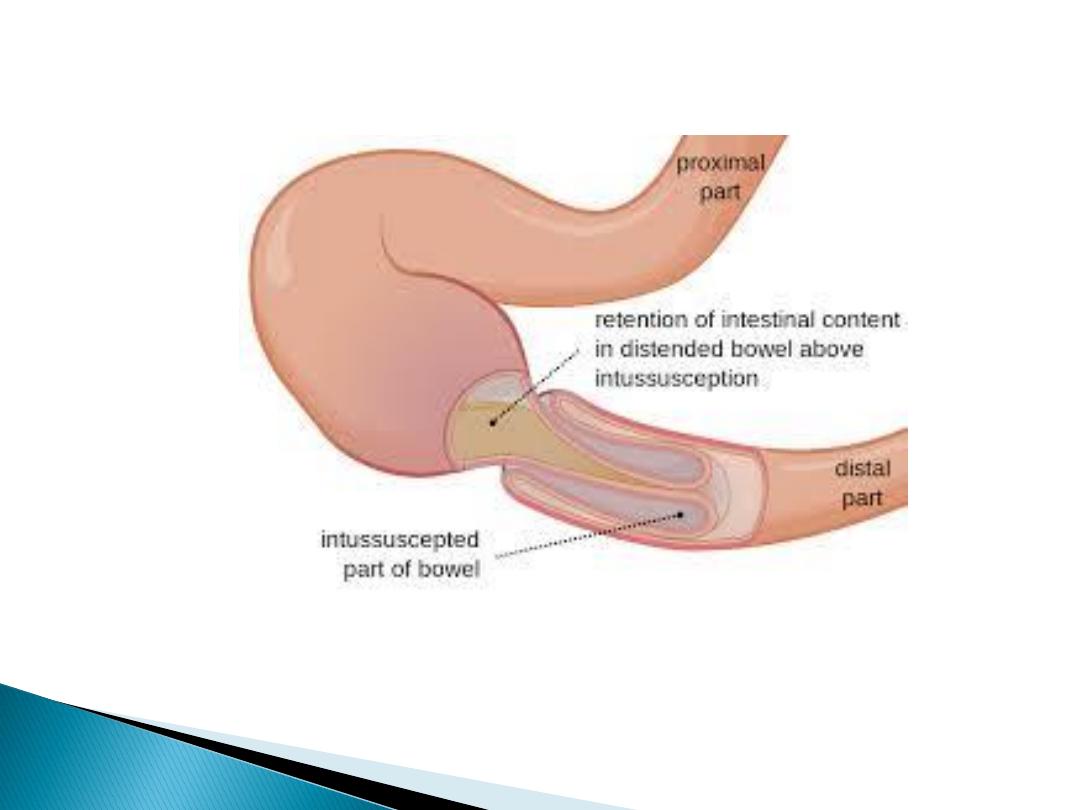
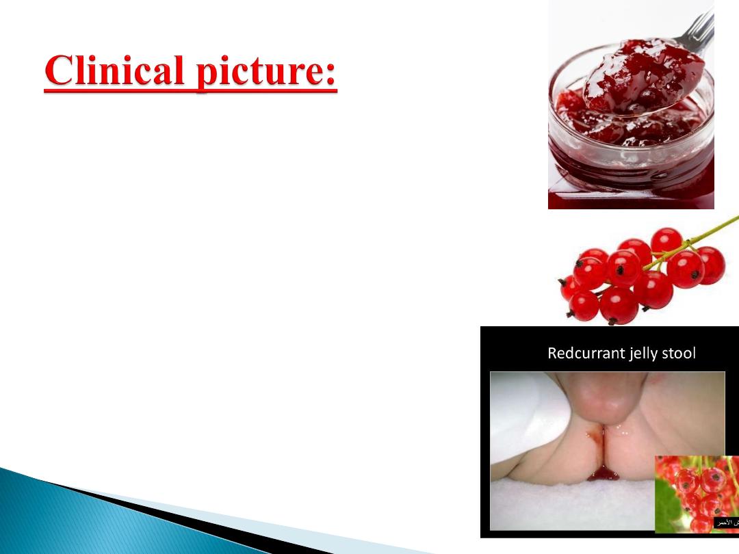
Acute intussusception
one portion of the gut invaginates into an immediately
adjacent segment.
Most in children, peak five and ten months.
Causes:
- idiopathic(most common)(associated upper respiratory
tract infection or gastroenteritis may precede the
condition) (hyperplasia of Peyer’s patches in the
terminal ileum )
-
leading point could be Meckel’s diverticulum, polyp,
duplication, Henoch–Schönlein purpura or appendix
occur in older age.
Adult cases are invariably associated with a lead point,
which is usually a polyp (e.g. Peutz–Jeghers
syndrome), a submucosal lipoma or other tumour.
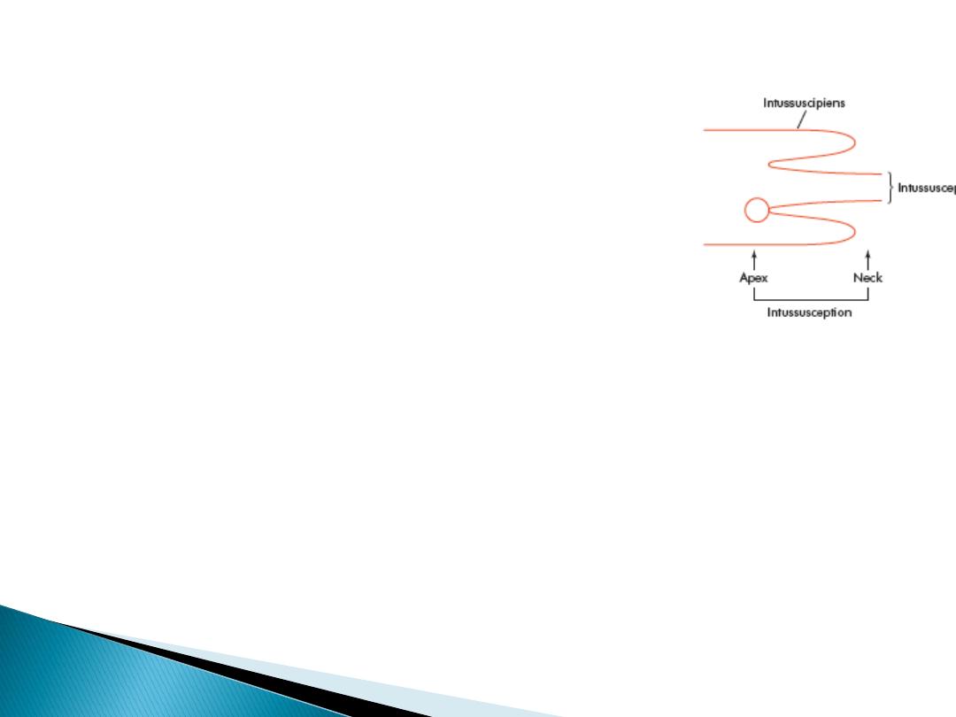
Pathology
is composed of three parts :
• the entering or inner tube (intussusceptum);
• the returning or middle tube;
• the sheath or outer tube (intussuscipiens).
The part that advances is the apex
the mass is the intussusception
the neck is the junction of the entering layer with the
mass.
In most children, the intussusception is ileocolic.
In adults, colocolic intussusception is more common .
Paroxysms of crampy abdominal pain
(screaming)and intermittent vomiting.
Between attacks, the infant may act
normally, but as symptoms progress,
increasing lethargy develops.
Bloody mucus (“red currant jelly ”
stool) may be passed per rectum.
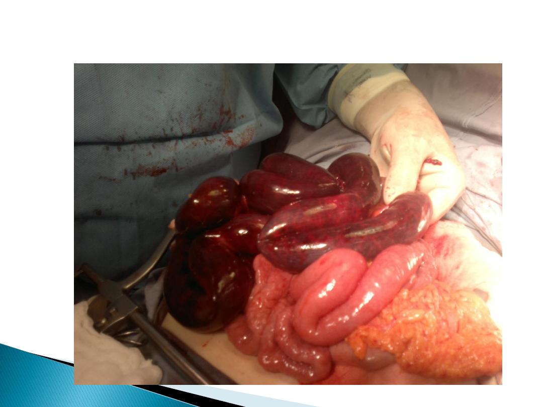
if reduction is not accomplished,
gangrene of the intussusceptum occurs,
and perforation Peritonitis
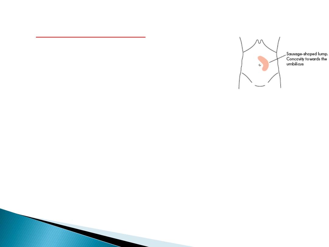
Physical examination:
- mass
in the right upper
quadrant or epigastrium
- absence of bowel
in the right lower quadrant
(Dance’s sign).
Rarely, the apex of intussusception may pass the colon
to protrude through anus
The mass may be seen on plain abdominal x-
ray/US/CT
(target sign
) but is more easily
demonstrated on air or contrast enema.

Treatment
NPO, IV fluid, IV antibiotics
absence of peritonitis radiographic(pneumatic)
reduction(air/barium enema is diagnostic and curative)
Peritonitis or systemically ill child, ileoileal,
pathological leading point urgent laparotomy
Reduction
(by gentle distal pressure, where the intussusceptum
is gently milked out of the intussuscipiens)
+
Non viable bowel resected
and primary anastamosis
+ Appendectomy(for ileocolic)
A fistula is defined as an abnormal communication
between two epithelialized surfaces.
1- internal fistula
: The communication occurs between
two parts of the GI tract or adjacent organs (e.g.,
enterocolonic fistula or colovesicular fistula).
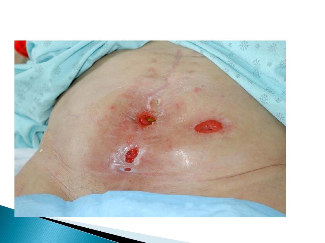
2- An external fistula
(e.g., enterocutaneous fistula or
rectovaginal fistula) involves the skin or another
external surface epithelium.
low-output fistulas
: Enterocutaneous fistulas that drain
less than 500 mL of fluid per day.
high-output fistulas
that drain more than 500 mL of
fluid per day .

1- Over 80% iatrogenic: complications of enterotomies
or intestinal anastomotic dehiscences( inadvertent small
bowel injury at the time of abdominal closure).
2- Trauma: gunshot wounds, stabbing or motor vehicle
accident.
3- Spontaneously without antecedent iatrogenic injury
are caused by:
-Crohn’s disease
- Cancer.
- Radiotherapy.
Iatrogenic enterocutaneous fistulas occurs between fifth and
tenth postoperative days.
1- Fever, leukocytosis.
2- prolonged ileus.
3- abdominal tenderness.
4- wound infection are the initial signs.
The diagnosis becomes obvious when
drainage of enteric material through the abdominal wound or
through existing drains occurs.
These fistulas are often associated with intra-abdominal abscesses.
Low-resistance enteroenteric fistulasmalabsorption.
Enterovesical fistulas recurrent urinary tract infections.
Enterocutaneous fistulas are irritating to the skin and cause
excoriation.
High-output fistulas originating from the proximal small
intestine dehydration, electrolyte abnormalities, and
malnutrition.

1. Stabilization.
Fluid and electrolyte
resuscitation .
Nutrition
(TPN), parenteral route initially.
Sepsis is controlled
with
antibiotics
and
drainage
of
abscesses.
The skin is protected
from the fistula effluent with ostomy
appliances.
The somatostatin analogue octreotide??
2. Investigation. The anatomy of the fistula is defined using the CT
scanning, or fistulogram
3. Definitive management: if 2-3 months of conservative therapy fails
then definitive surgical procedure should be performed .(Resection of
the fistula tract + resection of intestinal segment from which the fistul
arise)
4. Rehabilitation.

Over 50% of intestinal fistulas close spontaneously.
Factors inhibiting spontaneous closure(FRIENDS)
Fistulas have the potential to close spontaneously. Causes of failure to
close include:
1-malnutrition, immune suppression , steroids.
2- sepsis
3- inflammatory bowel disease(crohn’s)
4- cancer
5- radiation
6- obstruction of the intestine distal to the origin of the fistula
7- foreign bodies,
Gastric, Duodenal fistula, High output, short fistulous tract (<2 cm)
and epithelialization of the fistula tract are less likely to
closespontaneously.
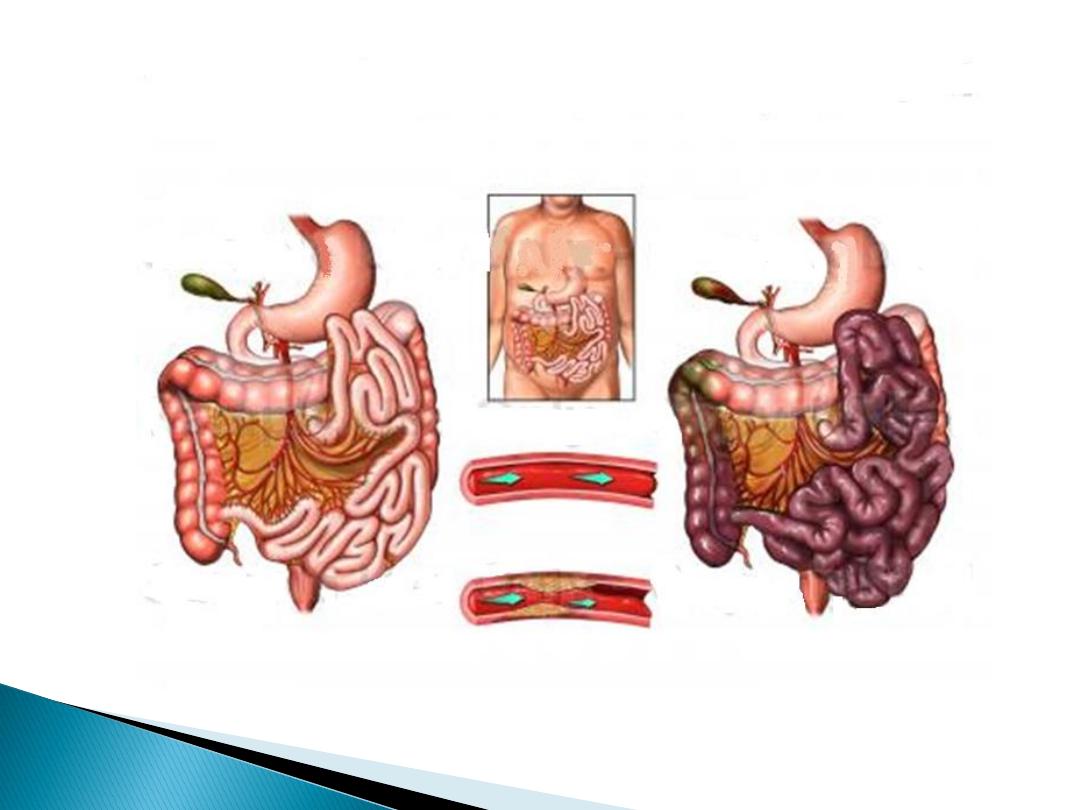
two distinct clinical syndromes:
1- acute mesenteric ischemia (embolus or thrombus)
2- chronic mesenteric ischemia.
Four distinct pathophysiologic mechanisms can lead to
acute mesenteric ischemia:
1. Arterial embolus(acute): most common, (left atrial(AF) or
ventricular thrombi or valvular lesions), occlude the
superior mesenteric artery(mid , distal).
2. Arterial thrombosis(acute or chronic): (proximal)
mesenteric arteries.
3. Vasospasm (also known as nonocclusive mesenteric
ischemia[NOMI])
, result of vasospasm from vasospastic
drugs.
4. Venous thrombosis: 10% of cases of acute mesenteric
ischemia and involved the superior mesenteric vein.
Sudden onset of
Severe mid-abdomen pain, out of
proportion to the degree of tenderness on examination
, is
the hallmark of acute mesenteric ischemia.
in patients with
underlying cardiac or atherosclerotic
disease
Associated symptoms can include nausea, vomiting, and
diarrhea.
Physical findings are characteristically
absent early
in the
course of ischemia.
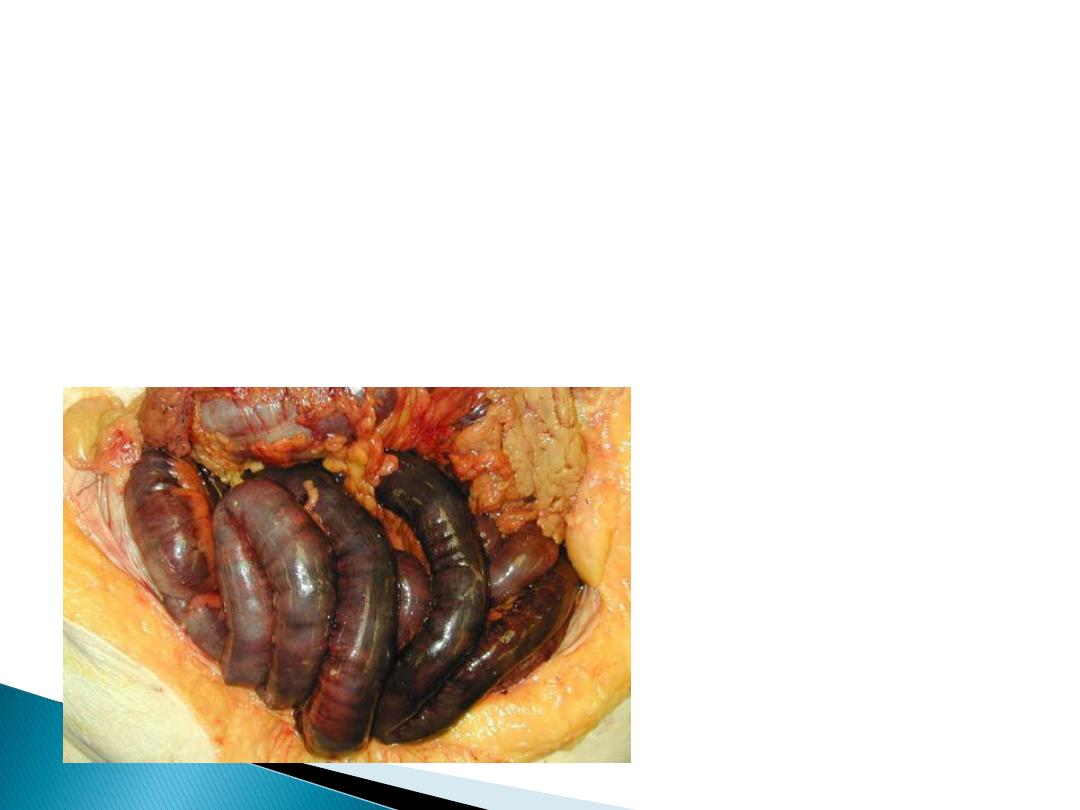
Fever, passage of bloody stools, Diffuse abdominal
tenderness, rebound, and rigidity
are late signs and usually
indicate bowel infarction and necrosis.
presents
insidiously
(because of collateral).
Postprandial abdominal pain
is the most prevalent symptom,
(“
food fear
”)
weight loss.
Persistent nausea and occasionally diarrhea may coexist
Usually misdiagnosed.
Chronic mesenteric venous
thrombosis
asymptomatic
, because of extensive collateral venous drainage.
incidental finding
on imaging studies.
some patients present with
bleeding
from esophagogastric
varices.

an elderly + multiple comorbiditie
s +
digitalis or
vasoconstrictor such as epinephrine.
70% abdominal pain.
30% no abdominal pain , progressive abdominal
distention , acidosis impending infarction.

Laboratory evaluation is not sensitive not specific
1-hemoconcentration and leukocytosis.
2-Metabolic acidosis.
3-Elevated serum amylase.
4- in the late stages :increased lactate levels,
hyperkalemia, and azotemia.
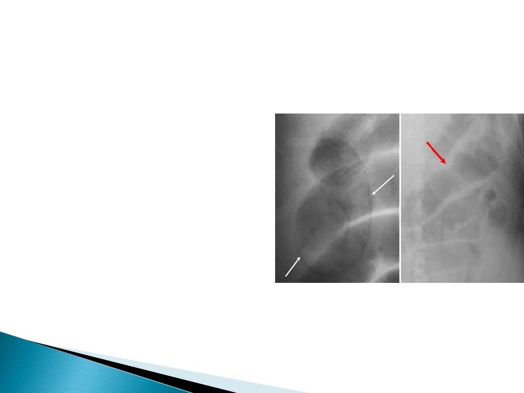
Plain abdominal radiographs
- to exclude other causes of abdominal pain
-Pneumoperitoneium , pneumatosis intestinalis, and gas in
the portal vein may indicate infarcted bowel.
-ileus with a gasless abdomen.
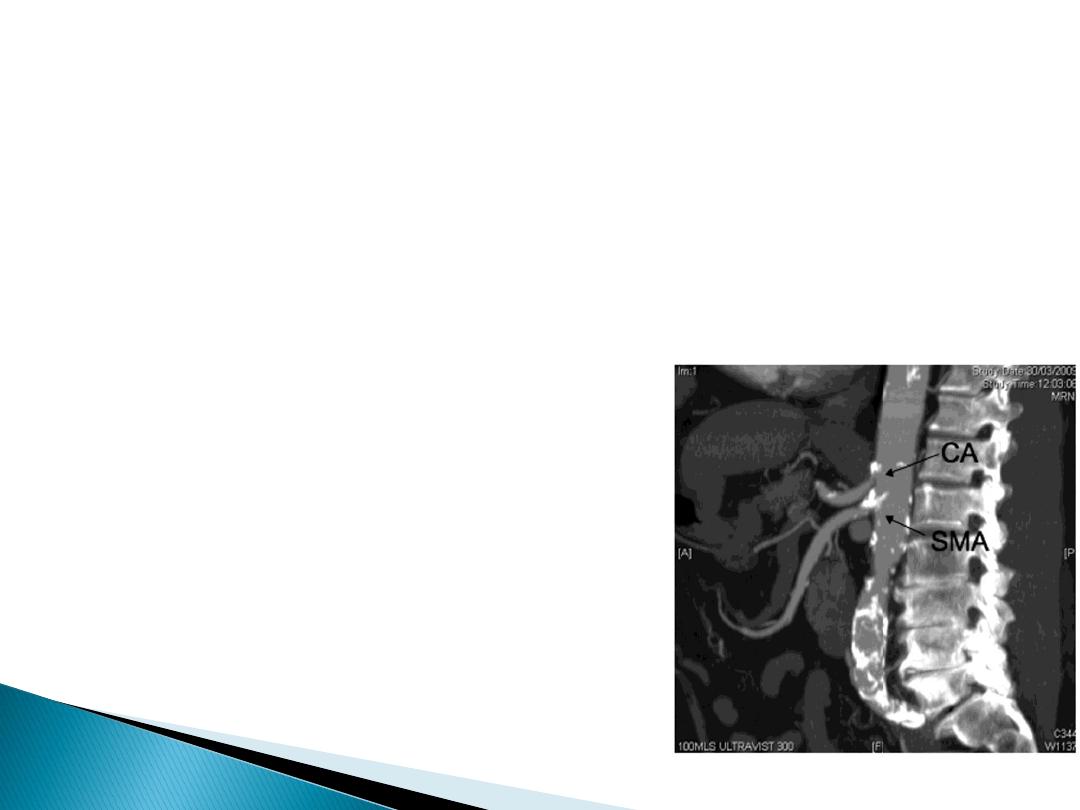
Duplex ultrasonography
CTA and MRA
Mesenteric arteriography
(definitive diagnosis)
Has therapeutic role,
infusion of vasodilating agents,
such as papaverine, thrombolytic??
chronic intestinal angina develop acute abdomen and
peritonitis
immediate exploration
+ assessment of
intestinal viability and vascular reconstruction is the
best choice.
(arteriography is time consuming)

IV fluid
resuscitation
systemic anticoagulation with
heparin
Significant metabolic acidosis not responding to fluid
resuscitation should be corrected with
sodium
bicarbonate
.
A central venous catheter and Foley catheter
antibiotics
immediate
surgical exploration
, avoiding the delay
required to perform an arteriogram
Surgery :
Arteriotomy+ embolectomy+ an assessment of
intestinal viability+ nonviable bowel must be
resected.
A second-look procedure(24 to 48 hr)in many patients
to reassess the remaining bowel viability.

Same preoperative management
Surgery: SMA bypass graft may originate from either
the aorta or iliac artery
Chronic Mesenteric Ischemia.
Endovascular Balloon dilatation or stent placement
Surgical: transaortic endarterectomy or mesenteric
artery bypass.

mesenteric arterial catheterization and infusion of
vasodilatory agents, such as tolazoline or papaverine.
cessation of other vasoconstricting agents
intravenous heparin
Surgical exploration is indicated if the patient develops
signs of continued bowel ischemia or infarction
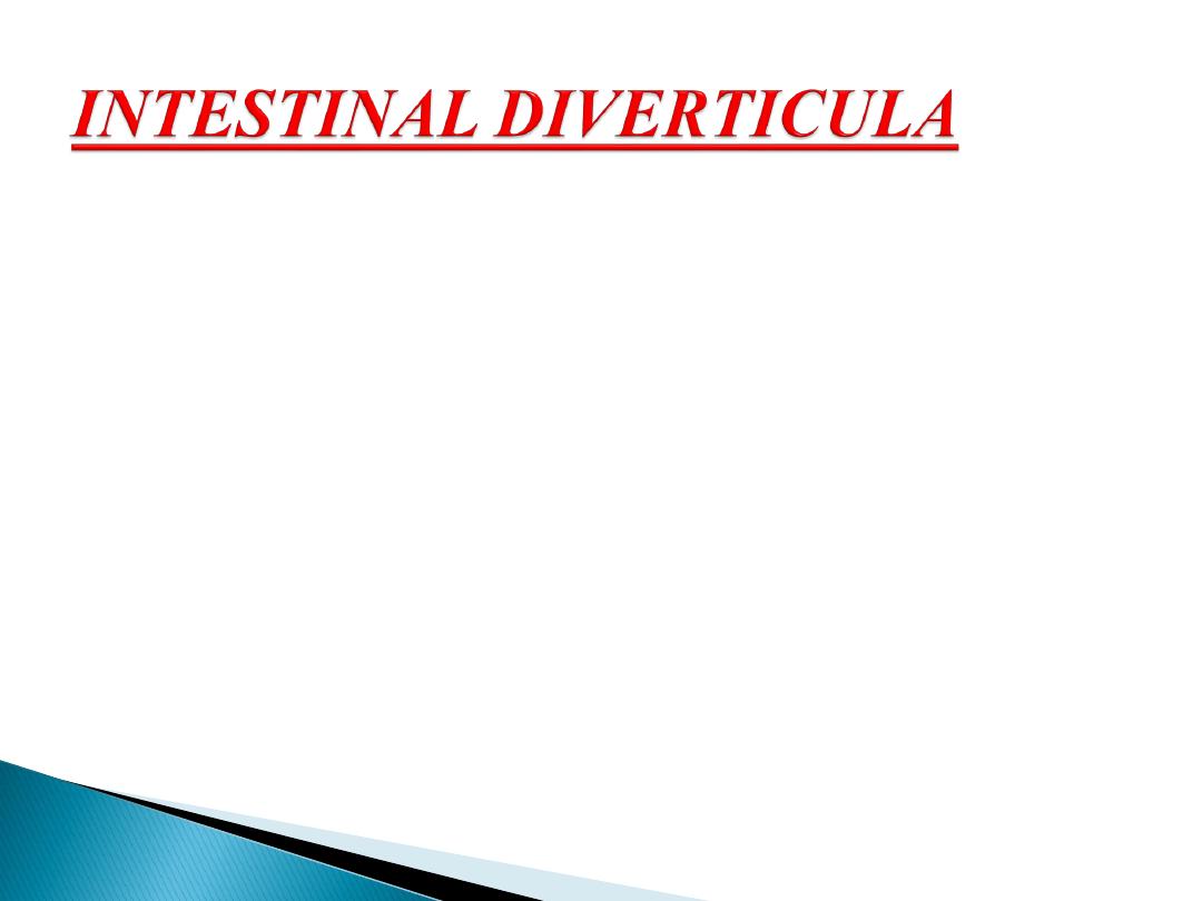
Diverticula (hollow out-pouchings) are a common
structural abnormality that can occur from the
oesophagus to the rectosigmoid junction (but not
usually in the rectum).
They can be classified as:
• Congenital. All three coats of the bowel are present in
thewall of the diverticulum, e.g. Meckel’s diverticulum.
• Acquired. There is no muscularis layer present in the
diverticulum, e.g. sigmoid diverticula.

mucosal herniation at the point of entry of the blood vessels.
vary in size and are often multiple.
Presentation:
1- Asymptomatic (incidental finding at surgery or on
radiological imaging )
2- Malabsorption, as a result of bacterial stasis
3- Acute abdominal emergency if they become inflamed or
perforate.
4- Bleeding from a jejunal diverticulum is a rare .
Treatment :
Asymptomatic need no treatment
Elective resection of an affected small bowel segment that is
causing malabsorption.
If perforated jejunal diverticulitis is found at emergency
laparotomy, a small bowel resection + anastomosis /stoma
formation.
Extensive jejunal diverticulosis can be very difficult to treat.
Jejunal diverticula
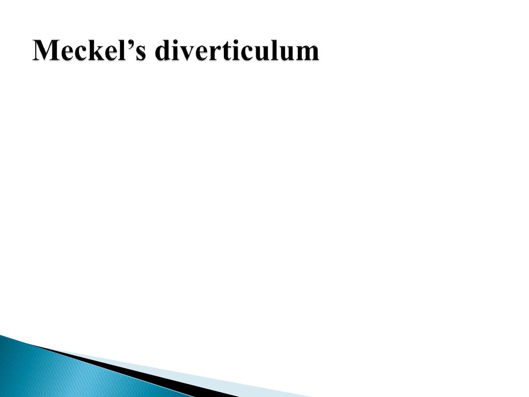
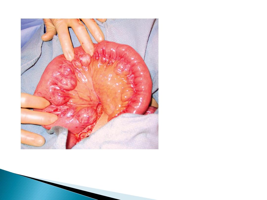
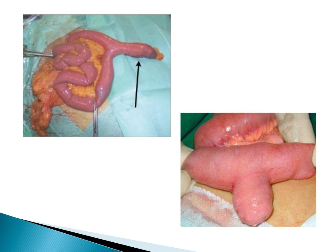
A Meckel’s diverticulum is a persistent remnant of the
vitellointestinal duct and is present in about 2 per cent of
the population.
- on the
antimesenteric side
of the ileum
-
60 cm from the ileocaecal valve
-
5 cm long
.
- contains
all three coats
of the bowel wall and has its own
blood supply.
- In around 20 per cent the mucosa of a Meckel’s
diverticulum contains heterotopic epithelium of gastric,
colonic or pancreatic type.

A Meckel’s diverticulum can present clinically in the following ways:
1- Asymptomatic(mostly)
2- Haemorrhage
If gastric mucosa is present, peptic ulceration can occur and present as melaena.
3- Diverticulitis
presents like appendicitis.
4- Intussusception
It can be the lead point for ileoileal or ileocolic intussusception.
5- Chronic ulceration
Pain is felt around the umbilicus, as it is midgut in origin.
6- Intestinal obstruction
A band between the apex of the diverticulum and the may cause obstruction
directly or by a volvulus around it.
7- Perforation
. may resemble a perforated duodenal ulcer.
The finding of a Meckel’s diverticulum in an inguinal or femoral hernia has been
described as Littre’s hernia.

Usually diagnosed incidentally(intraoperatively)
- Radionuclide scans (99mTc-pertechnetate)
- Angiography can localize the site of bleeding
Incidental finding of Meckel’s can safely be left if it has
a wide mouth and is not thickened. When there is doubt, it
can be resected.
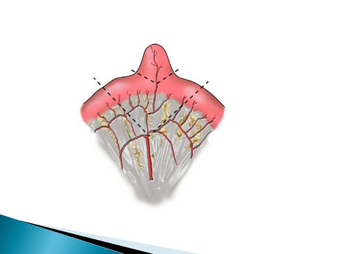
If symptomatic: Excise the diverticulum
(by resecting it and suturing the defect at its base, or with a
linear stapler-cutter)
limited small bowel resection of the involved segment +
anastomosis,
1-If the base of the diverticulum is indurated , inflamed or
perforated.
2- in bleeding
3- if the divertic. contains a tumor .
rare and <10 per cent of gastrointestinal neoplasia.
Benign
Most small bowel neoplasms are benign:
adenomas, lipomas, haemangiomas and neurogenic tumours.
frequently asymptomatic and identified incidentally,
May present with:
- intussusception
- small bowel obstruction
- bleeding that may cause anaemia or may even be overt.
Diagnosis:
- CT
- small bowel contrast studies do not show them easily.
- Capsule endoscopy or small bowel endoscopy
Treatmen:
Symptomatic lesions can be treated by small bowel resection and
anastomosis.
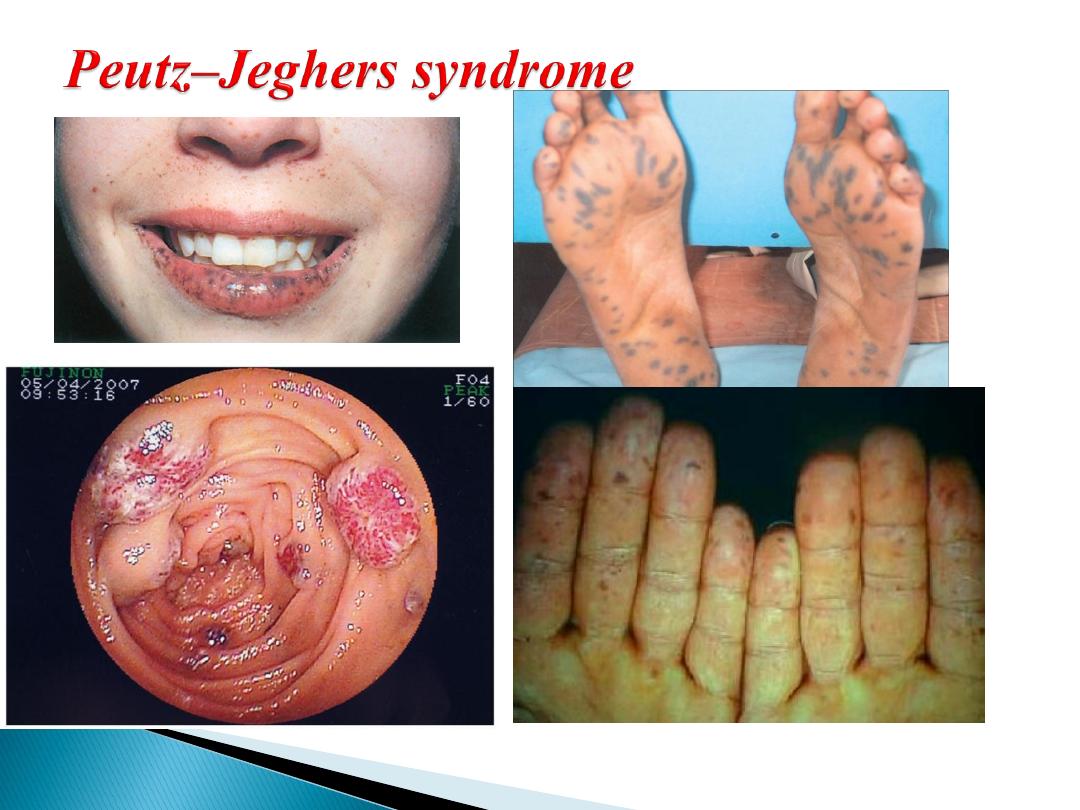
autosomal dominant
melanosis of the mouth and lips + multiple hamartomatous
polyps in the small bowel and colon .
Melanin spots on digits and perianal skin.
Malignant change in the polyps rarely occurs and, in
general the polyps can be left alone.
Resection may be indicated for heavy and persistent or
recurrent bleeding or intussusception.
Polyps may be removed by
- enterotomy or laparotomy
- snared via a colonoscope introduced via an
enterotomy.
Heavily involved segments of small intestine may
occasionally be resected.

rare and present late, most often diagnosed after
surgery for small bowel obstruction.
Adenocarcinoma
carcinoid tumours
lymphomas
mesenchymal tumours (gastrointestinal stromal
tumours(GIST)
more in jejunum
more with Crohn’s disease, coeliac disease, familial
adenomatous polyposis (FAP) and Peutz-Jeghers syndrome.
They present with anaemia,
gastrointestinal bleeding
intussusception or obstruction.
Prognosis is poor as tumours often present late
the surgical treatment:
Resection of small bowel and the affected mesentery.
A right hemicolectomy for tumours of the distal ileum.

2- Carcinoid tumour
most in appendix, ileum and rectum in decreasing order .
arise from Kulchitsky cells
Small +/- significant lymph node metastases
may be multiple.
produce a number of vasoactive peptides, most commonly
5-hydroxytryptamine (serotonin), but also histamine,
prostaglandins and kallikrein.
When they metastasise to the liver, the carcinoid syndrome can
become evident, because the vasoactive substances escape the
filtering actions of the liver.
The clinical syndrome itself consists of:
- reddish-blue cyanosis
- flushing attacks(induced by
alcohol)
- diarrhoea, borborygmi
- asthmatic attacks
- pulmonary and tricuspid
stenosis .

octreotide scanning
detect primary and secondary
tumours.
Plasma markers chromogranin A concentration
(markers of recurrence and prognostic value).
primary disease
Surgical resection
(significant
recurrence).
metastatic disease
hepatic resection+octreotide
(a
somatostatin analogue).

Primary or more common
secondary to systemic lymphoma
.
more common in patients with
Crohn’s disease
and
immunodeficiency
syndromes.
Hodgkin’s lymphoma(rare ) to affect the small bowel and most
western-type lymphomas are
non-Hodgkin’s B-cell lymphomas
.
Clinical presentation: -anaem -anorexia
-weight loss -Bleeding - perforation
Coeliac disease
T-cell lymphoma .
North Africa and the Middle East
Mediterranean lymphoma
(widespread ).
Burkitt’s lymphoma
can aggressively affect the ileocaecal region,
particularly in children.
Treatment :
Chemotherapy
obstruction, perforation or bleeding surgery .
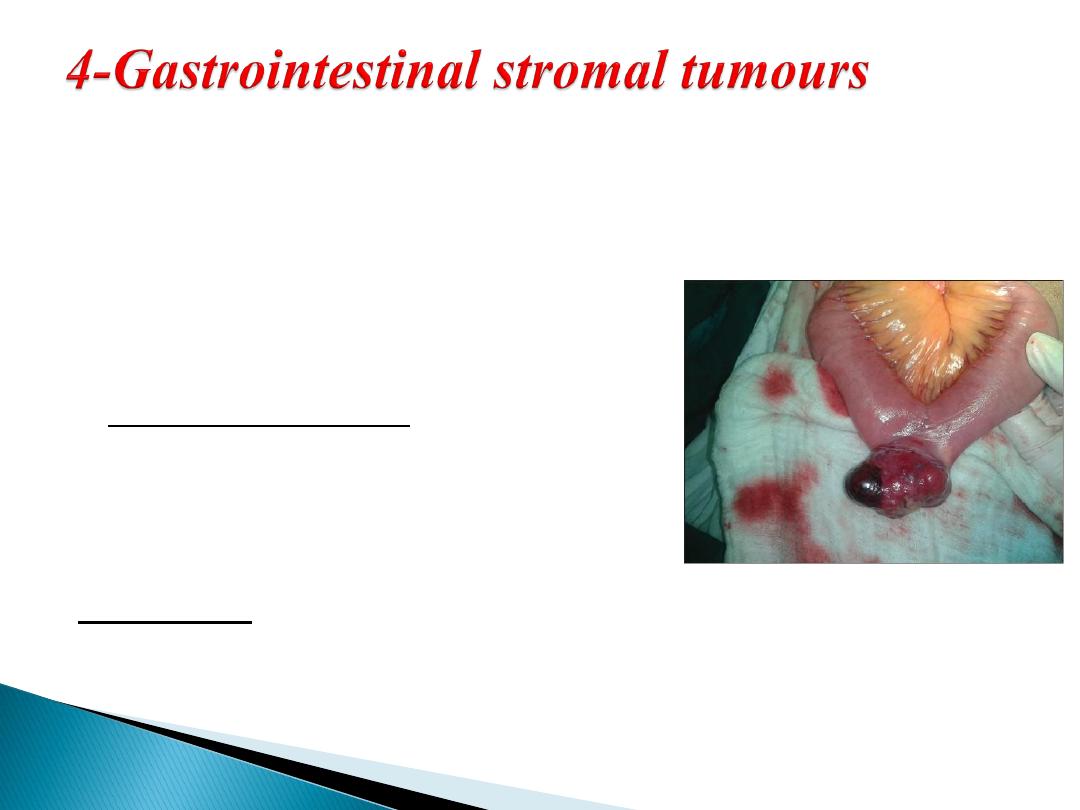
These are mesenchymal tumours
benign or malignant
.(difficult to distinguish)
Increased
size
and high levels of
c-kit (CD117)
staining
malignant potential.
most commonly in the
stomach
, but can be found in other parts
of the gut.
50- to 70-year
age group.
Patients may be asymptomatic.
Symptoms include: lethargy
pain
nausea
haematemesis
or melaena.
Treatment :
Surgical excision
Glivec (imatinib)(adjuvant)
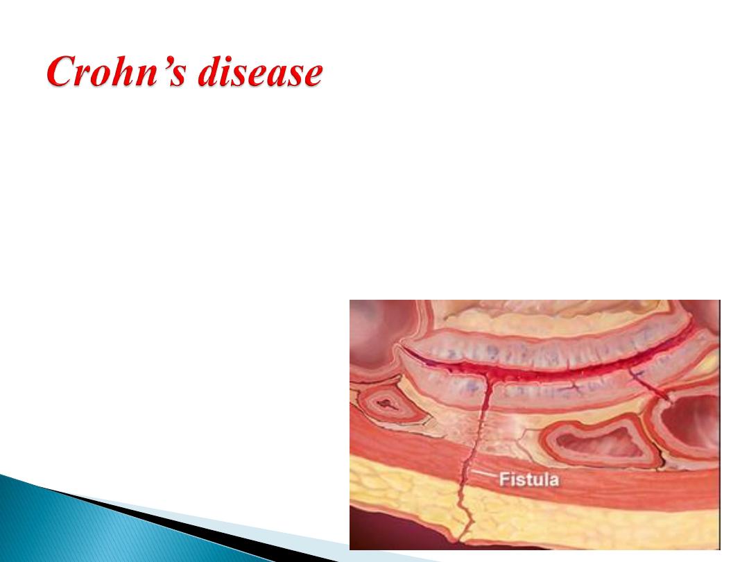
is a chronic, idiopathic segmental transmural
inflammatory disease with a propensity to affect the
distal ileum
any part of the alimentary tract can be involved.
small bowel affected in 80%
Both genetic and
environmental factors
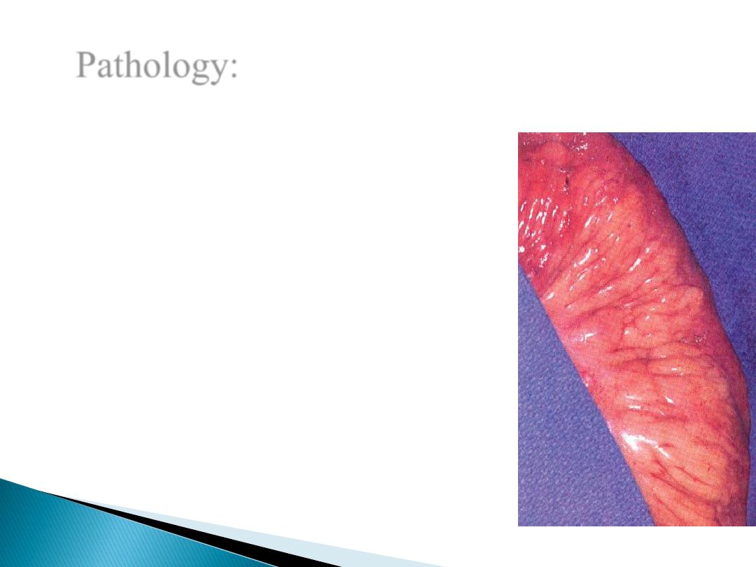
Pathology:
transmural inflammation of the intestine
aphthous ulcer.
Noncaseating granulomas
multiple ulcers in intestinal mucosa
cobblestoned appearance of the mucosa
Serosal involvement , adhesion to other
loops of bowel or other adjacent organs
fibrosis with stricture formation, intra-
abdominal abscesses, fistulas, and, rarely,
free perforation.
“skip lesions”
fat wrapping(pathognomonic)
Risk for malignant transforamtion

(a) fibrostenotic disease
(b) fistulizing disease
(c) aggressive inflammatory disease.
Abdominal pain(RIF mimcking appendicitis) ,
diarrhea, and weight loss
waxing and waning course
Constitutional symptoms (weight loss and fever, or
growth retardation in children)
Complication (obstruction, fistula, abscess,
perforation, perianal abscess or fistula)
Extraintestinal manifestation
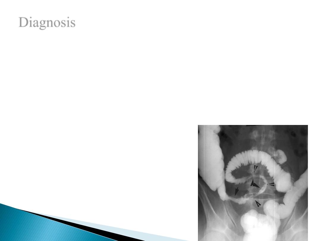
Diagnosis
Radiographic, Endoscopic, and Pathologic
Colonoscopy with intubation of terminal ileum,
Esophagoscopy, capsule endoscopy
ulcerations, cobblestone appearance, Skip areas
Contrast examination strictures , ulcers, fissures
CT scanning abcsess , free perforation
Biopsy with endoscopy
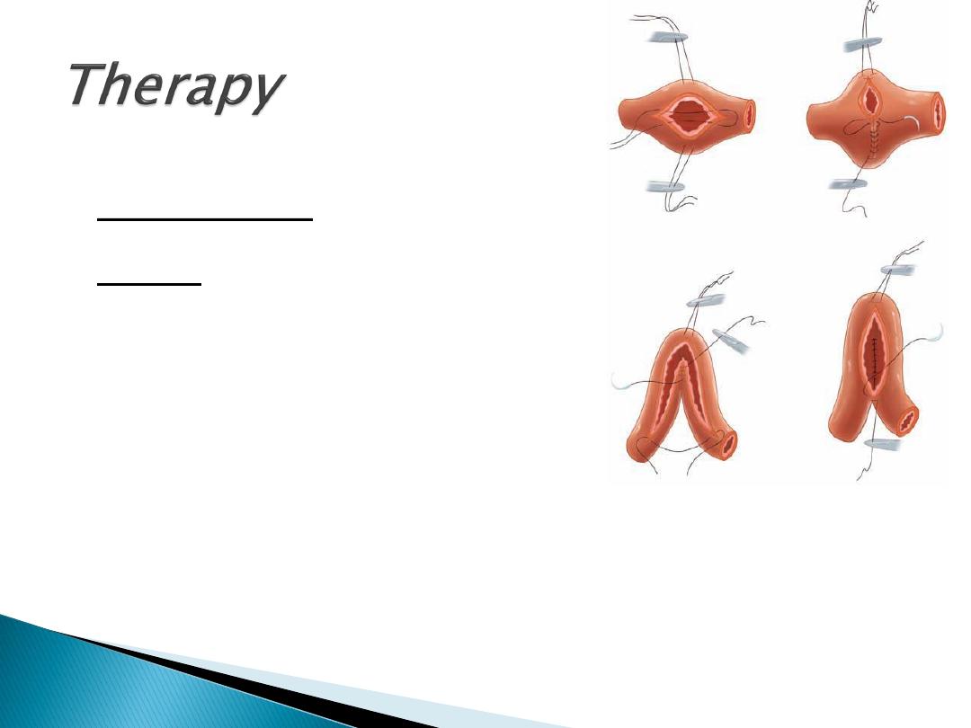
no curative, palliate symptoms
Medical therapy :
induce and maintain remission.
Surgery -Complication
obstruction
perforation
complicated fistulas
Haemorrhage
Malignancy
-Failure of medical therapy
Conserve as much as you can of the Bowel (open/laparoscopy)
Segmental intestinal resection of gross disease +primary anastomosis
stricturoplasty

Thank you