Cell structure and function Chapter (3) / part (1) / Lec (8)
Huda Ayad - Monday 21/12/2015-Objectives:-
- In the end lecture , you should be able to :-
1- Define of the cell
2- Describe the structure of the cell
3- Identify the component of human cell and state its function
4- Distinguish between the structure of prokaryotic cell and Eukaryotic cell
5- Definition how Eukaryotic cell evolved from prokaryotic cell
What Is a Cell?
cell:- are the basic building blocks of the living things
It is basic structural and functional unit of any living thing
The human body is composed of trillions of cells
The Cell Theory: A cell is the basic unit of life
All living things are made up of cells.
New cells arise only from pre-existing cells
The light microscope, invented in the seventeenth century
Figure3.1 cells vary in structure and function
Cell Size
A few cells, such as a hen’s egg or a frog’s egg, are large enough to be seen by the naked eye.
In comparison, a human egg cell is around 100 μm in size
Most cells are much smaller.
Most cells are small and can be seen only under a microscope.
The small size of cells means that they are measured using the smaller units of the metric system, such as the micrometer (μm).
A micrometer is 1/1,000 millimetre.
Most human cells are about 100 μm in diameter, about the width of a human hair
μm = 1×10⁻³ mm
nm = 1×10⁻⁹ m 1 / 1,000,000,000 1×10⁻⁹
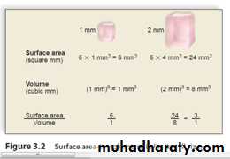

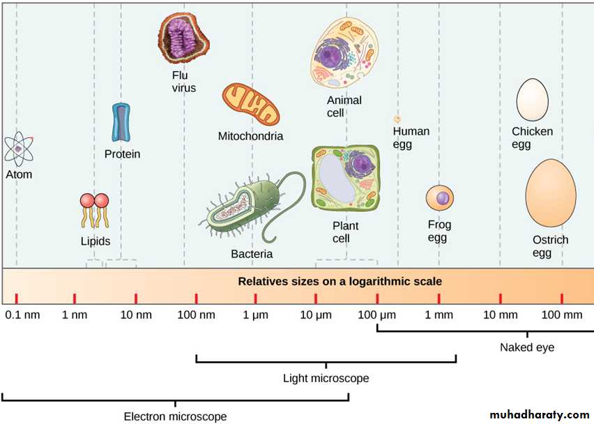
Figure 3.a Relative size of the cells organisms on logarithmic scales
Figure 3. b Shows surface area-to- volume
The small size of cells is explained by considering the surface area-to-volume ratio of cells.
Nutrients enter a cell—and waste exits a cell—at its surface.
Therefore, the greater the amount of surface, the greater the ability to get material in and out of the cell.
A large cell requires more nutrients and produces more waste than a small cell.
Yet, as cells become larger in volume, the proportionate amount of surface area actually decreases.
How Cells Are Organized Cells
are classified into two broad categories:
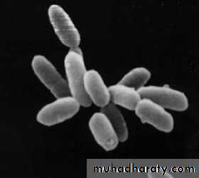
The prokaryotes : like bacteria
The eukaryotes: like cells of animals, plants, fungi, and some single-celled organisms

The ARMAN are a new group of archaea recently discovered in acid mine drainage
Evolutionary History of the Eukaryotic Cells
The first cells to arise were prokaryotic cells.Prokaryotic cells today are represented by the bacteria and Archaea, which differ mainly by their chemistry.
Bacteria are well known for causing diseases in humans, but they also have great environmental and commercial importance.
The Archaea are known for living in extreme environments that may mirror the first environments on Earth. These environments are too hot, too salty, and/or too acidic for the survival of most cells.
The eukaryotic cell is believed to have evolved from the Archaea, How?
The internal structure of eukaryotic cells is believed to have evolved, as shown in the figure 3.5.
The nucleus could have formed by invagination of the plasma membrane, a process whereby a pocket is formed in the plasma membrane.
The pocket would have enclosed the DNA of the cell, thus forming its nucleus.
Surprisingly, some of the organelles in eukaryotic cells may have arisen by engulfing prokaryotic cells.
The engulfed prokaryotic cells were not digested; rather, they then evolved into different organelles.
One of these events would have given the eukaryotic cell a mitochondrion
Mitochondria are organelles that carry on cellular respiration.
Another such event may have produced the chloroplast.
Chloroplasts are found in cells that carry out photosynthesis.
This process is often called endosymbiosis
Symbiogenesis or endosymbiotic theory
It is an evolutionary theory which explains the origin of eukaryotic cells from prokaryotes
It states that several key organelles of eukaryotes originated as symbiosis between separate single-celled organisms.
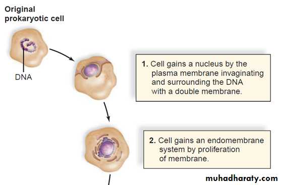
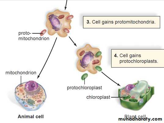
Figure3.5 The evolution of eukaryotic cells
Early prokaryotic organisms, such as the Archaeans, were well adapted to life on the early Earth.The environment that they evolved in contained conditions that would be instantly lethal to life today.
The atmosphere contained no oxygen; instead, it was filled with carbon monoxide and other poisonous gases; the temperature of the planet was greater than 200؛F (93.3 C); and there was no ozone layer to protect organisms from damaging radiation from the sun.
Despite these conditions, prokaryotic life survived and in doing so gradually adapted to Earth’s environment.
In the process, most of the archaea bacteria went extinct. However, we now know that some are still around and can be found in some of the most inhospitable places on the planet, such as thermal vents and salty seas.
The study of these ancient bacteria is still shedding light on the early origins of life.
Cell organization of prokaryotes and eukaryotes
There is difference in cell organization between the two types of cells
Plasma membraneIt is an outer membrane that regulates what enters and exits a cell.
- It is a phospholipid bilayer. that looks like a “sandwich”, made of two layers of phospholipids.
Their polar phosphate molecules (hydrophilic) form the top and bottom surfaces of the bilayer, and the no polar lipid lies in between (hydrophobic ).
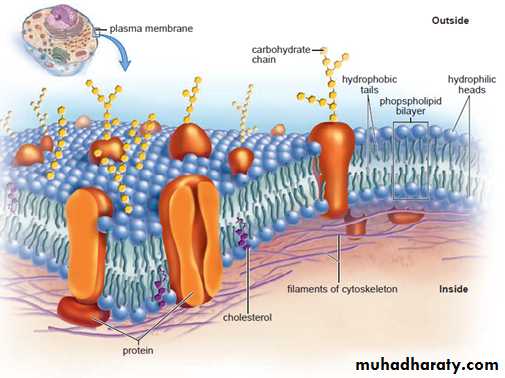
Figure3.6 Organization of the plasma membrane
The phospholipid bilayer is selectively permeableThis means it allows certain molecules—but not others—to enter the cell.
Proteins scattered throughout the plasma membrane play important roles in allowing substances to enter the cell.
Cytoplasm
Also, all types of cells contain cytoplasmCytoplasm: It is a semifluid medium that contains water and various types of molecules suspended or dissolved in the medium.
The presence of proteins accounts for the semifluid nature of the cytoplasm.
The cytoplasm contains organelles.
Organelles
Eukaryotic cells have many different types of organelles.Originally, the term organelle referred to only membranous structures (in the textbook, the term will be used for any well-defined subcellular structure).
Many organelles are surrounded by a membrane, which allows compartmentalization of the cell.
This keeps the various cellular activities separated from one another.
Internal Structure of Eukaryotic Cells
The most prominent organelle within eukaryotic cell is :
Nucleus : a membrane-enclosed structure in which DNA is found.
Prokaryotic cells, such as bacterial cells, lack a nucleus .
Although the DNA of prokaryotic cells is centrally placed within the cell, it is not surrounded by a membrane
The Nucleus and Endomembrane System
The nucleus and several organelles are involved in the production and processing of proteins.The endomembrane system is a series of membrane organelles that function in the processing of materials for the cell
The Nucleus
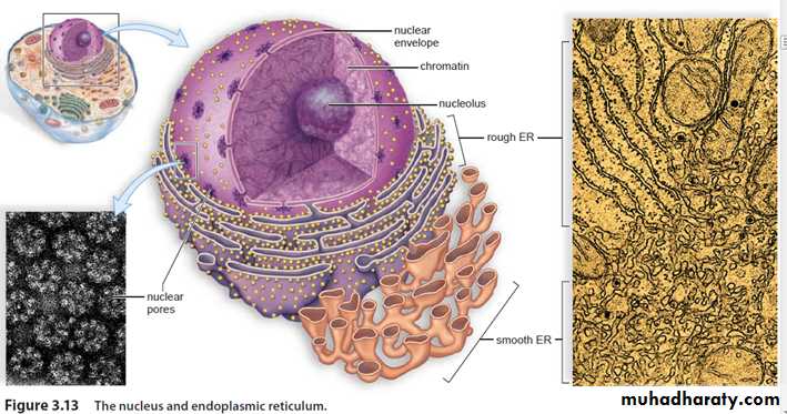
The nucleus, a prominent structure in cells, stores genetic information.
Every cell in the body contains the same genes.
Genes are segments of DNA that contain information for the production of specific proteins.
Each type of cell has certain genes turned on and others turned off.
DNA, with RNA acting as an intermediary, specifies the proteins in a cell.
Proteins have many functions in cells, and they help determine a cell’s specificity.
Chromatin
Chromatin is the combination of DNA molecules and proteins that make up the chromosomes.
Chromatin can coil tightly to form visible chromosomes during meiosis (cell division that forms reproductive cells in humans) and mitosis (cell division that duplicates cells).
Chromatin is immersed in a semifluid medium called the nucleolus.
A difference in pH suggests that nucleoplasm has a different com This is also where rRNA joins with proteins to form the subunits of ribosomes. position from cytoplasm.
The nucleus is separated from the cytoplasm by a double membrane known as the nuclear envelope.
This is continuous with the endoplasmic reticulum (ER), a membranous system of saccules and channels discussed in the next section.
The nuclear envelope has nuclear pores of sufficient size to permit the passage of ribosomal subunits out of the nucleus and proteins into the nucleus.
Ribosomes
Ribosomes are organelles composed of proteins and rRNA.
Protein synthesis occurs at the ribosomes.Ribosomes are often attached to the endoplasmic reticulum; but they also may occur free within the cytoplasm, either singly or in groups called polyribosomes.
Proteins synthesized at ribosomes attached to the endoplasmic reticulum have a different destination from that of proteins manufactured at ribosomes free in the cytoplasm
The Endomembrane System
The endomembrane system consists of the nuclear envelope, the endoplasmic reticulum, the Golgi apparatus, lysosomes, and vesicles.
This system compartmentalizes the cell so that chemical reactions are restricted to specific regions.
Vesicles
Tiny membranous sacs.The vesicles transport molecules from one part of the system to another.
The Endoplasmic Reticulum
The endoplasmic reticulum has two portions.Rough ER is studded with ribosomes on the side of the membrane that faces the cytoplasm.
Here, proteins are synthesized and enter the ER interior, where processing and modification begin .
Some of these proteins are incorporated into membrane, and some are for export.
Smooth ER, continuous with rough ER, does not have attached ribosomes.
Smooth ER synthesizes the phospholipids that occur in membranes and has various other functions, depending on the particular cell.
In the testes, it produces testosterone.
In the liver, it helps detoxify drugs.
The Golgi Apparatus
The Golgi apparatus is named for Camillo Golgi, who discovered its presence in cells in 1898. The Golgi apparatus consists of a stack of slightly curved saccules, whose appearance can be compared to a stack of pancakes.
Here, proteins and lipids received from the ER are modified.
For example, a chain of sugars may be added to them.
This makes them glycoproteins and glycolipids, molecules often found in the plasma membrane.
Lysosomes
Lysosomes, membranous sacs produced by the Golgi apparatus, contain hydrolytic enzymes.Lysosomes are found in all cells of the body but are particularly numerous in white blood cells that
engulf disease-causing microbes.
When a lysosome fuses with such an endocytic vesicle, its contents are digested by lysosomal enzymes into simpler subunits that then enter the cytoplasm. In a process called autodigestion, parts of a cell may be broken down by the lysosomes .
Some human diseases are caused by the lack of a particular lysosome enzyme.
Tay–Sachs disease occurs when an undigested substance collects in nerve cells, leading to developmental problems and death in early childhood.
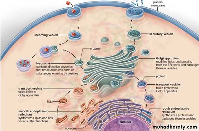
Figure 3.14 The endomembrane system
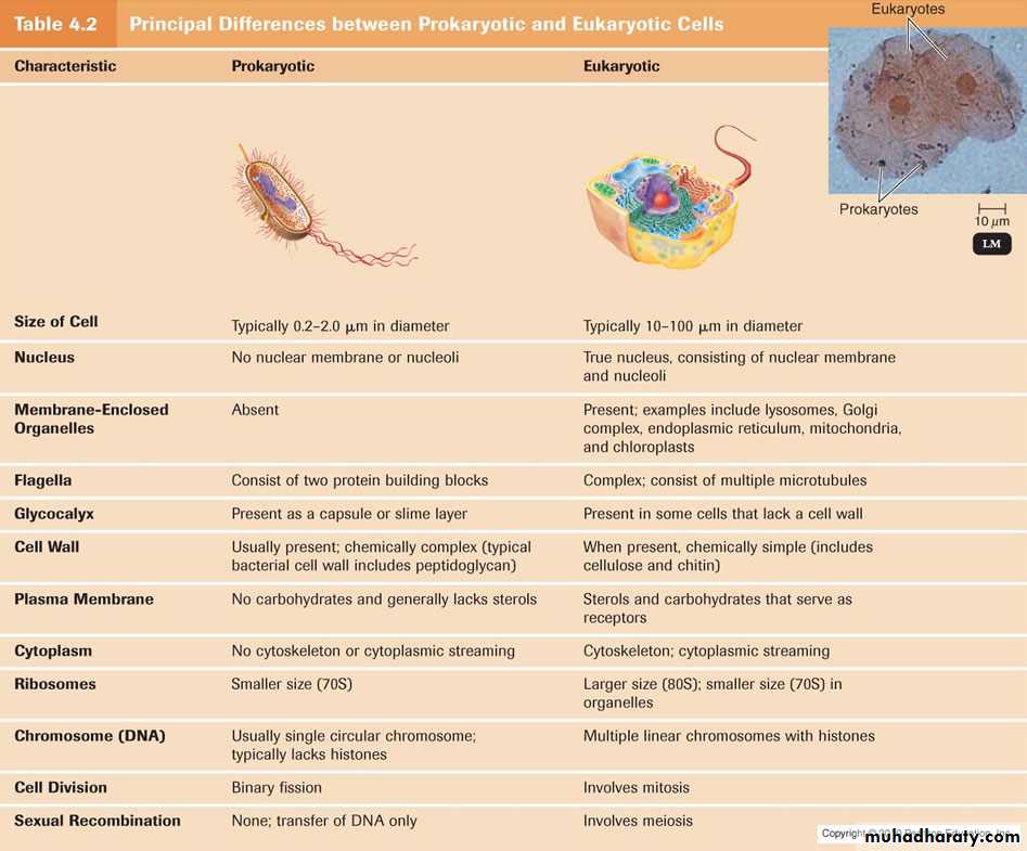
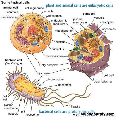
Figure (C) : differences between prokaryotic and eukaryotic cells
References
Mader S and Windelspecht M (2012) Human Biology. 12 edition.
Microscope. 18th century microscopes from the Musée des Arts et Métiers, Parishttp://en.wikipedia.org/wiki/Microscope
Chapter 3. Cell Structure and Function
http://courses.lumenlearning.net/biology/chapter/chapter-3-cell-structure-and-function/
Transmission electron microscopy
http://en.wikipedia.org/wiki/Transmission_electron_microscopy