


1
Eyelids Dr Najah
Baghdad medical college 2015-2016
Anatomy:
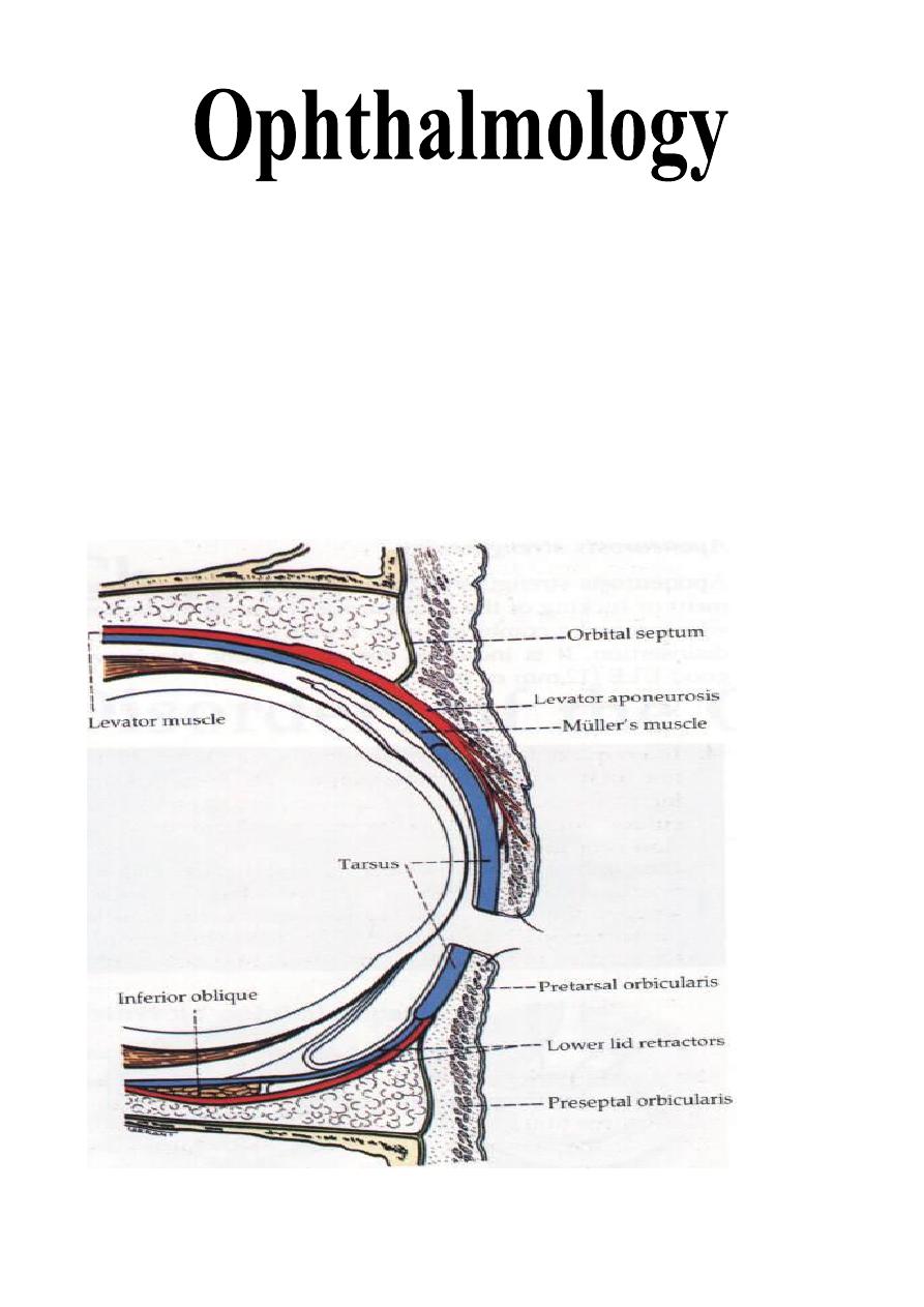
Eyelids are thin movable curtains composed of skin on their anterior surface
and mucus membrane (conjunctiva) on the posterior surface, they contain:
1- Smooth muscles (Müller's muscles).
2- Striated muscles (Orbicularis oculi and Levator palpebrae superioris
"LPS").
3- Dense fibrous plates (Tarsal plates).
4- Glands.
5- Nerves and blood vessels.
Lecture: 12 & 13
2
The contents of the lid are distributed as follows: the anterior surface is made
of skin which has a round edge with the lid margin, the subcutaneous tissue,
muscular layer, the submuscular (areolar tissue) layer, the orbital septum which
end as a tarsal plate (that forms the architecture of lid) and finally the
conjunctiva (palpebral) which is situated most posterior.
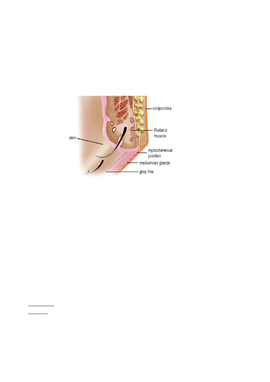
The free margin of the eyelids contains:
1- The lashes (Cilia).
2- Grey line.
3- Orifices of Meibomian glands.
4- Mucocutaneous junction
5- Superior and inferior puncti of Naso-Lacrimal System (NLS).
Muscles of the eyelids:
1- Orbicularis oculi muscle:
It is a thin oval sheet of concentric striated fibrous surrounding the palpebral
fissure.
It can be divided into:
a- Peripheral (orbital) part: This is involved in forceful closure of lids.
b- Central (palpebral) part: This is involved in involuntary blinking and
participates in forceful closure with the orbital part.
c- Muscle of Rioland's: this part is represented by the gray line of lid margin.
d- lacrimalis muscle: that attached to the fundus of lacrimal sac. This part is
involved in pumping lacrimal drainage system.
Nerve supply:
Sensory: Ophthalmic branch of trigeminal nerve
Motor: Facial nerve.
2- Levator palpebrae superioris muscle:
It is originates from the periosteum covering the lesser wing of sphenoid bone
at the apex of the orbit.
The aponeurosis inserts into:

3
a- Skin of the upper eyelid, so it forms skin creases on the eyelid.
b- Upper edge and anterior surface of the tarsal plate.
c- Medial and lateral palpebral ligaments.
Function: To keep the palpebral fissure open against gravity.
Nerve supply:
Sensory: Ophthalmic branch of trigeminal nerve
Motor: Oculomotor nerve.
3- Superior palpebral muscle (Müller's muscle or superior tarsal muscle):
It is a small sheet of smooth muscle originated from the under surface of the
LPS muscle and inserted to the upper edge of the upper tarsal plate.
Nerve supply: Sympathetic nerves.
Function: Like LPS, is to keep the palpebral fissure open against gravity.
Glands in the eyelids:
1- Meibomian glands (Tarsal glands):
Modified sebaceous gland located in the tarsal plate. The upper tarsus
contains 30-40 glands, while the lower tarsus contains 20-30 glands.
Function: It secret the lipid forms the outer layer of the tear film.
2- Zeis glands:
Modified sebaceous gland opened with lash follicles.
3- Glands of Moll:
They are modified sweat gland whose ducts also open into lash follicle or
directly in the anterior lid margin between the lashes.
Congenital anomalies of eyelids:
1- Ablepharon: Absence of the eyelids.
Treatment:
Reconstructive plastic surgery.
2- Ankyloblepharon: Imperfect separation of the eyelids.
Treatment:
Open the adhesion in between.
3- Coloboma of the eyelids: Common congenital anomaly in which there is
failure of a portion of the lid to develop leading to notch in the lid margin.
Treatment:
Plastic surgery.
4- Blepharophimosis: Narrowing of the palpebral fissure.
Treatment
: Plastic surgery at pre-school age.
5- Epicanthus: Common anomaly. There is a vertical skin fold in the medial
canthal region, which conceals the medial angle and caruncle giving a picture
simulates convergent squint (pseudosquint).
Abnormalities in shape and position:
1- Entropion:
It is an inward rolling of the lid margin causing continuous rubbing of the
cornea, which leads to conjunctival congestion, painful watery eye (tears

4
production due to stimulation of cornea) and sometimes epithelial defect and
even corneal ulceration. So, it is a vision threatening disease.
Causes (Types) of entropion:
a- Congenital: which is very rare.
b- Spastic entropion: secondary to any condition that causing severe ocular
irritation (irritation leads to overriding of Orbicularis oculi muscle fibers),
e.g.: conjunctivitis, keratitis and ocular surgery.
Treatment
:
of underlying cause and taping of lid (turned outward).
c- Senile entropion (involutional entropion): affecting elderly patients and
usually involves the lower lid.
Treatment
: is surgical correction.
d- Cicatricial entropion: secondary to any condition causing scaring and
shrinkage of conjunctiva, e.g.:
i- Chlamydial conjunctivitis.
ii- Chemical conjunctivitis (whether it is acidic or alkaline).
iii- Autoimmune conjunctivitis. e.g.
Cicatricial pemphigoid
and
Stevens-
Johnson syndrome
iv- Irritation conjunctivitis.
2- Ectropion:
It is an outward rolling of the lid margin causing exposure of the palpebral
and bulbar conjunctiva. This exposure leads to exposure conjunctivitis,
exposure keratitis and watery eye. Long-standing cases lead to:
i- Dry and thick conjunctiva and even keratinization.
ii- Corneal ulceration. So, it is vision threatening disease too.
iii- Redness, crusting and dermatitis of skin due to?
Causes (types) of ectropion:
a- Congenital ectropion.
b- Paralytic ectropion (Atonic): seen in cases of facial nerve palsy.
Treatment
: we should wait for 6 months for spontaneous recovery e.g. (Bell's
palsy) then lateral tarsorrhaphy is indicated.
c- Senile ectropion (involutional): due to atrophy of orbicularis oculi muscle
and tarsal plate.
Treatment
: surgical correction.
d- Cicatricial ectropion: which is occur secondary to:
i- Chemical burns of skin.
ii- Infection of skin.
iii- Radiation of skin.
5
iv- Improper suturing of lid wound.
v- Trauma to the lid.
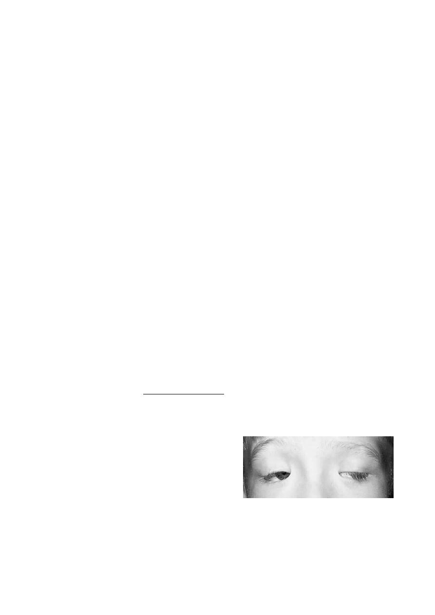
3- Blepharoptosis:
It is an abnormal low position or dropping of the upper eyelid. It could be
unilateral or bilateral, and both of them could be partial or complete. Usually
the upper lid covers only 2 mm from cornea. If more, is called blepharoptosis.
Causes of blepharoptosis:
a- Congenital blepharoptosis: the LPS muscle is very poor and there is
absence of skin creases.
b- Neurogenic blepharoptosis:
i- Oculomotor nerve palsy: causing severe blepharoptosis.
ii- Horner's syndrome: causing mild blepharoptosis.
iii- Marcus Gunn Jaw-winking syndrome: it is a congenital anomaly in
which, nerve to LPS is a branch from nerves to mastication muscle (i.e. from
trigeminal nerve).
iv- 3
rd
nerve misdirection: either acquired or congenital.
* Why it is severe in (i) and mild in (ii)?
c- Myogenic blepharoptosis:
i- Myasthenia gravis: there is a defect in the neuro-muscular junction
(damage to acetyl choline receptors).
Treatment :
medical treatment.
ii- Myotonic dystrophy.
iii- Ocular myopathy (localized defect similar to myasthenia gravis).
iv- Simple congenital myogenic blepharoptosis.
v- Blepharophimosis syndromes: it is a
syndrome in which, there are bilateral
ptosis,
bilateral
palpebral
fissure
narrowing, bilateral ectropion, and
increase in the distance between the two
medial canthi (telecanthus).
d- Aponeurotic blepharoptosis:
i- Involutional (senile).
ii- Post operative.

6
e- Mechanical blepharoptosis:
There is deformity and increases weight of the eyelid:
Causes:
i- Trachoma, VKC and eyelid tumor.
ii- Cicatricial (due to LPS and superior rectus muscles fibrosis).
iii- Trauma (collection of fluid).
iv- Iatrogenic by surgeons.
v- Lack of support (phthisical or nanophthalmos): it is not a real ptosis, but
the lid drops as there is no support below it due to small size of eyeball.
Treatment of ptosis: The treatment of all types is surgical except in
myasthenia gravis, where the treatment is medical. Surgical procedures are:
a- Levator resection.
b- Frontalis brow suspension (Sling operation).
c- Tarso-conjunctival resection (Fasanella servete procedure).
Choice of one of these three types depends on:
a- Aetiology of ptosis.
b- Severity of ptosis: Mild, moderate or severe.
c- Function of LPS: Good, fair or poor.
4- Trichiasis:
Backward misdirection of the eyelashes (cilia), sometimes it is associated
with entropion and it is called pseudotrichiasis.
Causes:
a- Any cause leads to entropion of the eyelid, so, it is Pseudo-trichiasis.
b- Trachoma without or with entropion, so it is True or pseudo-trichiasis
respectively.
c- Chronic blepharitis: which causes True trichiasis.
Treatment
: For isolated misdirection cilia (true trichiasis)
a- Epilation: Repeated every few weeks.
b- Electrolysis: Destruction to hair follicles by cauterization.
c- Cryosurgery: Destruction to hair follicles by freezing.
d- Laser ablation: Destruction to hair follicles by laser.
For pseudo-trichiasis: done by correction of entropion surgically.
5- Blepharospasm:
Involuntary sustained closure of the eyelids which occurs spontaneously
(essential) or by sensory stimuli (reflex).

7
Treatment:
Is to treat the underline cause and in sever, and resistance cases Botulinum
toxin injection is indicated. It is temporary measure and can be repeated
frequently.
6- Madarosis:
It is a decrease in number or complete loss of lashes.
Causes:
Local Causes: chronic blepharitis, burns, radiation and infiltrating tumor.
Systemic causes: generalized alopecia, psoriasis, SLE, syphilis and leprosy.
Benign nodules and cysts:
1- Chalazion (Meibomian cyst):
It is a chronic lipogranulomatous inflammatory lesion caused by blockage of
meibomian gland orifices and stagnation of sebaceous secretions and there is
NO infection.
It is more frequent and multiple in patients with acne rosacea or seborrhoeic
dermatitis and other sebaceous gland dysfunctional diseases.
Presentation: Painless nodule and occasionally blurred vision (Why?)
Signs:
a- Round, firm lesion in the tarsal plate.
b- Polypoid granuloma on eversion of lid (i.e. seen from the palpebral part of
conjunctiva).
c- Sometimes there is associated blepharitis.
Treatment:
a- Surgical:
The most common method, if the lesion is recurrent or multiple then biopsy
should be taken specially in old age patient (to exclude sebaceous gland
carcinoma which is extremely rare).
b- Steroid injection:
Good alternative to surgery, 0.1-0.2 ml triamcinolone infiltrated around the
lesion, the success rate is 80%. In unresponsive cases, another injection is
given two weeks later. Chalazion should be small in size to be treated with
steroid injection.
c- Systemic tetracyclines:
As prophylaxis, particularly in acne rosacea and seborrhoeic dermatitis
where chalazion is very frequently recurrent.

8
2- Internal Hordeolum:
It is a small abscess caused by an acute staphylococcal infection of
Meibomian glands.
Signs: Tender, inflamed swelling within the tarsal plate.
Treatment:
a- which is done through using topical antibiotics (ointments more important
than drops). (Why?). Systemic antibiotics are needed if there is associating lid
infection.
b- Incision and curettage for the residual nodule after the acute infection has
subsided.
3- External hordeolum (Stye):
It is an acute small staphylococcal abscess of a lash follicle and its associated
gland of Zeis.
Signs:
- Tender inflamed swelling in the lid margin, which points anteriorly through
the skin.
- More than one lesion may present.
- Occasionally minute abscess may involve the entire margin.
Treatment:
a- Hot compresses to produce vasodilatation of blood vessels: is to increase
the circulation in the area and also for liquefaction of sebum to prevent
stagnation in the duct of affected gland.
b- Topical antibiotics to prevent infection of neighboring glands (ointment is
a must).
c- Epilation (removal of eyelashes by a forceps) to enhance drainage of pus.
d- Systemic antibiotics if there is severe preseptal cellulitis.
Note: An infection anterior to the orbital septum (affecting anterior lamella of
eyelid) is "preseptal cellulitis" while infection posterior to orbital septum is
orbital cellulitis.

9
Marginal Chronic Blepharitis
Chronic blepharitis is a very common condition which is usually bilateral
symmetrical.
Types of chronic blepharitis:
1- Anterior: a- Staphylococcal infection.
b- Seborrheic dysfunction.
c- Mixed.
2- Posterior: a- Meibomianitis.
b- Meibomian seborrhea.
3- Mixed (any one of the anterior type plus any one of the posterior).
Pathogenesis of chronic blepharitis:
1- Anterior chronic staphylococcal blepharitis: due to staphylococcal infection.
2- Anterior chronic seborrhoeic blepharitis: is usually associated with
seborrhoeic dermatitis e.g. scalp, naso labial folds, retro auricular area and
sternum.
Neutral lipids are converted by Bacterial lipase into irritating fatty acids
responsible for increase of symptoms.
3- Posterior chronic blepharitis: Associated with dysfunction of Meibomian
gland (ocular rosacea) which is in some patients associated with acne rosacea of
the face.
Symptoms of chronic marginal blepharitis: (anterior and posterior)
They are: Burning, grittiness, mild photophobia, and crusting and redness of
the lid margin. The symptoms are characterized by remissions and
exacerbations. The symptoms are usually worse in the mornings.
Signs of anterior blepharitis:
a- The anterior lid margins show (by slit-lamp) hyperaemia and telangiectasia.
In longstanding cases the lid margin became scarred and hypertrophied (due to
recurrent infections), intrafollicular abscess may be present (staphylococcal
blepharitis).
b- Two types of scales: (important in differentiation)
i- Staphylococcal blepharitis: Are hard and brittle and are centered around
the lashses (collarettes).
ii- Seborrhoeic blepharitis: Are soft and greasy and located anywhere on lid
margin or on the lashes.
c- In longstanding cases, trichiasis, madarosis and occasionally poliosis
(whitening of the eyelashes) will occur.

10
Complications of anterior blepharitis:
a- External hordeolum (stye).
b- Tear film instability (due to the abnormal lipid secretion that affects the tear
film).
c- Hypersensitivity to staphylococcal exotoxins papillary conjunctival
reaction, punctuate epitheliopathy and marginal keratitis.
Treatment:
a- Lid hygiene: removing crusts and toxic products by scrubbing the lid
margins daily with either commercially lid scrub or a cotton bud tipped in 25%
solution of baby shampoo.
b- Topical antibiotic ointment: fusidic acid or chloramphenicol.
c- Weak topical steroids: e.g. fluorometholone to control hypersensitivity to
exotoxins.
d- Tear substitutes.
Signs of posterior blepharitis (Meibomian seborrhea or meibomianitis):
a- Small oil globules capping the meibomian gland orifices.
b- Pressure on tarsal plate results in expression of copious amounts of
meibomian oil.
c- The posterior lid margin shows diffuse or localized inflammation centered
around meibomian gland orifices.
d- Blockage of the main meibomian ducts gives rise to secondary cystic
dilatation and occasionally meibomian cysts.
Complications:
a- Tear film instability.
b- Papillary conjunctivitis plus punctuate epitheliopathy.
c- Internal hordeolum.
Treatment:
a- Systemic tetracyclines (Corynebacterium acnes) for 6-12 weeks: one of the
followings,
i- Tetracycline: Four times daily for 1 week, then twice daily for 5-11 weeks.
ii- Doxycycline: Twice daily for 1 week, then once daily for 5-11 weeks.
iii- Minocycline.
b- Erythromycin when (a) is contraindicated.
c- Lid hygiene.
d- Topical steroids.
e- Tear substitutes.
f- Warm compresses to melt solidified sebum and mechanical expression (to
evacuate meibomian glands from their contents).