sliding HH
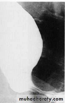
Para esoph. HHNormal extrinsic compression
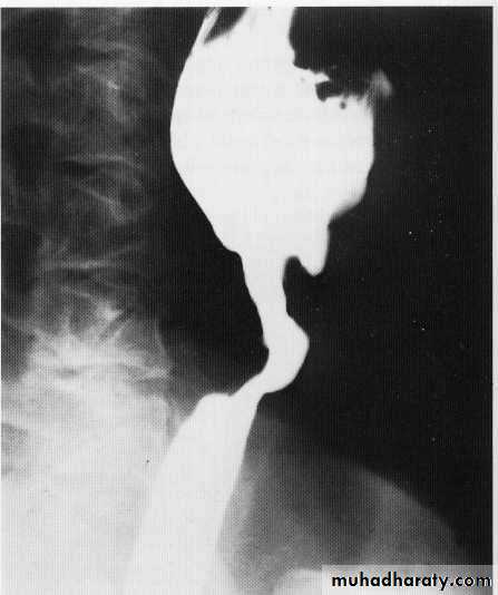
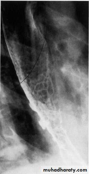
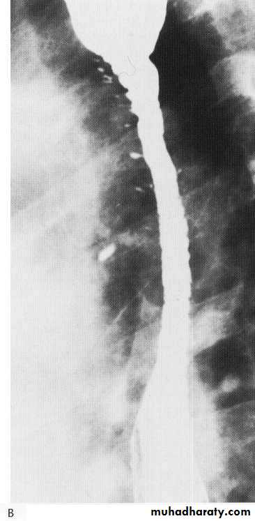
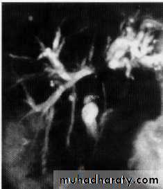
Enlarged cervical LAPCA esophagus with malignant stricture
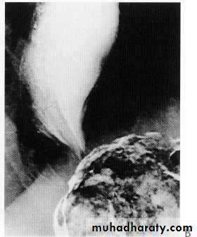
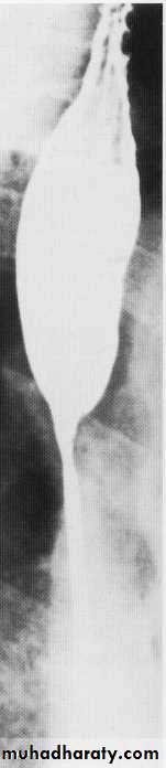
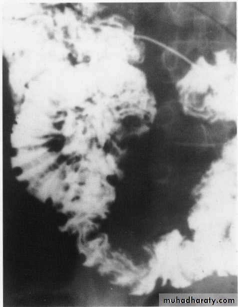
ACHLASIA
Scleroderma
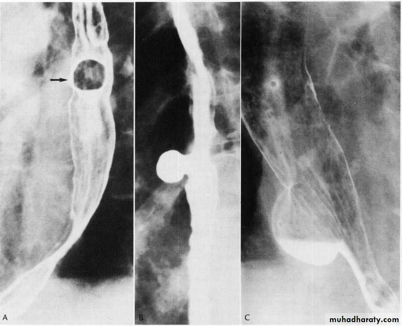
diverticulum
Benign strictures
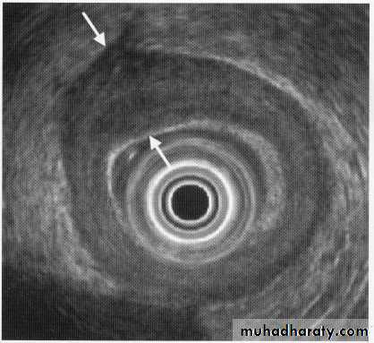
Esophageal tumours lyomyoma
Esophageal caWith or with out regional nodal infiltration
Local invasion
Distant mets
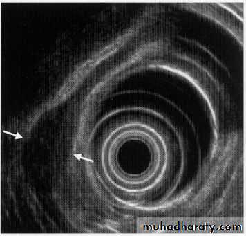
Leiomyoma EUS
Ca ESOPH.
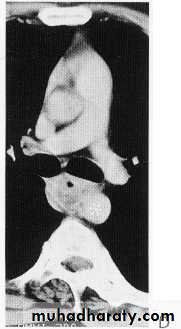
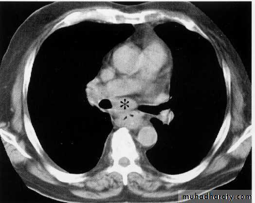
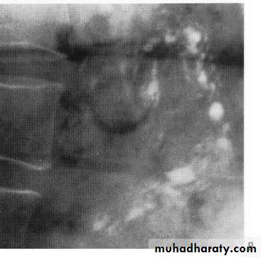
Ct scan carinal LAP
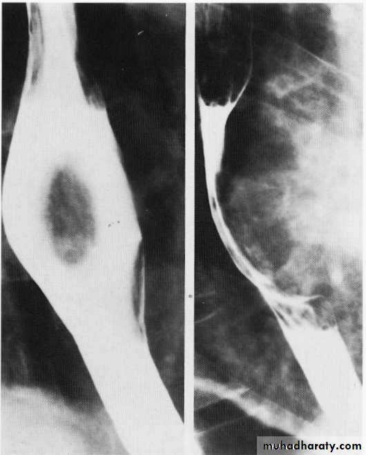
Leiomyoma Ba swallow
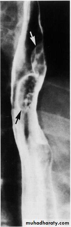
Esophageal diverticulae
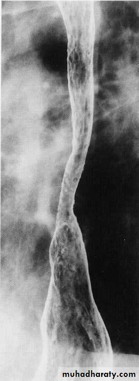
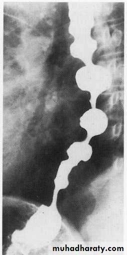
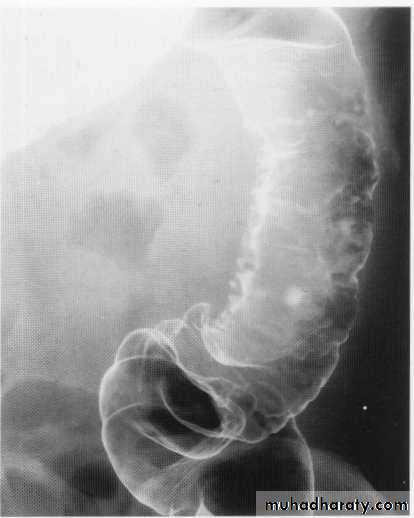
Tertiary contractions
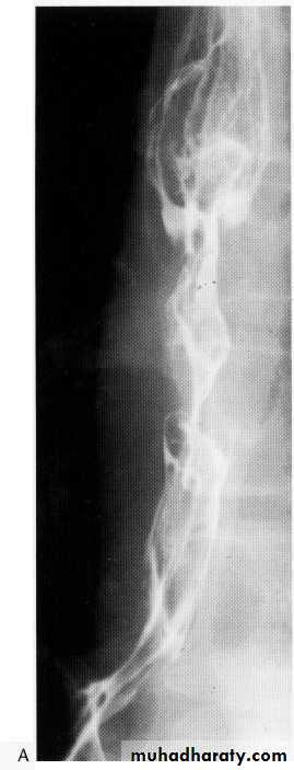
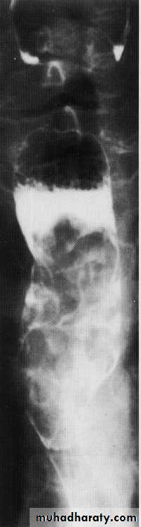
Advanced esophageal Ca
Stomach
Single contrastDouble contrast
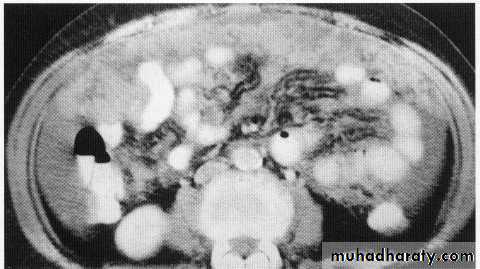
Ct scan with oral contrast
i.v contrast
Normal stomach
Normal du. cap
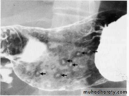
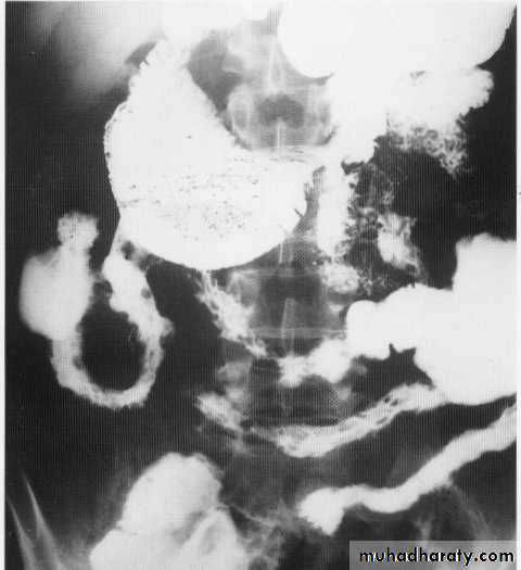
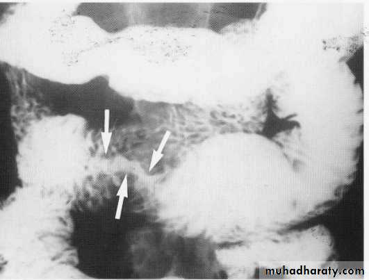
Multiple gastric ulcers
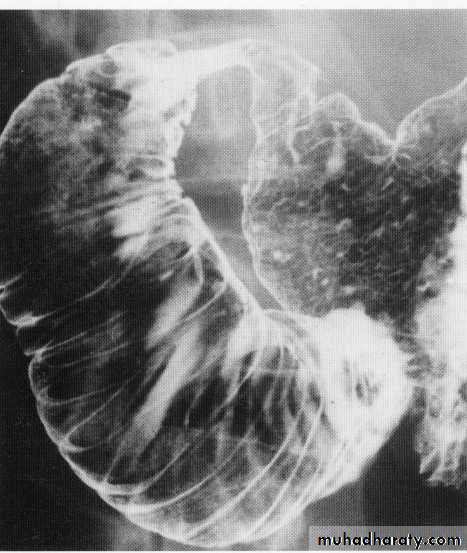
Chrons - du. cap
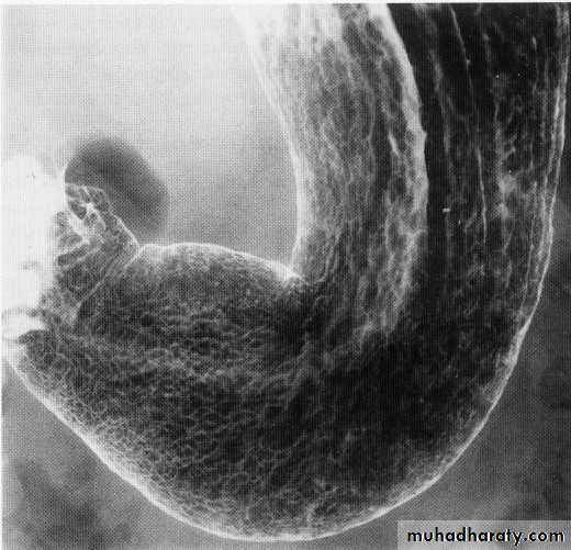
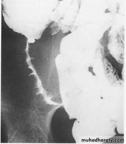
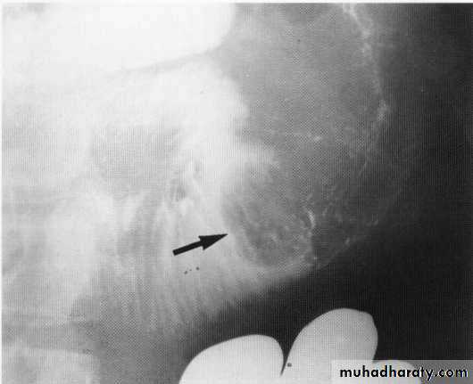
Gastric ulcer
Du. ulcer
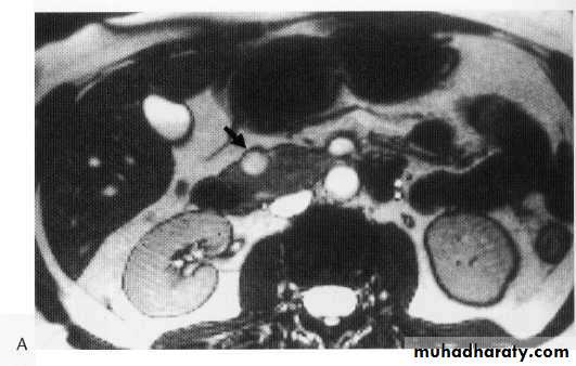
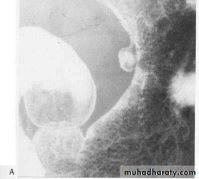
Benign du. Tumor arising from the medial wall
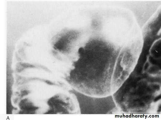
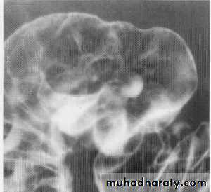
Benign gastric tumor
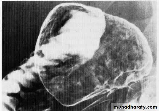
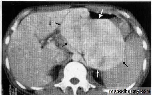
Gastric malig .tumor
Linilis plastica
Gastric tumor with regional LAP
Gastric lymphoma
Exophytic tumor

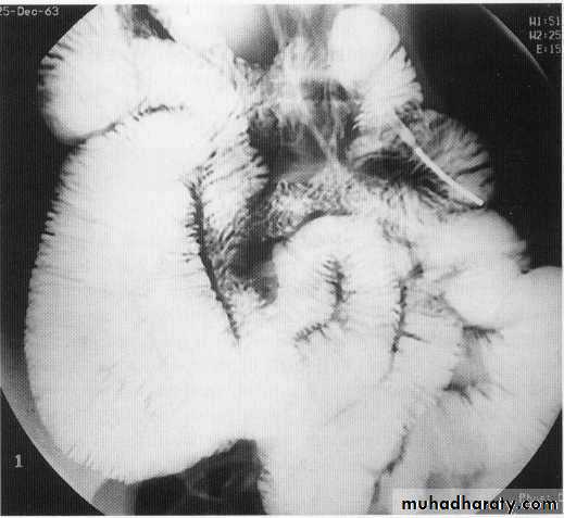
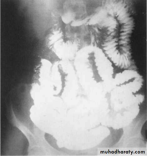
Small bowel contrast study
Barium follow throw x- rayCt scan with oral contrast
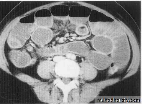
Ct scan with i.v contrast
Small bowel
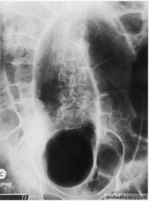
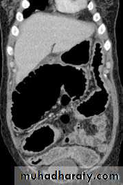
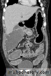
SB obstruct.
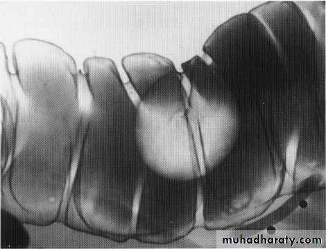
Chrons disease
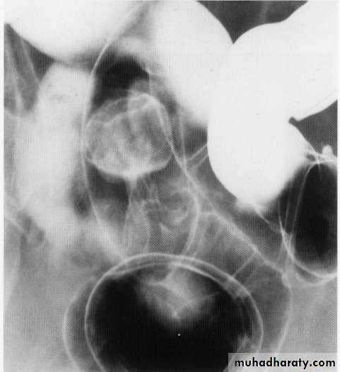
Chrons stricture
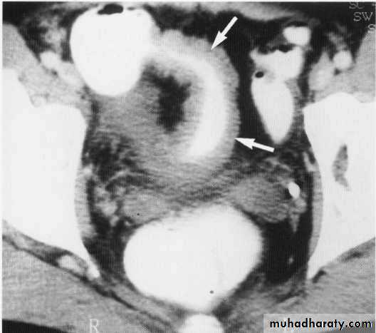
Stricture + pericolic abscess
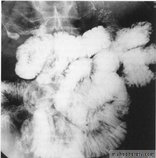
Small bowel tumor
Small bowel lymphoma
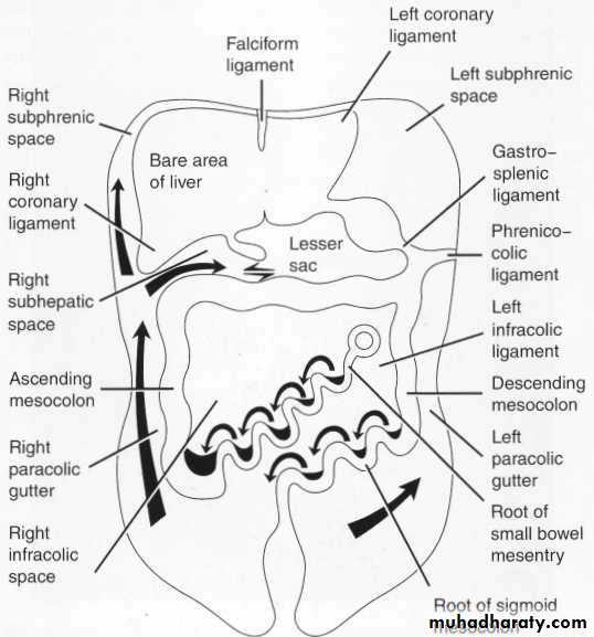
Peritoneal cavity
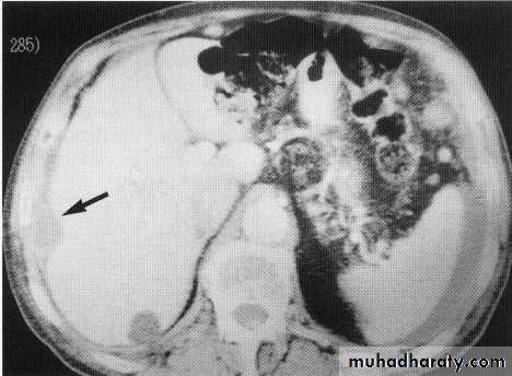
Hepatic secondaries , normal
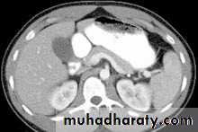
Peritoneal deposit
Large bowel
Filling defect lipoma
Pedanculated polyp
stricture
Multiple strictures
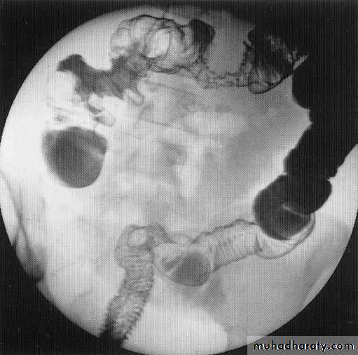
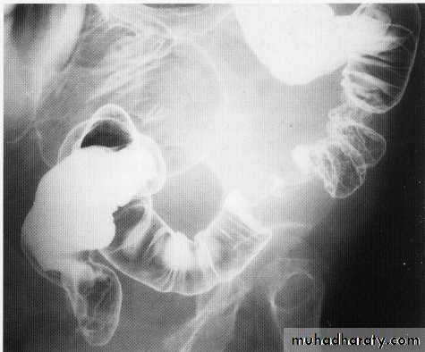
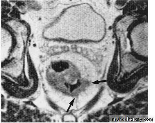
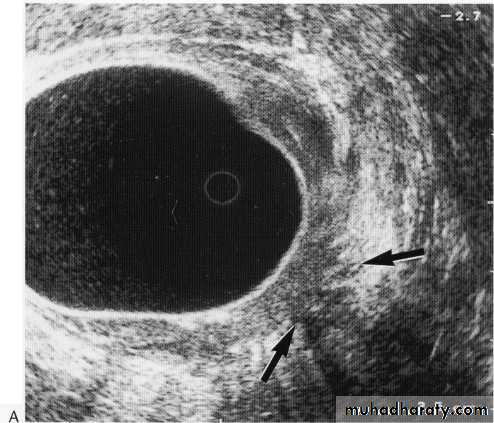
Rectal tumor with local invasion
Lymphoma infiltration
Colono-vesical fistula
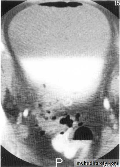
Abnormal haustra toxic megacolon
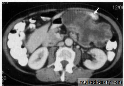
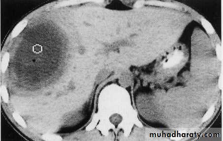
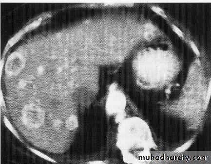
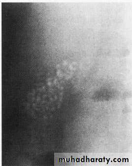
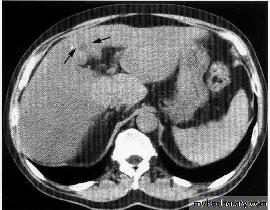
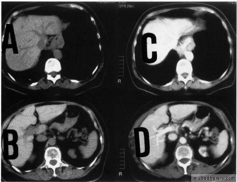
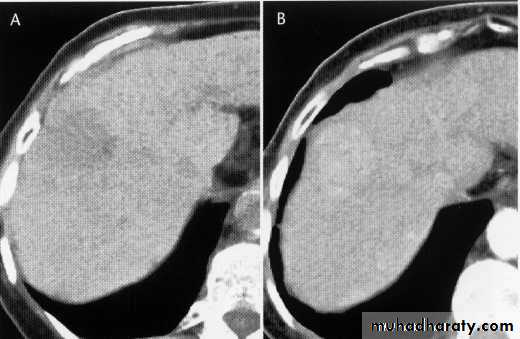
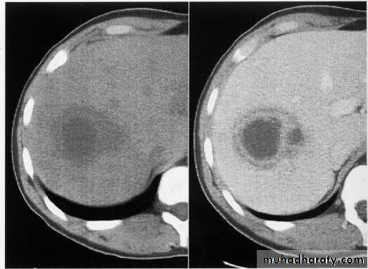
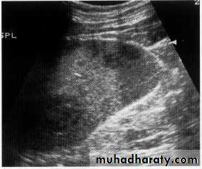
focal liver lesion
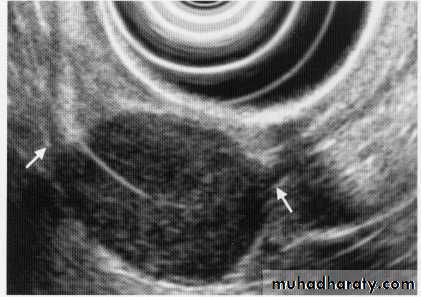
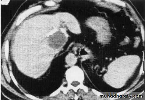
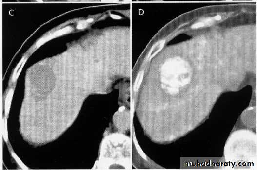
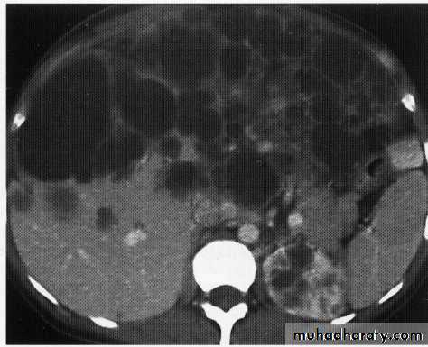
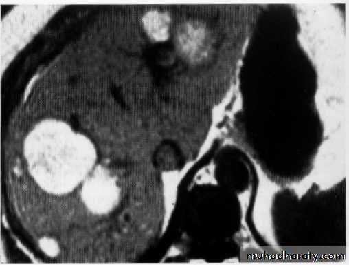
hydatid cyst
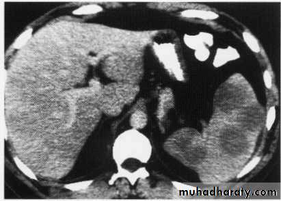
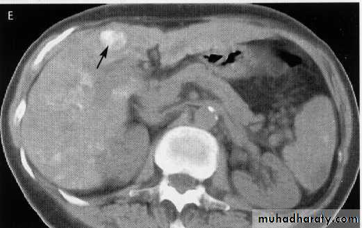
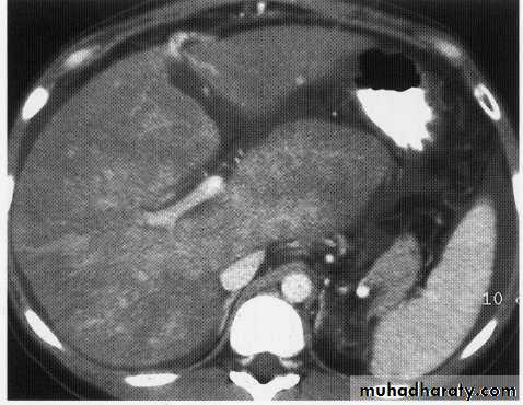
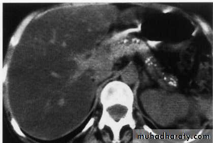
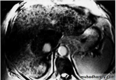
hepatic mets
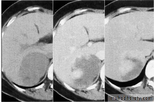
hepatic mets,arterial phase
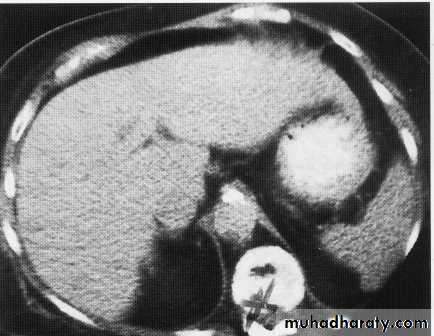
hepatic mets venous phase
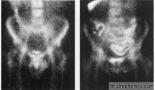
Isotope study of chrons
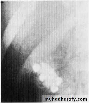
Gall stone
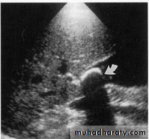
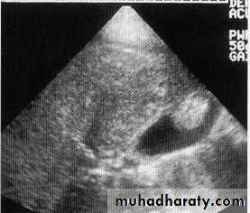
U/S of Gall stone ,acute cholecystitis and polyp
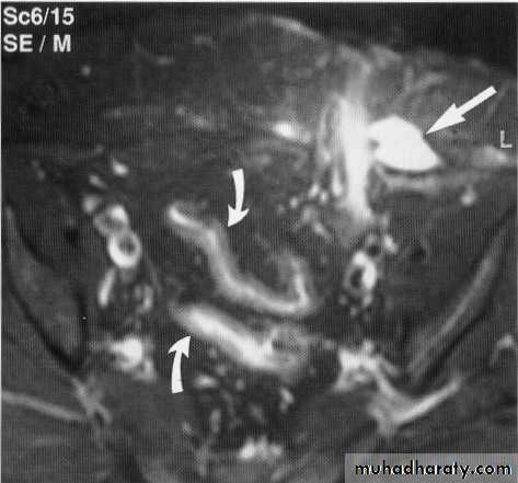
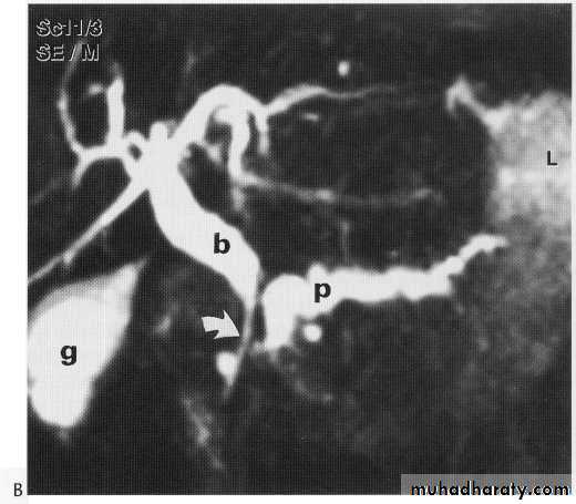
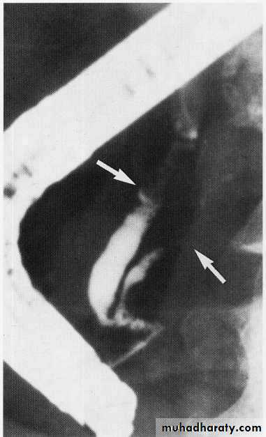
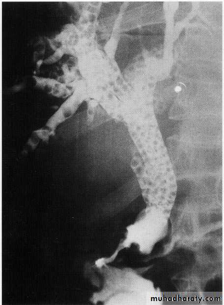
MRCP + ERCP
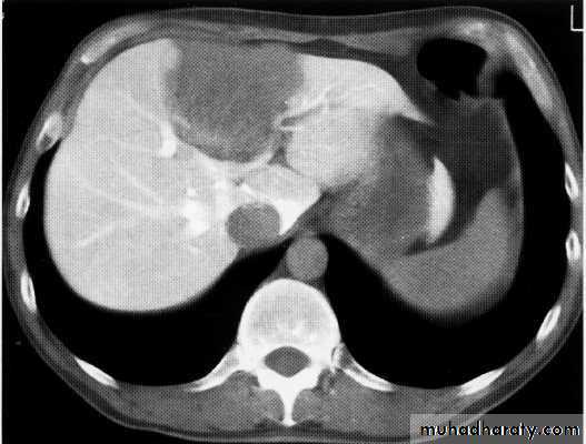
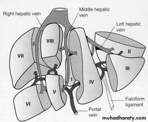
Normal liver , spleen
Enhanced + Unenhanced CT
Hepatoma + met.
Early cirrhosis + budd chiari
Innumerable calculi
Met. adenocarcinoma
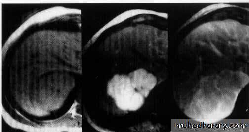
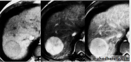
hemangeoma
Liver abscess
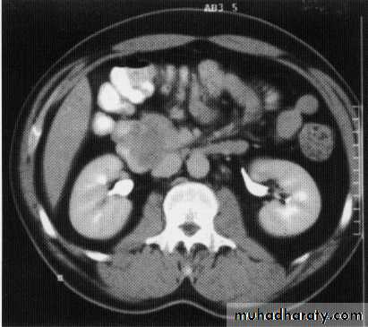
Hepatic , renal cyst
,Calcified hydatid , fatty liver
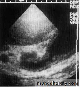
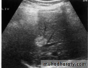
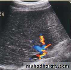
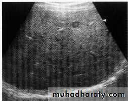
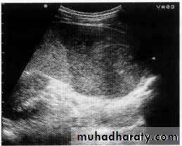
Normal liver U/S
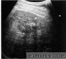
Hepatic mets.
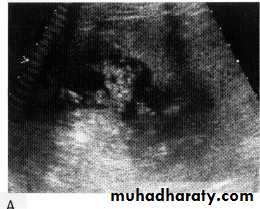
Splenic U/ S

FNH
HCC
Liver cirrhosis
Cholengio ca
Melanoma hepatic mets.
Pancreas calcific.
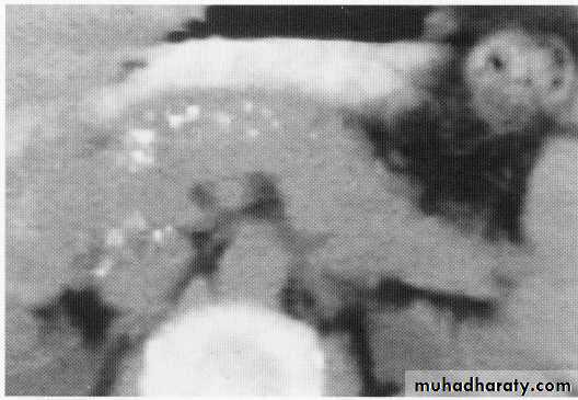
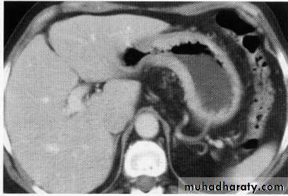
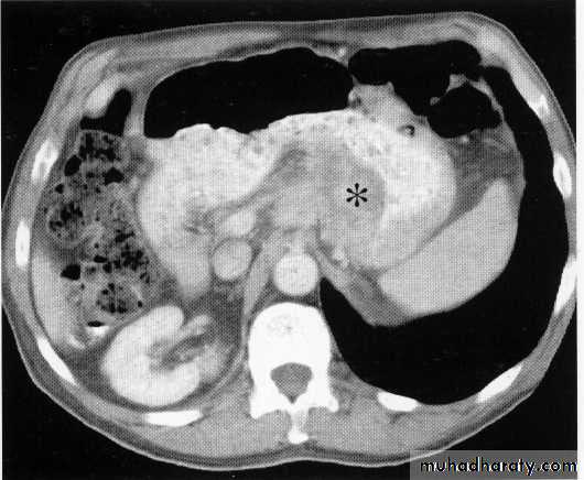
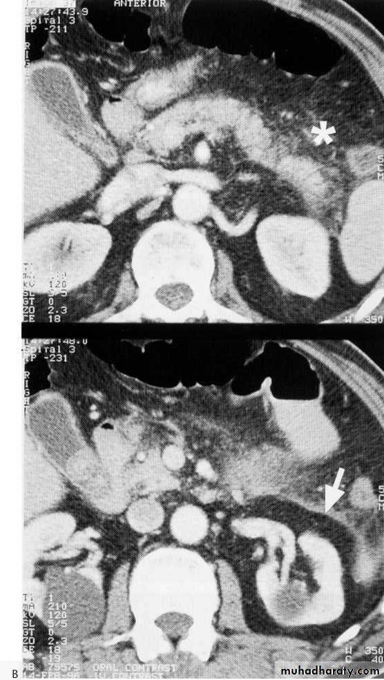
normal pancreas ca pancreas
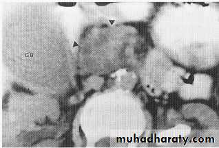

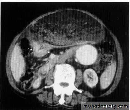
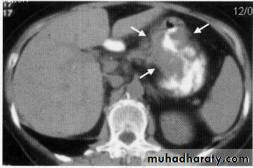
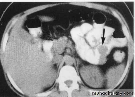
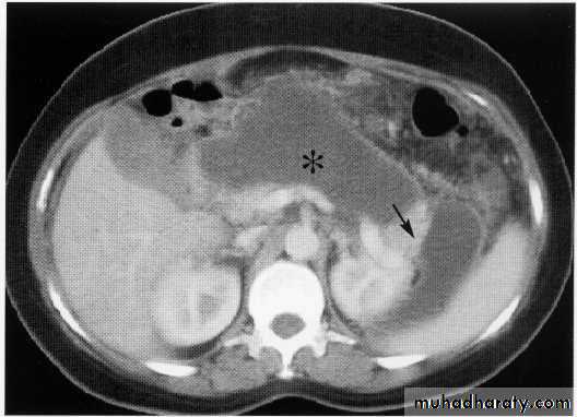
Acute pancreatitis
insulinoma