
Anatomy Lecture 3
Introduction to Bone
and Joints
Dr. Rana Al-tae
2015-2016

Anatomy of a Bone
• The structure of a bone is analyzed by
considering the parts of a long bone as
the humerus ( arm bone ) and the femur
( thigh bone). A long bone is the one that
has greater length than width. A typical
long bone consists of the following parts:
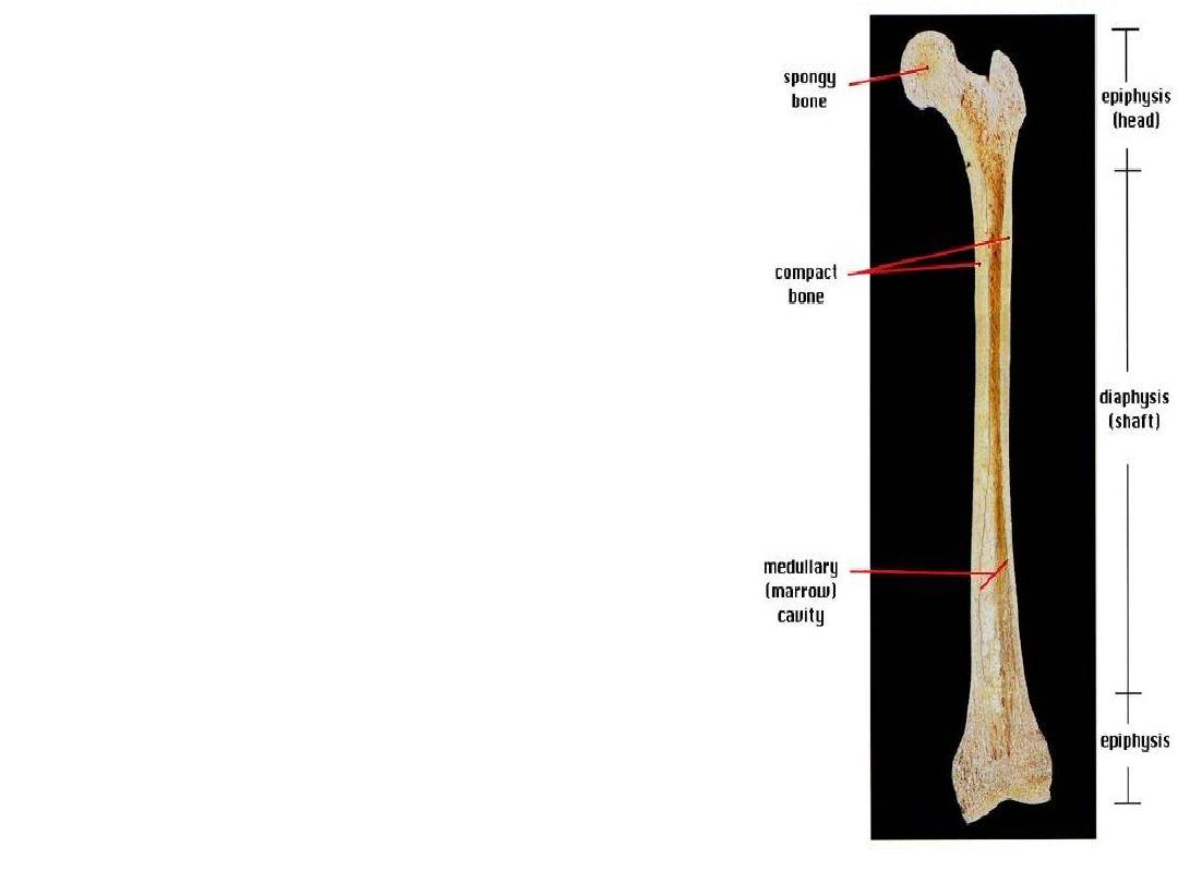
1.
The
diaphysis
is the bone's shaft or body-
the long, cylindrical, main portion of the
bone.
2.
The
epiphyses
are the proximal and distal
ends of the bone (the singular is epiphysis).
3.
The
metaphyses
are the regions in a mature
bone where the diaphysis joins the
epiphyses ( the singular is metaphysis). In a
growing bone the metaphyses are regions
that include the
epiphyseal plate
which is a
plate of hyaline cartilage that allow the
growth of the diaphysis in length but not in
width. When the bone growth in length
stops, the cartilage in the epiphyseal plate is
replaced by bone and the resulting bony
structure is known as the
epiphyseal line
.
4.
The
articular cartilage
is a thin layer of
hyaline cartilage that covers each epiphysis
where the whole bone forms an articulating
joint with another bone. Articular cartilage
reduces friction and absorbs shock at freely
movable joints.
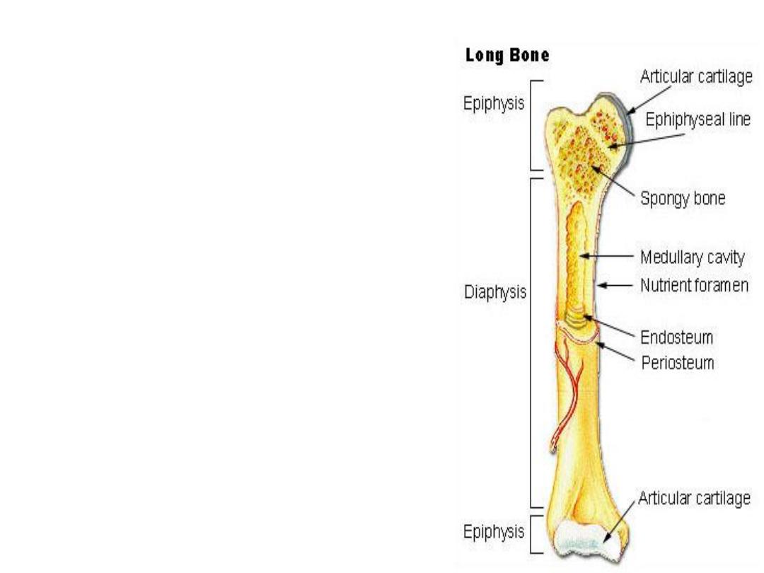
5. The
Periosteum
is a layer of
connective tissue that covers the
bone surface wherever there is
no articular cartilage. It helps the
bone to grow in width, protects
the bone and nourishes it, helps
in repair in fractures and serves
as an attachment point for
ligaments and tendons.
6. The
medullary cavity
or marrow
cavity is the space within the
diaphysis that contain the fatty
yellow bone marrow in adults.
7. The
Endosteum
is a thin
membrane that lines the
medullary cavity.

Joints
• Introduction
• Bones are too rigid to bend without being damaged.
Fortunately, flexible connective tissues forms joints
that hold bones together permitting some degree of
movement. A joint (articulation) is a point of contact
between two bones, between bone and cartilage or
between bone and teeth. When we say one bone
articulate with another bone we mean that the bones
form a joint. Because most movements of the body
occur at joints, You should appreciate their importance
if you imagine how a cast over your knee joint makes
walking difficult or how a splint on your finger limits
you ability to manipulate small objects.
• Some joints permit no movement, others permit slight
movement and others afford fairly free movement.

Joint classification
• The joints in the body are classified according to their
structure and to their function.
1. Structural classification
of joints depends on the type
of connective tissue that combines the bones
together and whether there is a space between the
articulating bones or not ( synovial cavity) and this
classification is as the following:
2. Fibrous joints
: the bones are held together by fibrous
connective tissue and the is no synovial cavity.
3. Cartilaginous joints
: the bones are held together by
cartilage and the is no synovial cavity.
4. Synovial joints
: the bones are held together by
capsule and ligaments and there is a synovial cavity.

• Functional classification
of the joints relates to
the degree of movement they permit as the
following:
1. Synarthrosis
: An immovable joint.
2. Amphiarthrosis
: A slightly movable joint.
3. Diarthrosis
: A freely movable joint.
• NOTE: all Diarthrosis are Synovial joints.
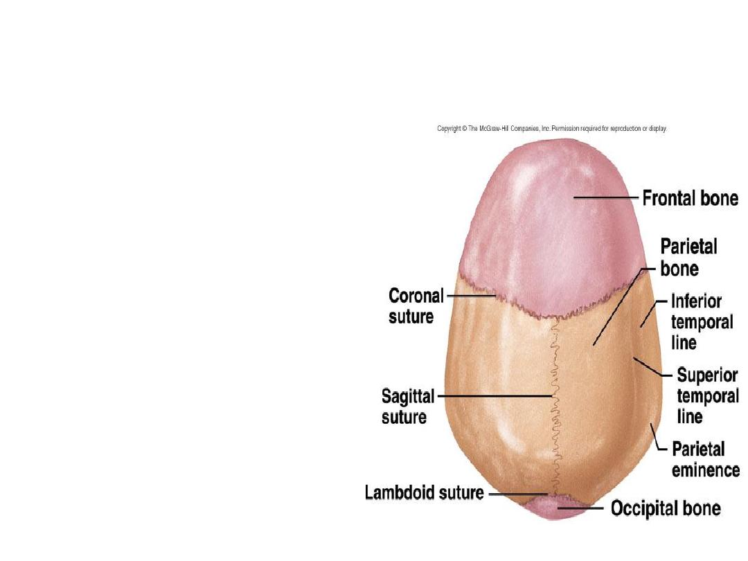
Types of fibrous joints :
1. Sutures
: as the
sagittal suture or the
coronal suture of the
skull which is
immovable so they
are classified
functionally as
synarthrosis
Types of fibrous joints :
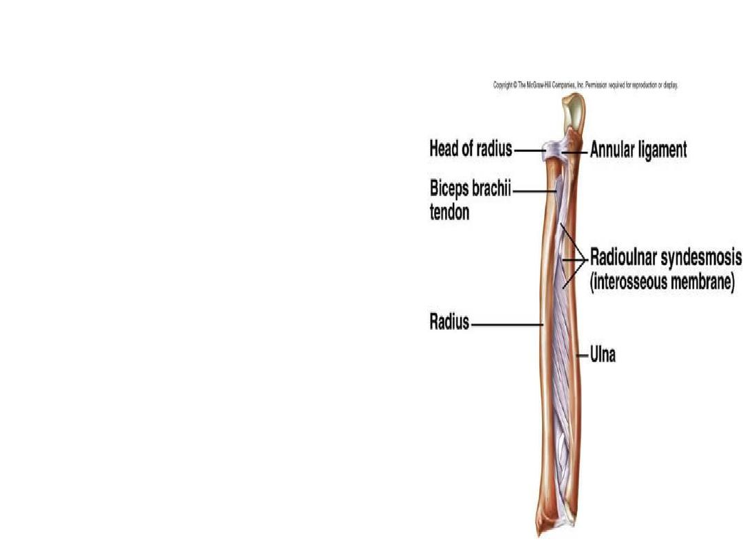
2. Syndesmoses
: (band or
ligament) is a fibrous joint in
which there is a greater
distance between the
articulating bones and more
fibrous connective tissue than
in sutures. Ex. The interossous
membrane between the
parallel borders of radius and
ulna bone ( middle radioulnar
joint)
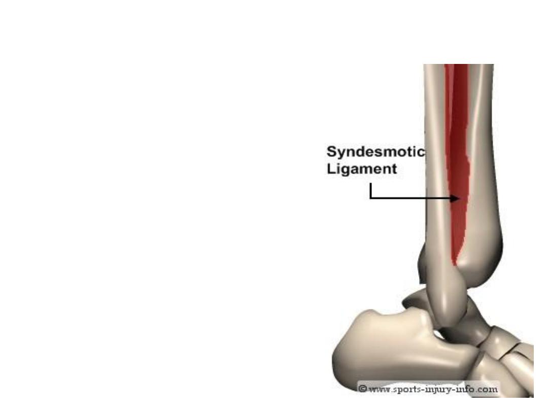
Types of fibrous joints :
• and between the parallel
borders of the tibia and fibula
( middle tibiofibular joint).
Because it allows slight
movement it is classified
functionally as an
amphiarthrosis.
Types of fibrous joints :
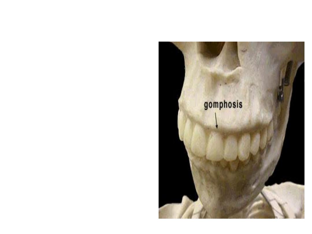
3. Gomphosis
: is a type of
fibrous joint in which a
cone-shape peg fits into
a socket. The only
example in the body is
the roots of teeth and
their sockets in the
maxilla and the
mandible bones. A
gomphosis is classified
functionally as a
synarthrosis, an
immovable joint.
Types of cartilaginous joints :
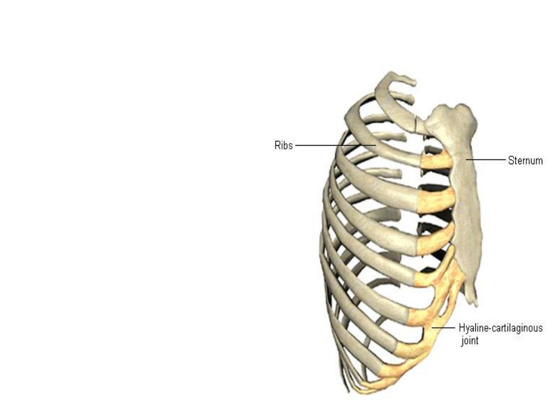
1. Synchondroses
: example
is the epiphyseal plate
that connects the
epiphysis with the
diaphysis of the long
bone. Other ex. Is the
joint between the first
rib and the sternum
bone. Functionally it is
classified as a
synarthrosis.
Types of cartiligenous joints :
2. Symphyses
: in which the ends
of the articulating bones are
covered by hyaline cartilage, but
the bones are connected by a
broad, flat disc of fibrocartilage.
All symphyses are present in the
midline of the body, ex: The joint
between the manibrium and the
body (parts ) of the sternum, the
joint between the two hip bones
called pubic symphyses, the
joints between the bodies of the
vertebrae forming the
intervertebral joints. A
symphysis is an amphiarthrosis,
slightly movable joint.
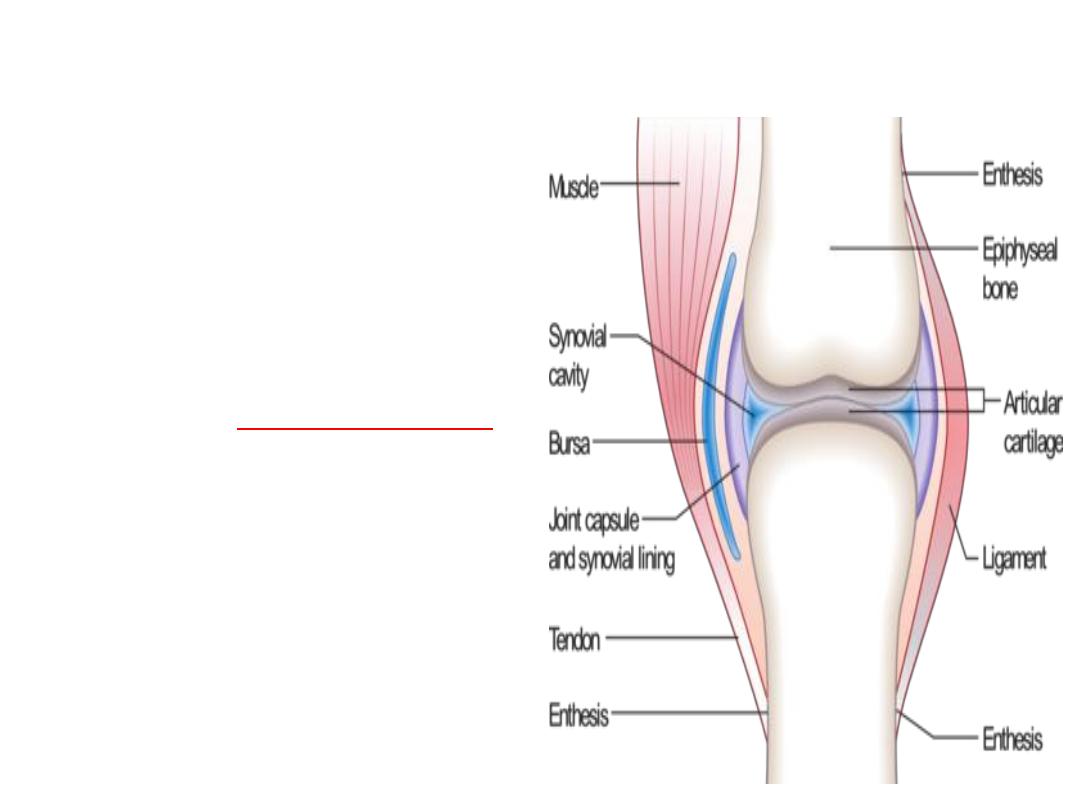
Synovial Joints
• They have a space called the
synovial (joint) cavity between
the articulating bones. The
structure of these joints allows
the bones to move freely, so
all of them are diarthrosis. The
bones at the synovial joint are
covered by
articular cartilage,
which is hyaline cartilage. This
cartilage provides a smooth,
slippery surface for the
articulating bones, but it does
not bind them together. It
reduces the friction between
the bones of the joint during
movement and helps to
absorb shock.
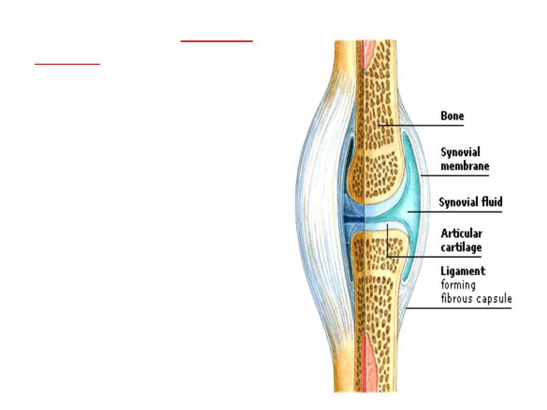
• There is also the
articular
capsule
surrounds a synovial
joint, encloses the synovial
cavity and unites the
articulating bones. It is
composed of two layers, an
outer fibrous capsule and an
inner synovial membrane.
The outer layer is fibrous,
tougher than the inner layer
and attaches to the
periosteum of the
articulating bones, while the
inner layer is areolar with
elastic fibers.

• The
synovial fluid
is secreted from the synovial
membrane, it is a thin film of fluid present in
the synovial (joint) cavity, viscous, clear or
pale yellow fluid similar in appearance and
consistency to uncooked egg white or
albumin. It consists of hyaluronic acid. Its
several functions are reducing friction,
lubricating the joint, supplying nutrients and
removing waste products and has phagocytic
cells that remove microbes and debris that
result from tear in the joint. When a synovial
joint is immobile the synovial fluid becomes
gel-like but as joint movement increases the
fluid becomes less viscous.

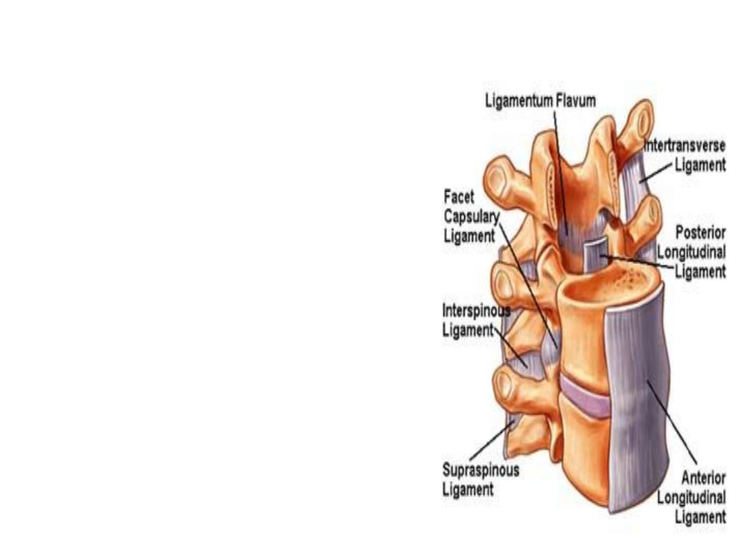
• Many synovial joints also contain
accessory
ligaments
called extra capsular ligaments and
intracapsular ligaments. The extra capsular
ligaments lies outside the articular capsule ,
while the intra capsular ligaments lies within
the articular capsule but are excluded from
the synovial cavity by folds of the synovial
membrane.

Nerve and Blood supply of the joint:
• The nerves that supply a joint are the same as those
that supply the skeletal muscles that move the joint.
Some nerve endings convey information about pain
in the joint, others are responsive to the degree of
movement and stretch at a joint.
• Also nearbying arteries are responsible for nourishing
the joint with oxygen and nutrients and the veins
carry out carbon dioxide and waste products from
the joint. The arteries send out numerous branches
that penetrate the ligaments and articular capsule.
We should know that the articulating portions of the
synovial joint receives its nourishment from the
synovial fluid whereas all other joint tissues are
supplied by blood capillaries.

Types of synovial joints:
• Although all synovial joints are similar in
structure, the shapes of the articulating
surfaces vary. Accordingly, synovial joints
are divided into six subtypes:
Planer,
Hinge, Pivot, Condyloid, Saddle, and ball-
and-socket joints.
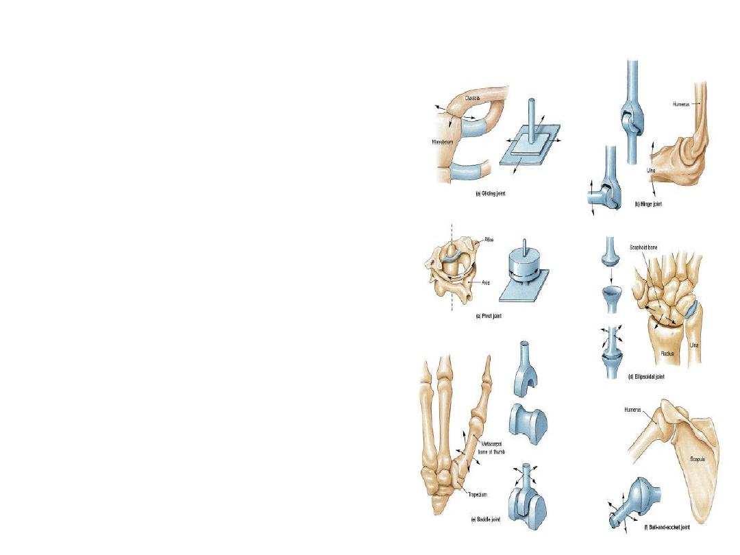
Types of synovial joints:
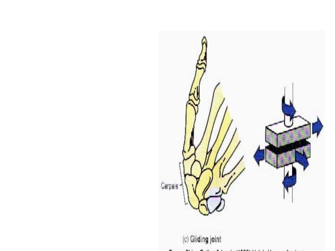
1.
Planer joints
: the articular surfaces
of bones are flat or slightly curved.
These joints permits side-to-side
and back-and-forth gliding
movements. These joints are said
to be nonaxial joints because the
motion they allow does not occur
around an axis or plane (line). Ex:
the intercarpal joints (between the
carpal bones at the wrist ) and the
intertarsal joints ( between the
tarsal bones of the ankle), the
sternoclavicular joint between the
manubrium part of the sternum
with the sternal end of the clavicle
bone, the sternocostal joints
between the body part of the
sternum and the ends of the costal
cartilages at the tips of the 2
nd
- 7
th
pairs of ribs, and the
vertebrocostal joints between the
heads of all ribs with the vertebrae
of the thoracic region.
Types of synovial joints:
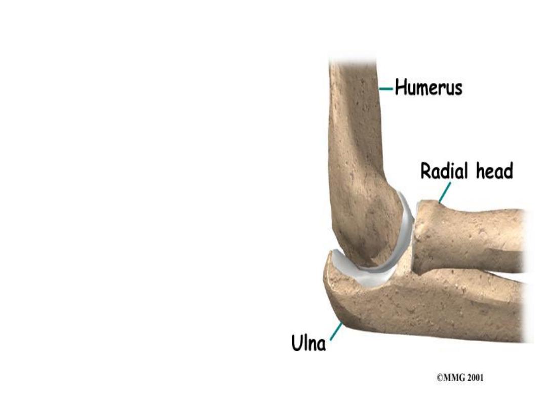
2. Hinge joints:
the convex
surface of one bone fits
into the concave surface of
the another bone. As the
name implied, hinge joints
produce an angular
opening and closing motion
like that of a hinged door.
Hinge joints are said to be a
monoaxial or uniaxial joints
because permits movement
in only one single axis. Ex.
Are the knee, elbow, ankle,
and interphalangeal joints.
Types of synovial joints:
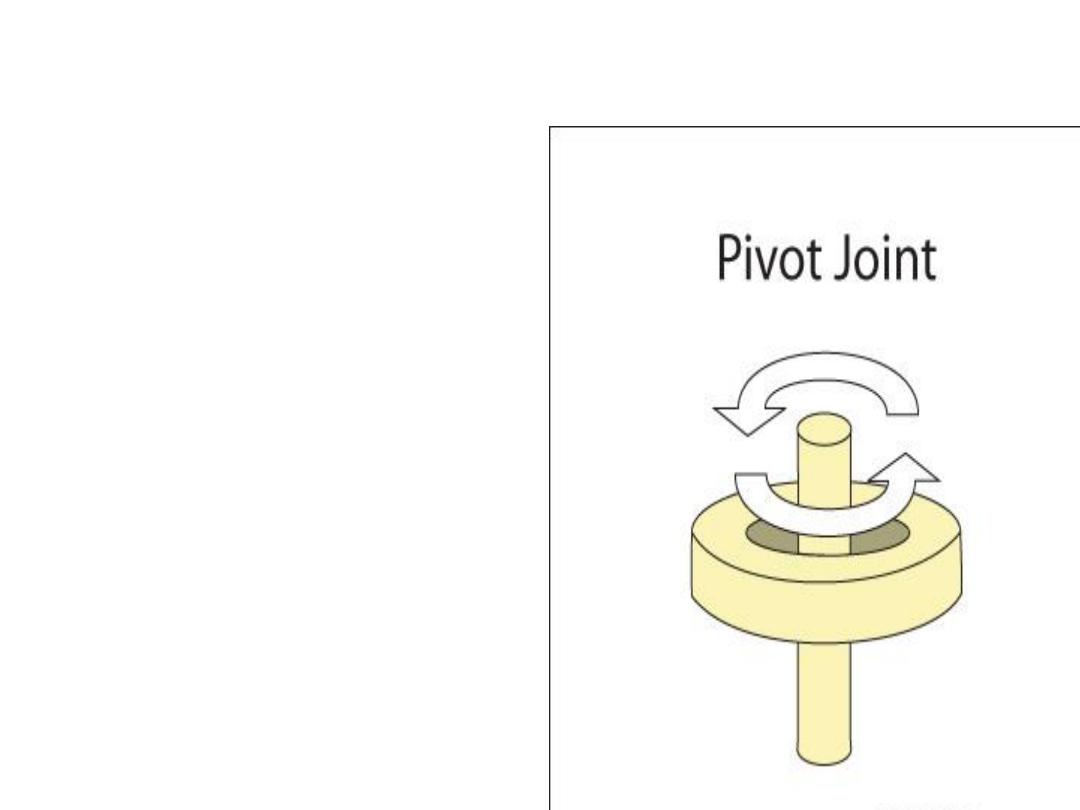
3. Pivot joints
: the rounded
or pointed surface of one
bone articulates with a
ring formed partly by
another bone and partly
by a ligament. A pivot joint
is a uniaxial because it
allows movement around
a longitudinal axis only.
EX: the atlantoaxial joint in
which the atlas bone
rotates around the axis
bone giving the" no"
movement of the head.
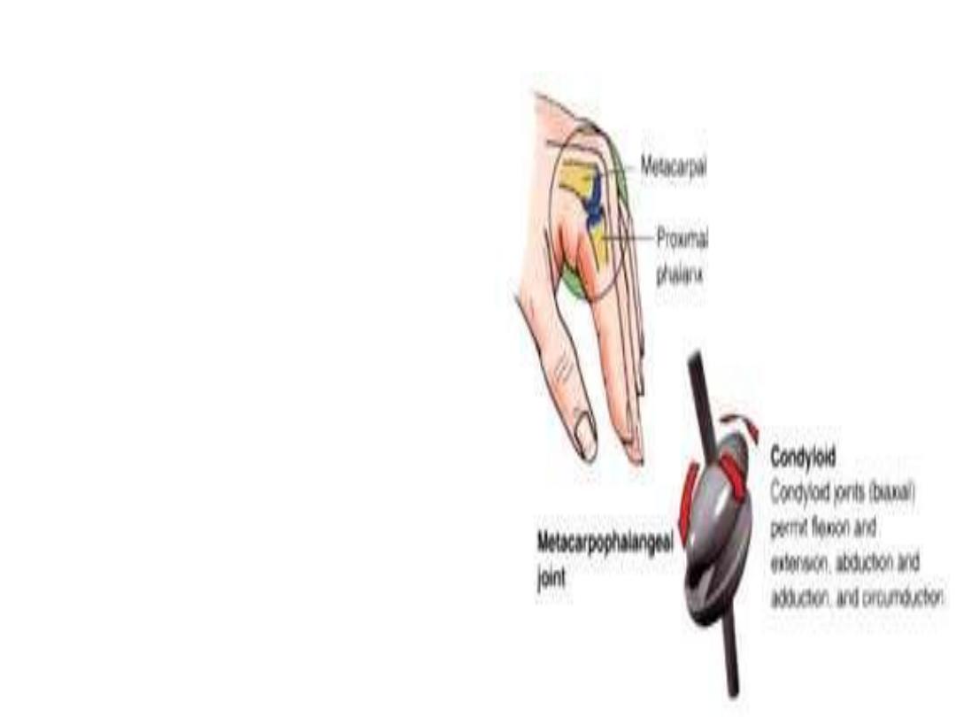
Types of synovial joints:
4. Conyloid joints :
(Ellipsoidal joint) a convex
oval surface of one bone
fits into a convex oval
surface of the other bone.
Ex: Metacarpophalangeal
joints of the 2
nd
-5
th
digits.
Condyloid joints are biaxial
joints because they allow
movements around two
axis. notice that your index
finger can be moved from
side-to-side and up and
down.
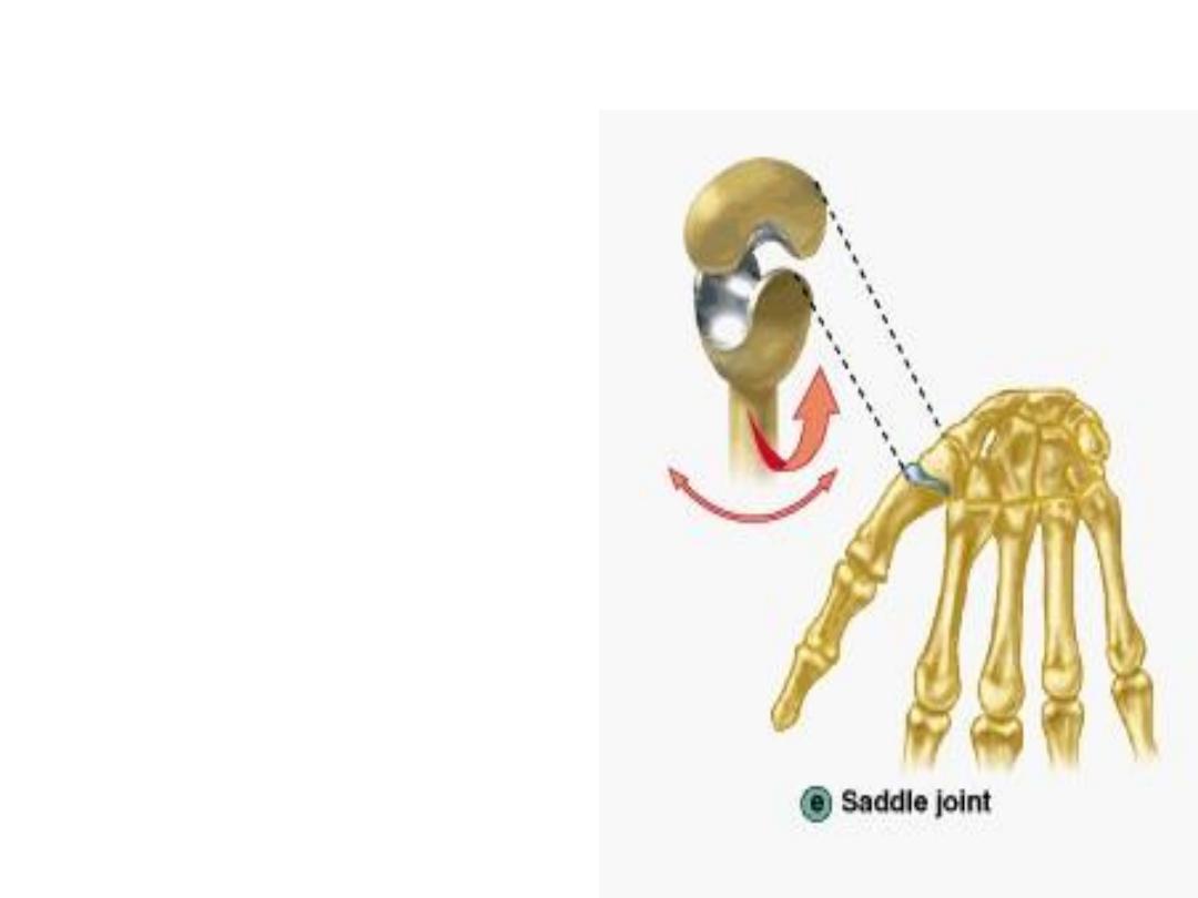
Types of synovial joints:
5. Saddle joints
: the articular
surface of one bone is
saddle shaped and the
articular surface of the
other bone fits into the
"saddle" as a sitting rider
would sit. a saddle joint is a
modified condyloid joint in
which the movement is
somewhat freer. They are
biaxial producing side-to-
side and up and down
movement. Ex: the joint in
the thumb between the
trapezium bone (one of the
carpal wrist bones) and the
metacarpal of the thumb.
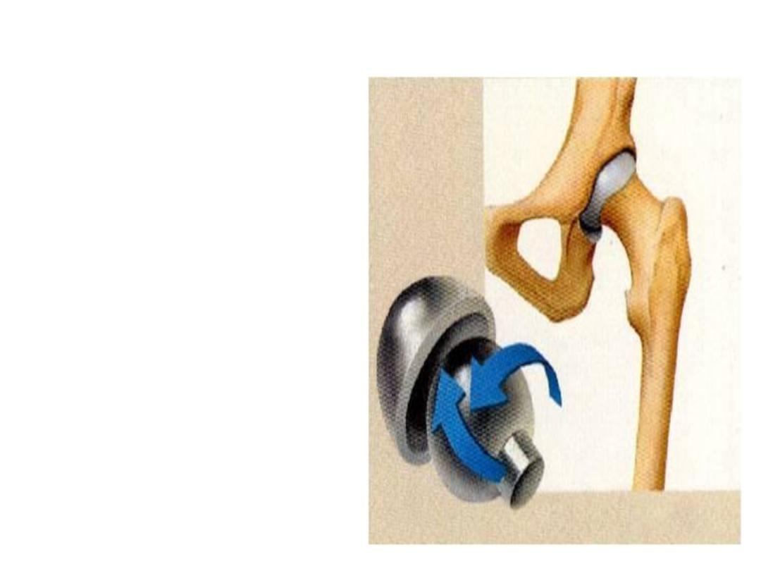
Types of synovial joints:
6. Ball-and –socket joints
:
consists of a ball-like
surface of one bone
fitting into a cuplike
depression of another
bone. Such joints are
multiaxial (polyaxial)
because they allow
movements in many
axis. Ex: shoulder joint
and Hip joint.

Types of Movements at synovial joints:
• Anatomists, Physical therapists use specific
terminology to design movements that can occur at
synovial joints. These precise terms may describe
the direction of movement or the relationship of
one part of the body to another. The movements are
grouped into four main categories:
1. Gliding
2. Angular movements.
3. Rotation.
4. Special movements.
• This last category includes movements that only
occur at certain joints.
Gliding
• Is a simple movement in
which the relatively flat
bone surfaces moves
from side to side, back
and forth with respect to
one another. This
movement is limited
according to the articular
capsule and the strength
of the surrounding
ligaments. Gliding occurs
at plane joints.

Angular movements
• There is an increase or decrease in the
angle between the articulating bones.
The principle angular movements are
flexion, extension, lateral flexion ,
hyperextension, abduction, adduction
and circumduction.
These movements
are discussed in respect to the body in
the anatomical position.
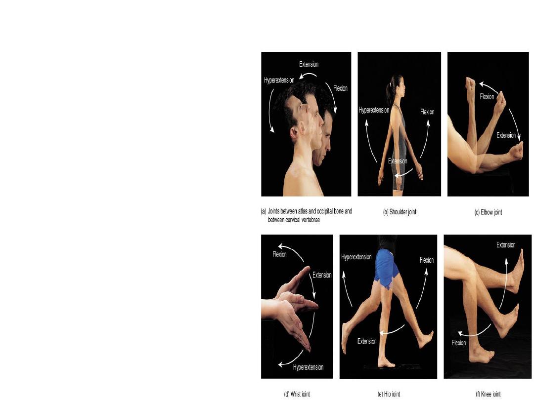
Angular movements
• Flexion
and
extension
are
opposite movements. in
flexion there is decrease in
angle between the articulating
bones while in extension there
is an increase in the angle.
(flexion=bend,
extension=strech out).
Extension usually restores the
part of the body to the
anatomical position after it
has been flexed. EX: tilting the
head downward to the chest
(flexion) and returning it back
to its normal position
(extension). Other EX:
(Homework).

Angular movements
• Lateral flexion
means
movement of the
trunk to the right or
left at the waist and
this involves the
intervertebral joints.
• Hyperextension
means continuation of
extension beyond the
anatomical position.
Ex: bending the head
backward at the
cervical intervertebral
joint. Other EX: (Home
work).
Angular movements
• Abduction
is the movement
away from the midline whereas
adduction
is the movement
toward the midline Ex: moving
the arm laterally at the shoulder
joint is abduction while
returning it back to its normal
anatomical position is
adduction. Other Ex: (Home
work).
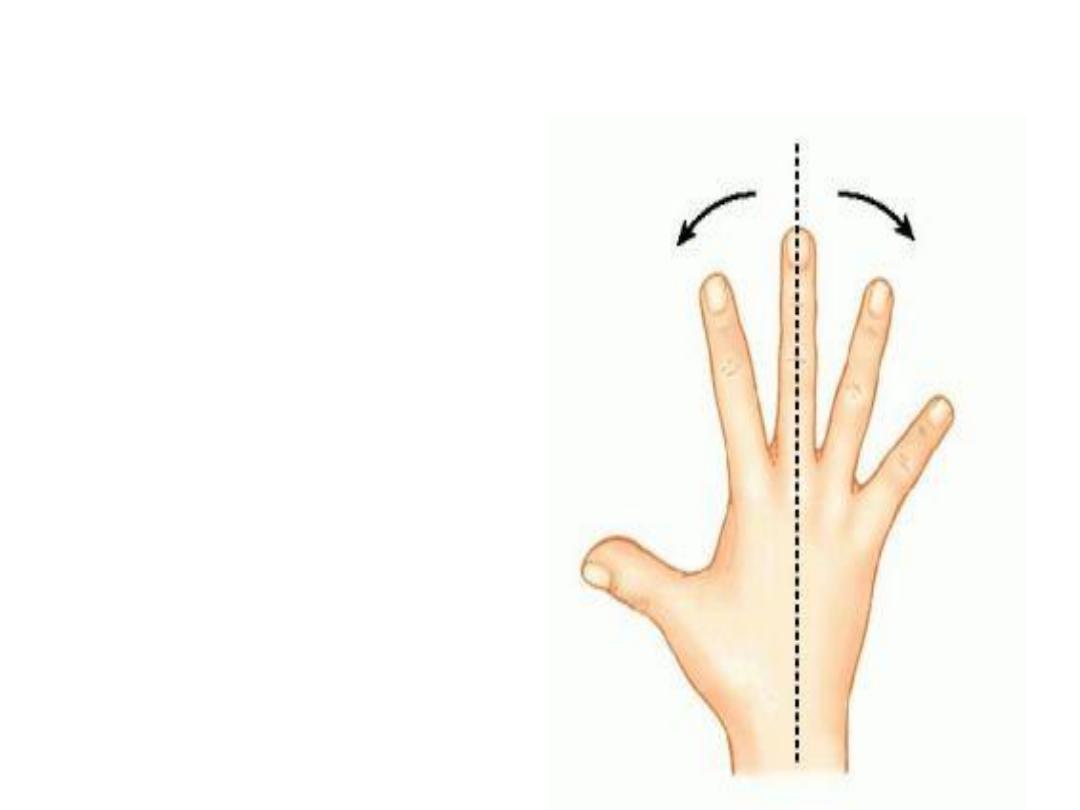
Angular movements
• Note that abduction and
adduction of the fingers
and toes are movements
away and towards an
imaginary line drawn
through the longest
middle finger in the hand
and the second toe in the
foot. So spreading out the
fingers is abduction while
returning them back to
their normal anatomical
position is adduction.
Angular movements
• Circumduction
is the
movement of the distal
end of the body around a
circle, it’s a result of a
continuous sequence of
flexion, abduction,
extension and adduction.
Ex: moving the arm in a
circle at the shoulder
joint. Other Ex: (Home
work).

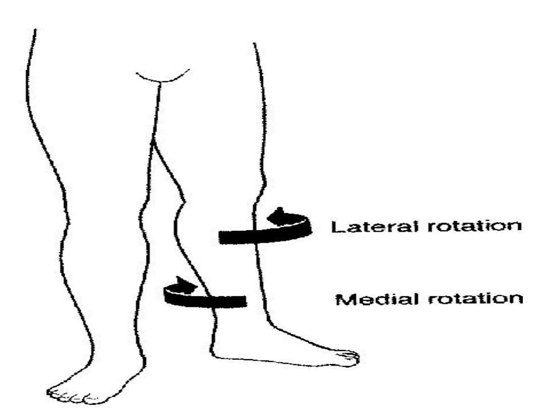
• Rotation
means that the bone revolves around
its own longitudinal axis EX: as in pivot joint,
and moving the trunk from side to side at the
intervertebral joints while keeping the hips
and the lower limbs in their anatomical
position. In the limbs , rotation is defined
relative to the midline and specific qualifying
terms are used. If the anterior surface of the
limb is turned toward the midline, the
movement is called
medial (internal) rotation
.
If the anterior surface of the limb is turned
away from the midline, the movement is
called
lateral (external ) rotation
.

• Special movements occurs only at
specific joints. They include
elevation,
depression, protraction, retraction,
inversion, eversion, dorsiflexion, planter
flexion, suppination, pronation, and
opposition.
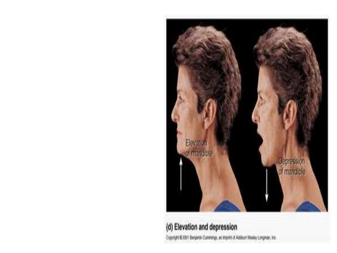
• Elevation
is an upward
movement of a part of the
body, such as closing the
mouth at the
tempromandibular joint
or shrugging the
shoulders at one of the
lateral joint of the clavicle.
• Depression
is a downward
movement of a part of a
body, such as opening the
mouth ton depress the
mandible or returning the
shrugged shoulders to
their anatomical position.
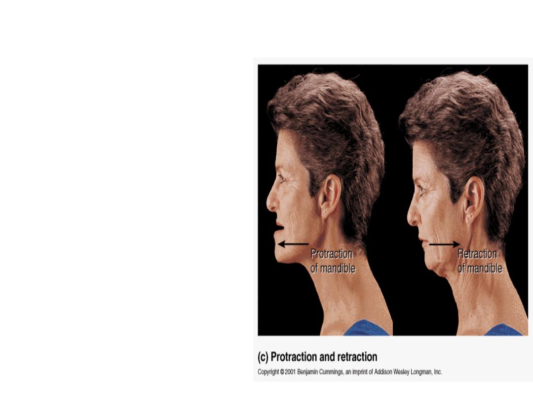
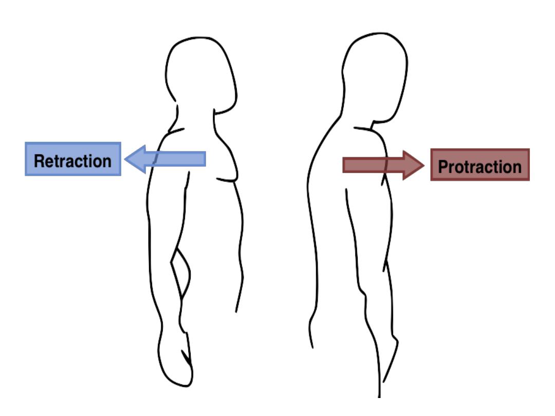
• Protraction
is a
movement of a part of
the body anteriorly (to
draw froth) as in protract
the mandible at the
tempromandibular joint
by thrusting it outward,
or protract your clavicles
by crossing your arms.
• Retraction
is the
movement of the
protracted part of the
body back to the
anatomical position. (to
draw back)
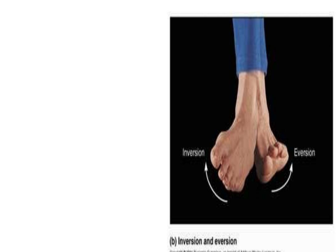
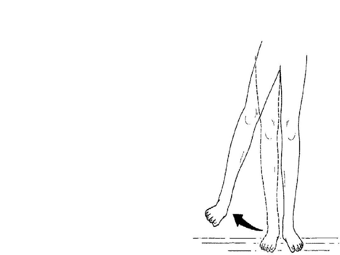
• Inversion
( to turn inward )
is movement of the soles
medially at the intertarsal
joints (between the
tarsals)
• Eversion
(to turn outward )
is the movement of the
soles laterally at the
intertarsal joints.
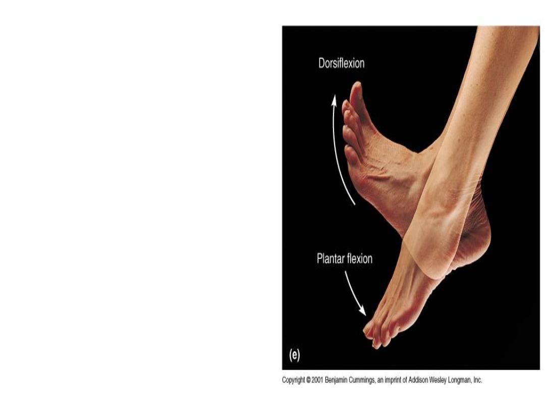
• Dorsiflexion
refers to
bending of the foot at the
ankle in the direction of the
dorsum (superior surface).
Dorsiflexion occurs when
you stand on your heels.
• Planter flexion
refers to
bending of the foot at the
ankle joint in the direction
of the planter or inferior
surface (sole), as when
standing on your toes.
• NOTE: dorsiflexion is true
flexion, whereas planter
flexion is true extension.
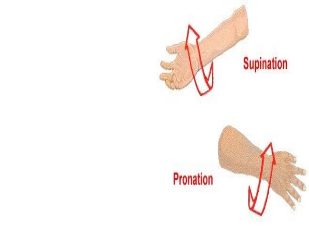
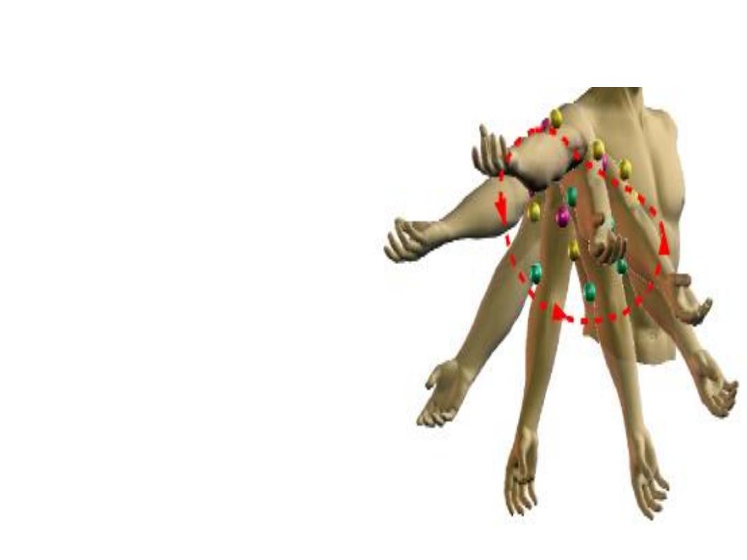
• Supination
is a movement
of forearm at the proximal
and distal radioulnar
joints in which the palm is
turned anteriorly or
superiorly. This position is
one of the defining
features of the anatomical
position.
• Pronation
is the
movement of the forearm
at the proximal and distal
radioulnar joints in which
the palm is turned
posteriorly or inferiorly.
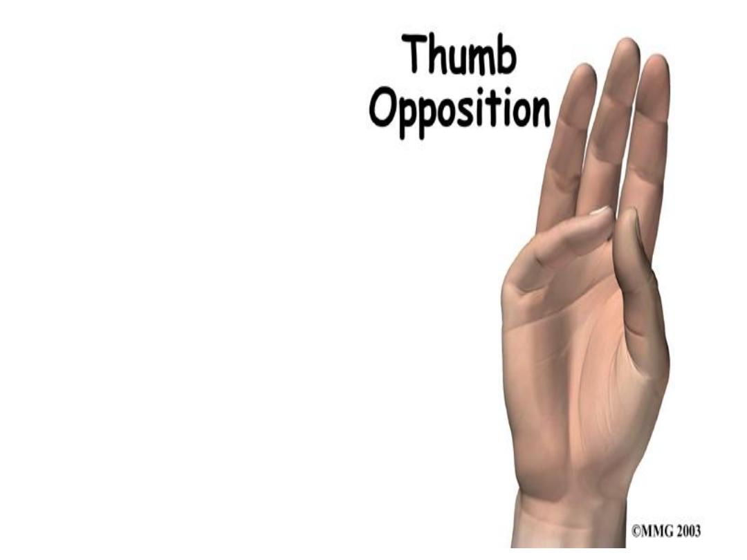
• Opposition
is the
movement of the thumb
at the carpometacarpal
joint in which the thumb
moves across the palm to
touch the tips of the
fingers on the same
hand. This gives the
ability to grasp and
manipulate objects very
precisely.

• The end….