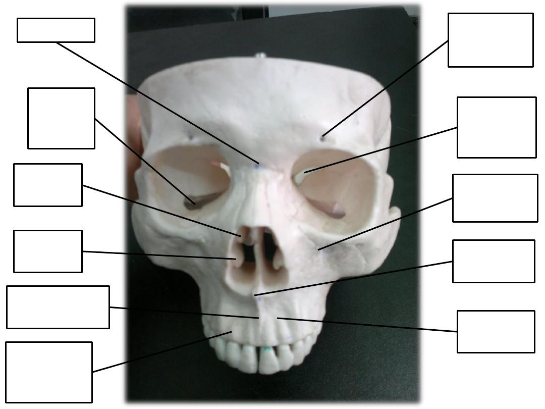
supraorbital
foramen
infraorbital
foramen
nasion
intermaxillary
suture
incesive
fossa
alveolar
process of
maxilla
anterior
nasal spine
middle
concha
inferior
concha
superior
orbital
fissure
inferior
orbital
fissure
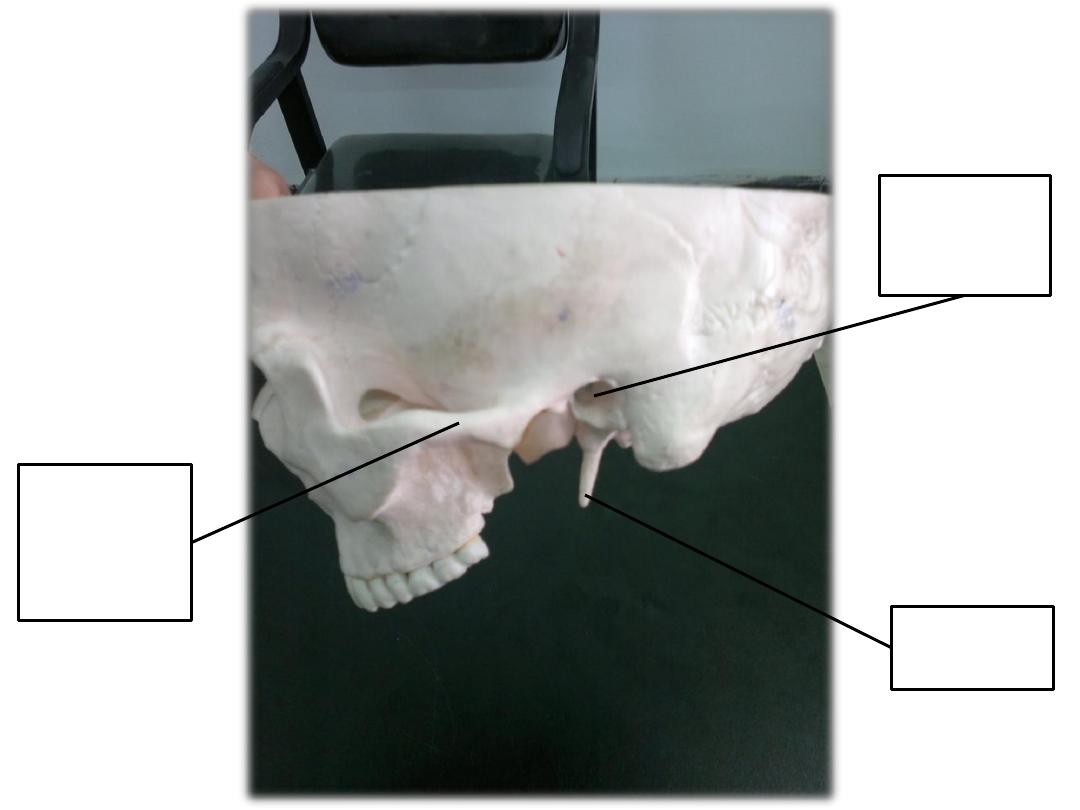
external
auditory
meatus
styloid
process
zygomatic
process of
temporal
bone
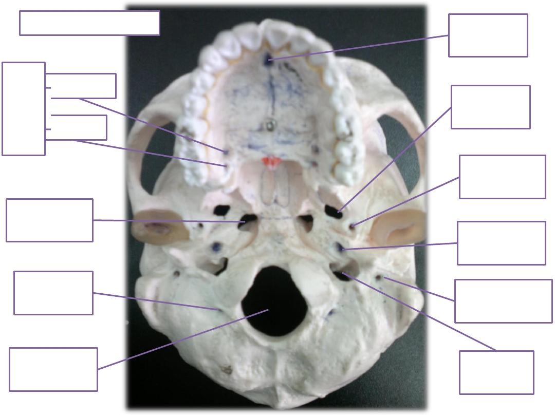
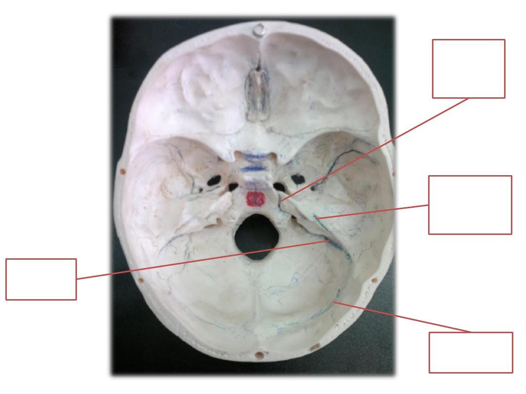
incisive
foramen
foramen
spinosum
jugular
foramen
foramen
magnum
greater
lesser
pala
tine
for
amen
foramen
ovale
carotid
canal
condylar
canal
stylomastoid
foramen
Foramen
lacerum
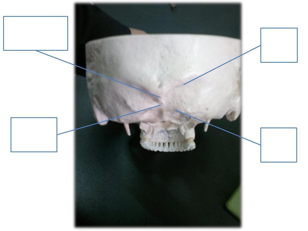
Just foramina here
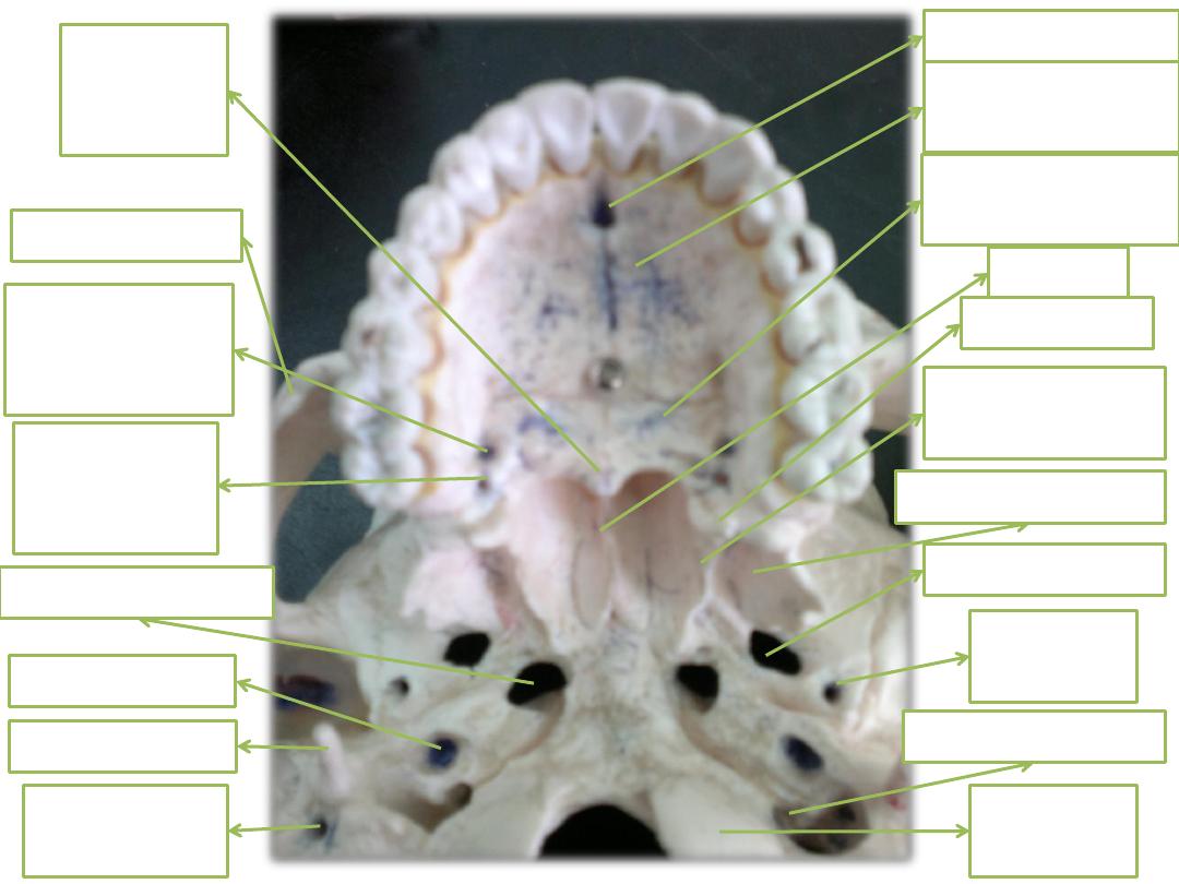
Incisive foramen
Palatine process
of maxilla
Horizontal plate
of palatine
vomer
Medial
petrygoid plate
Lateral petrygoid
hamulus
Foramen ovale
Foramen lacerum
Carotid canal
Foramen
spinosum
Occipital
condyle
Stylomastoid
foramen
Greater
palatine
foramen
Lesser
palatine
foramen
Jugular foramen
Zygomatic arch
Stylod process
posterior
nasal
spine
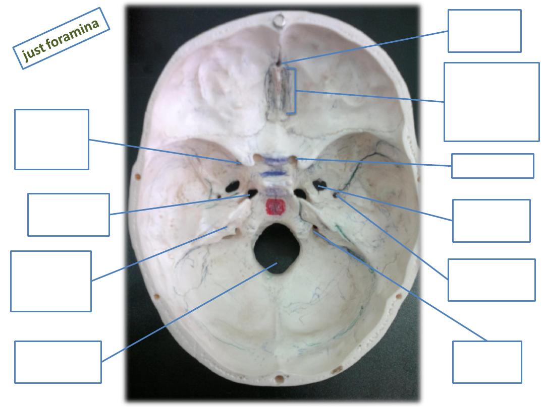
foramen
cecum
Olfactory
foramina
Cribriform
plate
optic canal
superior
orbital
fissure
jugular
foramen
foramen
spinosum
foramen
ovale
Lacerum
Foramen
internal
auditory
meatus
foramen
magnum
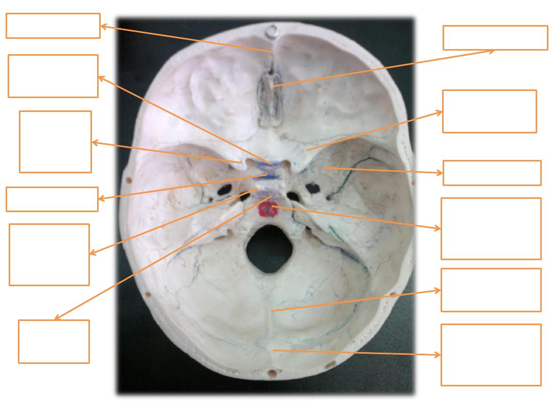
Crista galli
Lesser wing
of sphenoid
Frontal crest
Greater wing
Anterior
clinoid
process
Bailar part of
occipital
(clivus)
Tuberculum
sellae
Dorsum
sellae
Posterior
clinoid
proecess
Internal
occipital
protuberance
Internal
occipital crest
sella torcica
inferior
petrosal
sinus
superior
petrosal
sinus
sigmoid
sinus
transverse
sinus
superior
nuchal
line
external
occipital
protuberence
external
occipital
crest
inferior
nuchal
line
Frontal sinus
Cribriform plate of ethmoid
Sphenoidal sinus
Body of sphenoid
Vestibule
Hard palate
Incisive foramen
Intrinsic muscle of tongue
genioglossus muscle
Geniohyoid
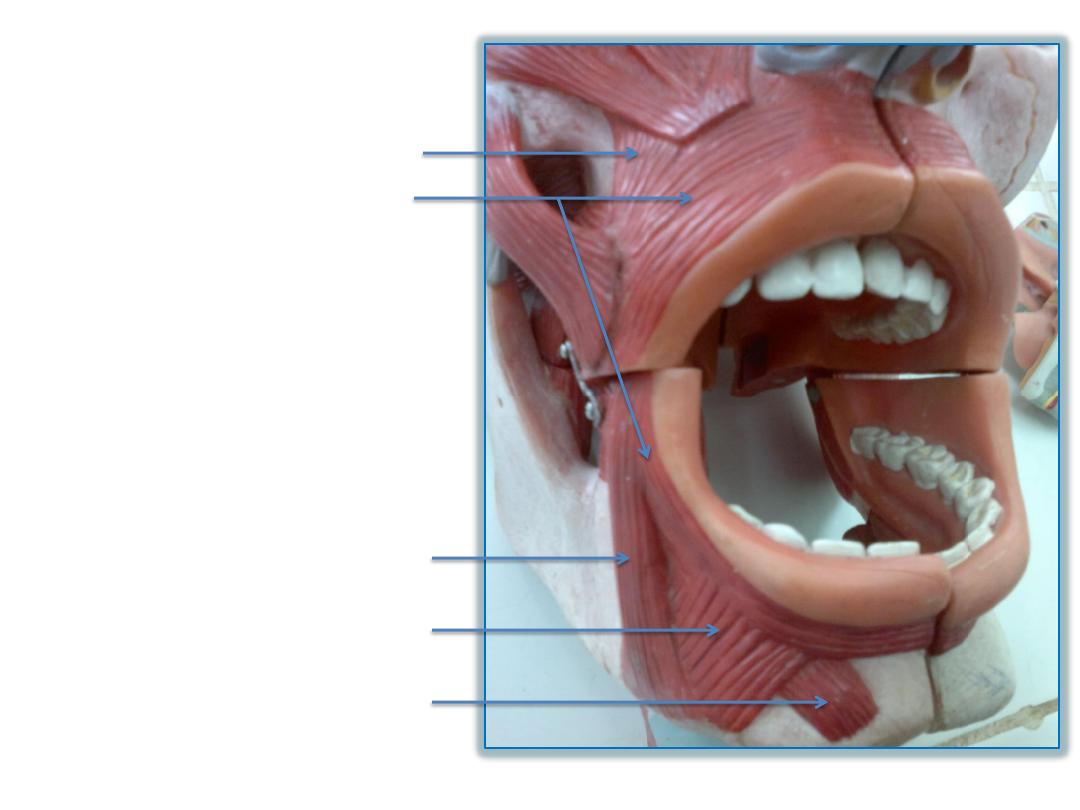
mentalis
Depressor labii inferioris
Depressor anguli oris
Orbicularis oris
Levator anguli oris
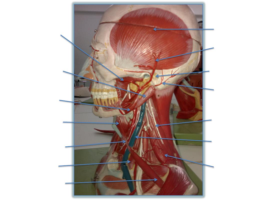
Auricularis
posterior
Posterior
auricular artery
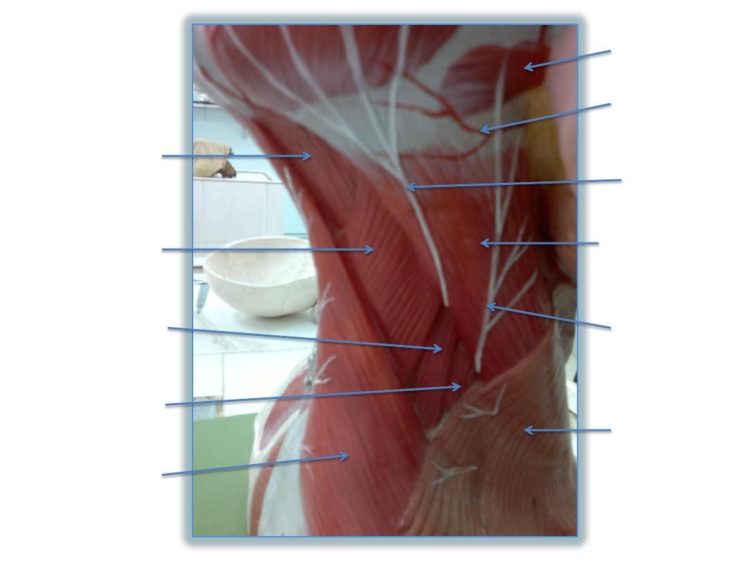
Greater
occipital
nerve
Sternogleidoma
stoid muscle
Lesser
occipital
nerve
Platysma
muscle
Trapezius
muscle
Scalenus
anterior muscle
Scalenus medius
muscle
Levator scapulae
muscle
Semispinalis
capitis muscle
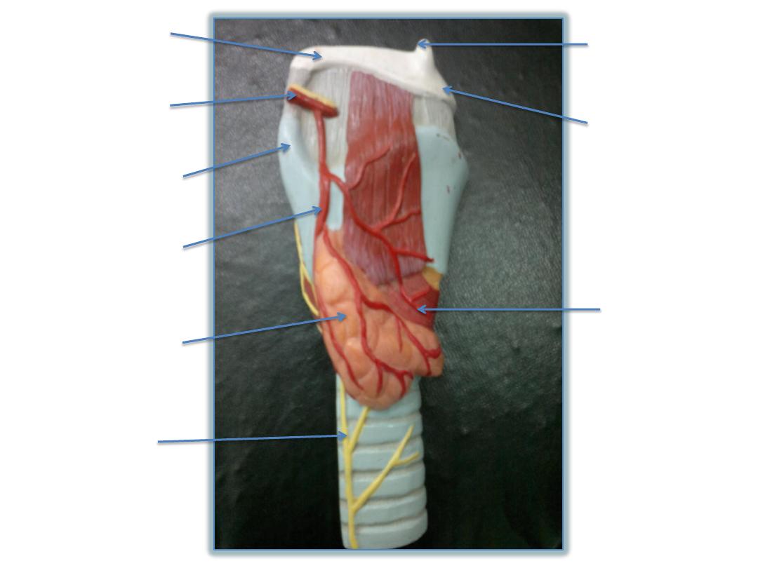
Greater cornu
of hyoid bone
Superior laryngeal
artery & internal
laryngeal nerve
Superior cornu of
thyroid cartilage
Superior thyroid
artery
Lobe of thyroid
gland
Recurrent
laryngeal nerve
Lesser cornu of
hyoid bone
Body of hyoid
bone
Cricothyroid
muscle
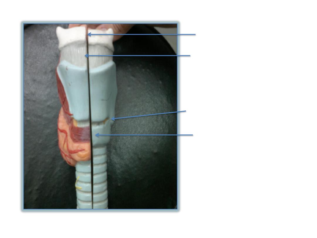
Body of hyoid bone
Median thyrohyoid ligament
Inferior cornu of thyroid cartilage
Arch of cricoid cartilage
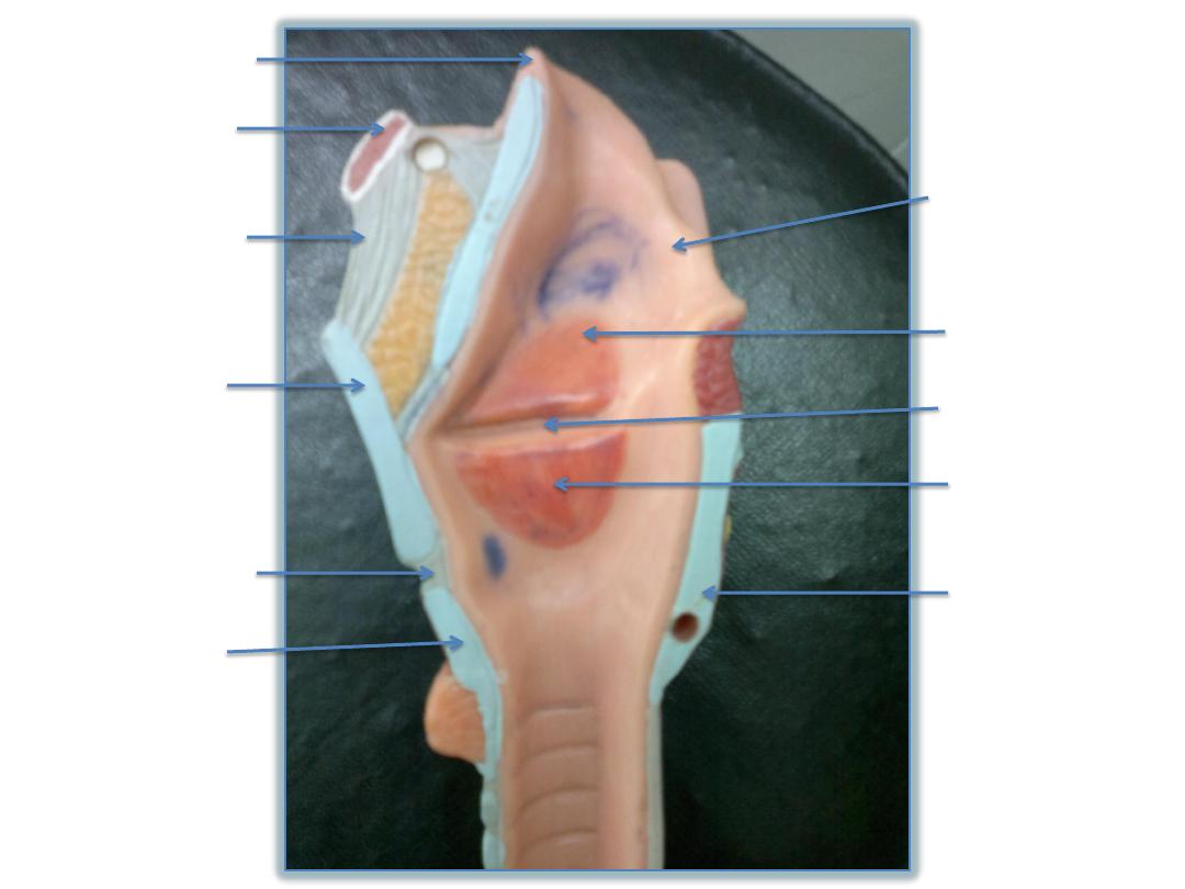
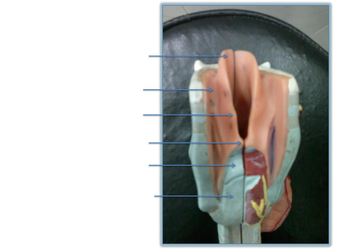
epiglottis
Hyoid bone
Thyrohyoid
membrane
Thyroid
cartilage
Cricothyroid
membrane
Cricoid
cartilage
Aryepiglottic
fold
Vestibular
fold
Laryngeal sinus
Vocal fold
Lamina of
cricoid
cartilage
epiglottis
Piriform fossa
Aryepiglottic fold within it
cuniform cartilage
Corniculate cartilage
Arytenoid cartilage
Cricoid cartilage
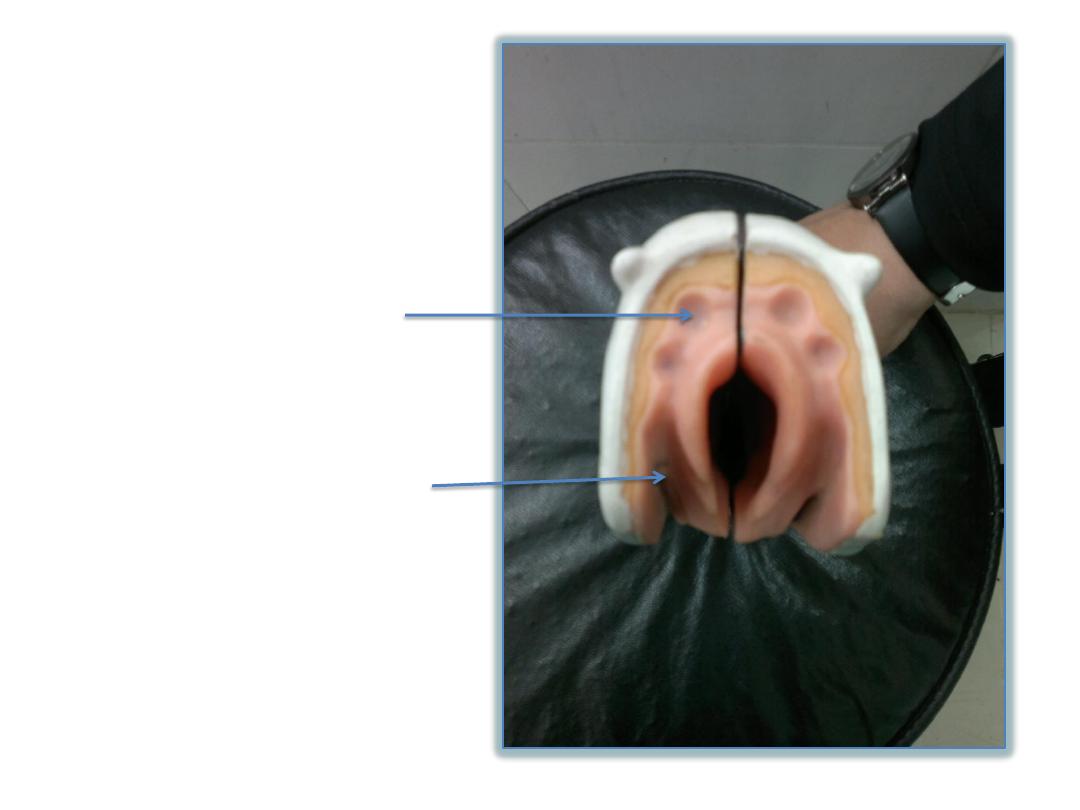
vallecula
Piriform fossa
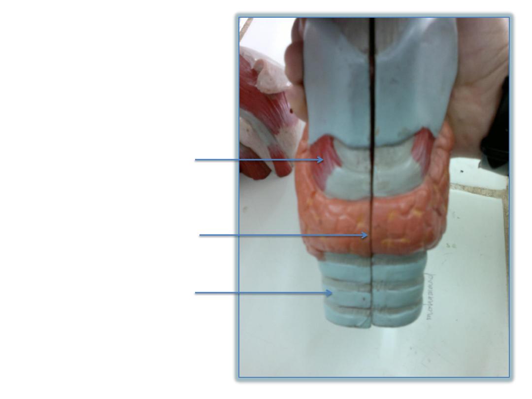
Cricothyroid muscle
Isthmus of thyroid gland
C- shaped cartilage of trachea
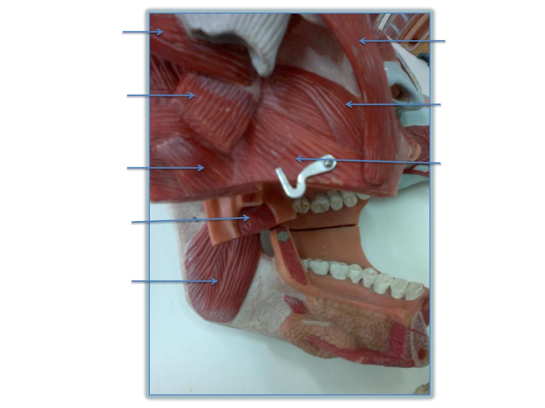
Lateral pterygoid
(inferior head)
Medial pterygoid
(superficial head)
ORIGIN
Palatine tonsils
Medial pterygoid
INSERTION
Zygomaticus
major
risorius
buccinator
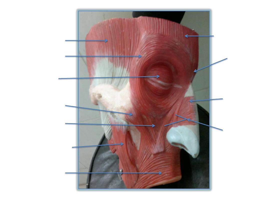
Temporalis
Papebral part of
orbicularis oculi
Orbital part of
orbicularis oculi
Zygomaticus minor
Levator labii
superioris
Zygomaticus major
Orbicularis oris
Levator
labii
superioris
alaque nasi
Compressor
naris
procerus
Frontal belly of
occipitofrontalis
Lateral glossoepiglottic fold
Median glossoepiglottic fold
Valleculla ( depression or
space between the 2 folds)
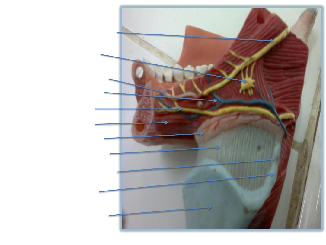
Lingual nerve
Submandibular ganglion
Submandibular duct
Lingual vessels
Hypoglossal nerve
Mylohyoid muscle
Hyoid bone
Thyrohyoid membrane
Lateral thyrohyoid ligament
Superior cornu of
thyroid cartilage
Lamina of thyroid cartilage
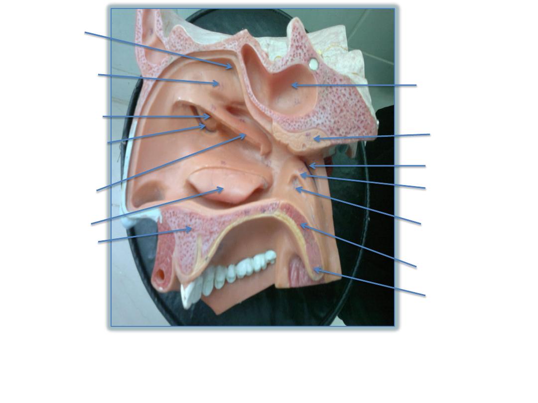
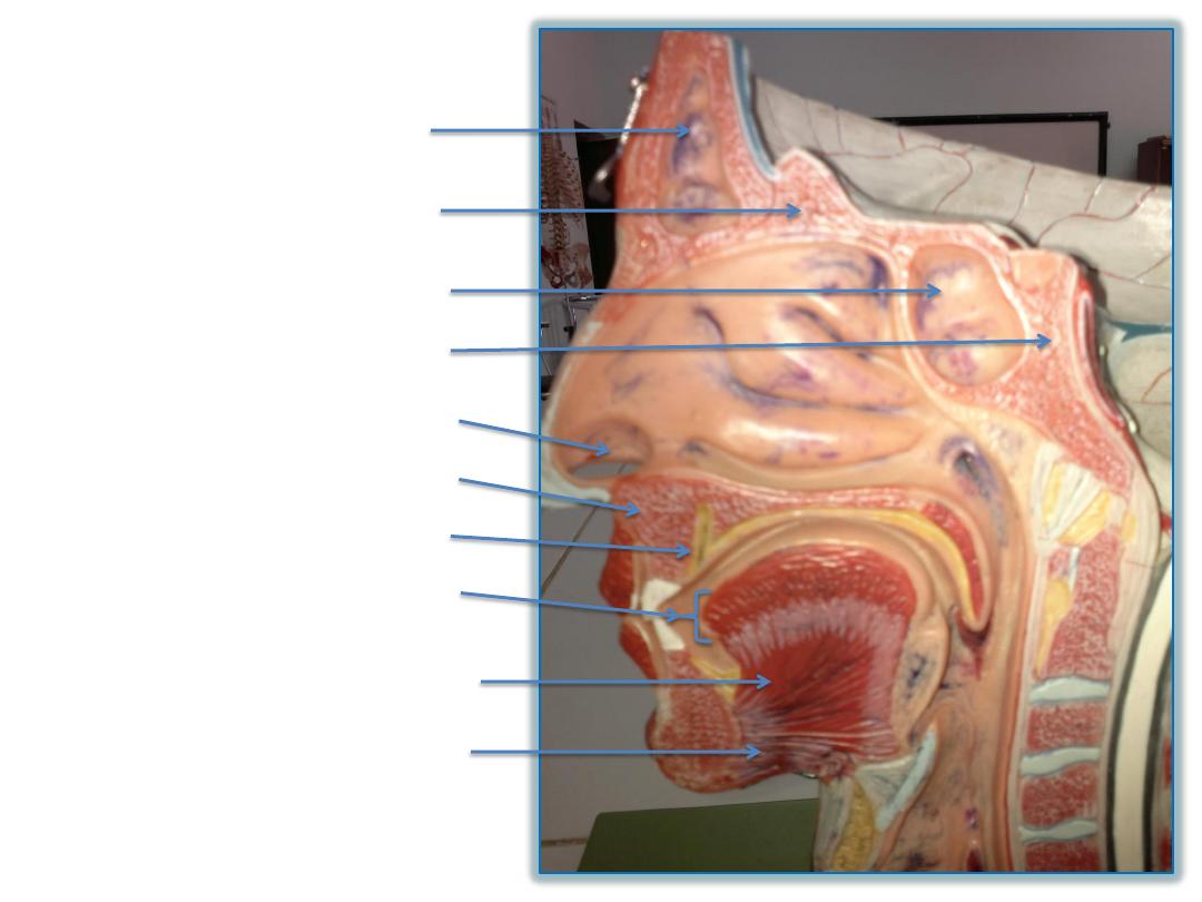
Spheno-ethmoial
recess
Superior
choncha
Inferior chonca
Hard palate
uvula
Soft palate
Opening of
auditory tube
Tubal elevation
Pharyngeal recess
Pharyngeal tonsil
Sphenoidal sinus
Middle chonca
Bulla ethmoidalis
Hiatus semilunaris
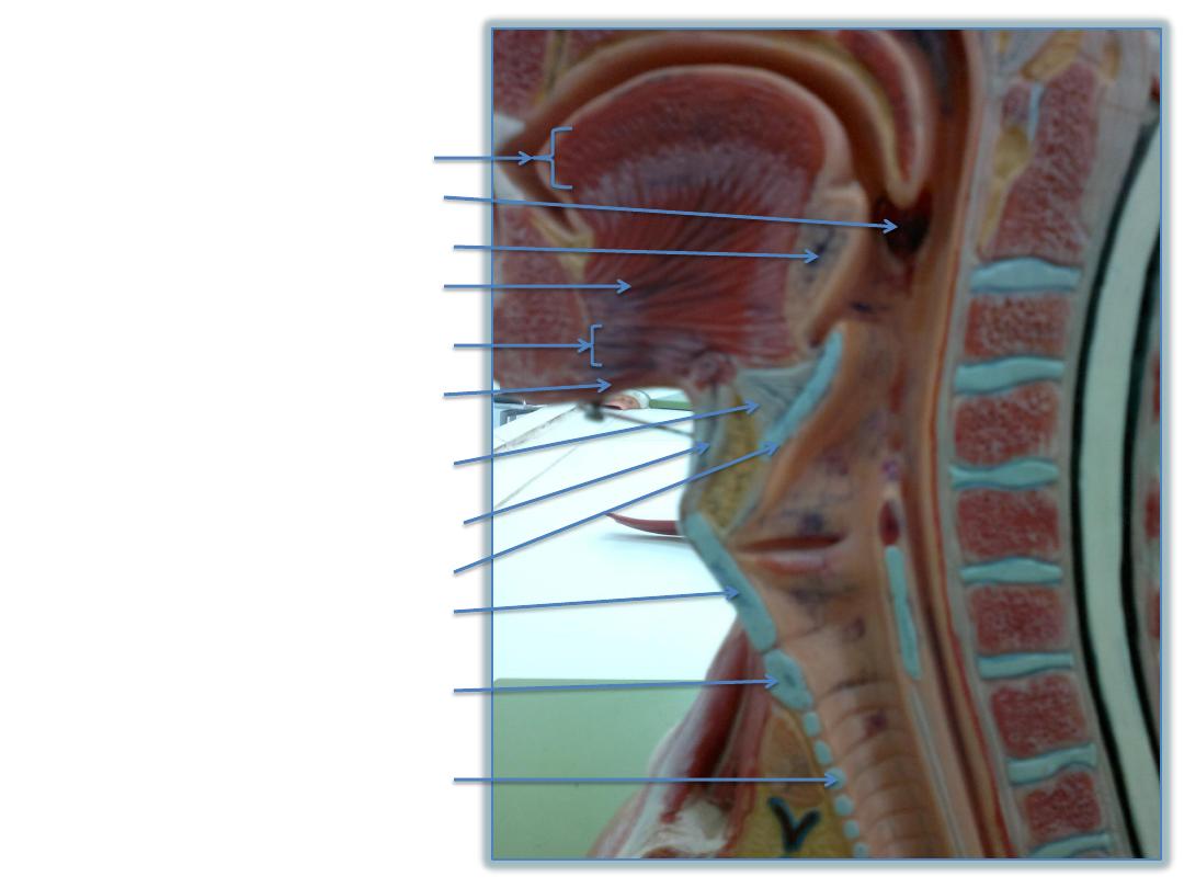
Intrinsic muscle of tongue
Lingual tonsil
Genioglossus (fan shaped)
geniohyoid
Mylohyoid (most superficial)
Hyoepiglottic ligament
Thyrohyoid membrane
epiglottis
Thyroid cartilage
Cricoid cartilage
Palatine tonsils
Ring of trachea
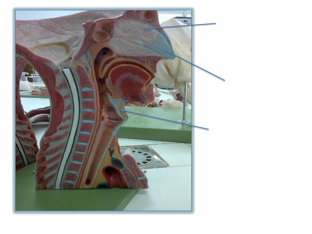
Bony septum of the
nasal cavity
Cartilaginous
septum of the nasal
cavity
Fatty tissue (the yellowish)
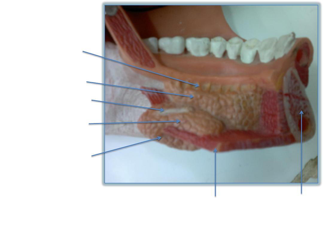
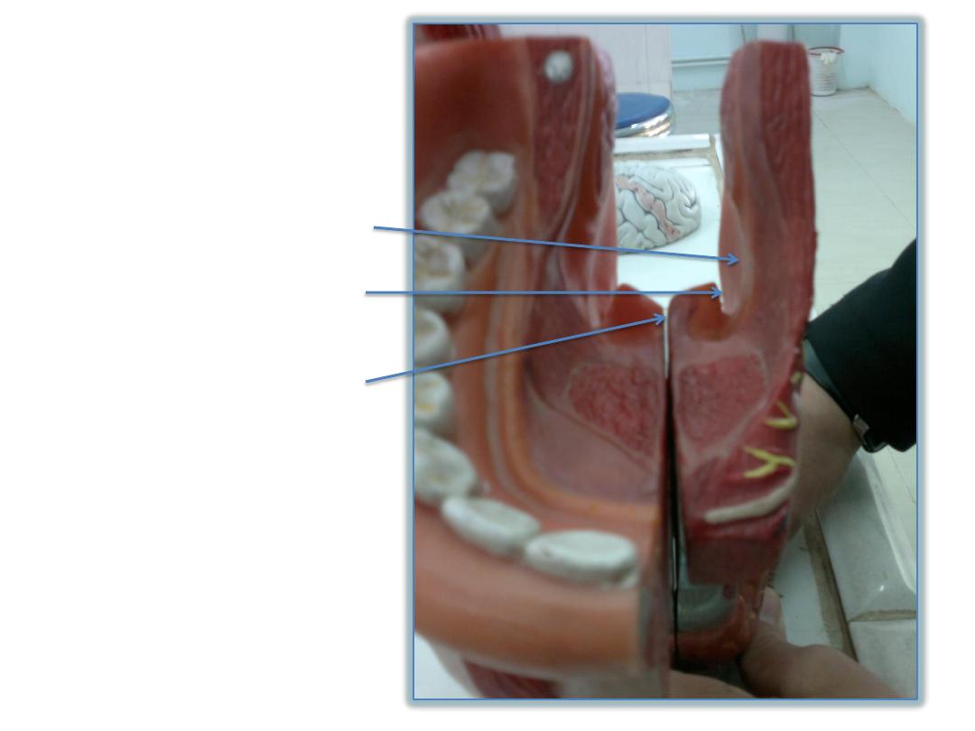
Sublingual ducts
Sublingual salivary gland
Submandibular duct
Deep part of
submandibular gland
Superficial part of
submandibular gland
Mylohyoid muscle
mandible
Medial pterygoid muscle
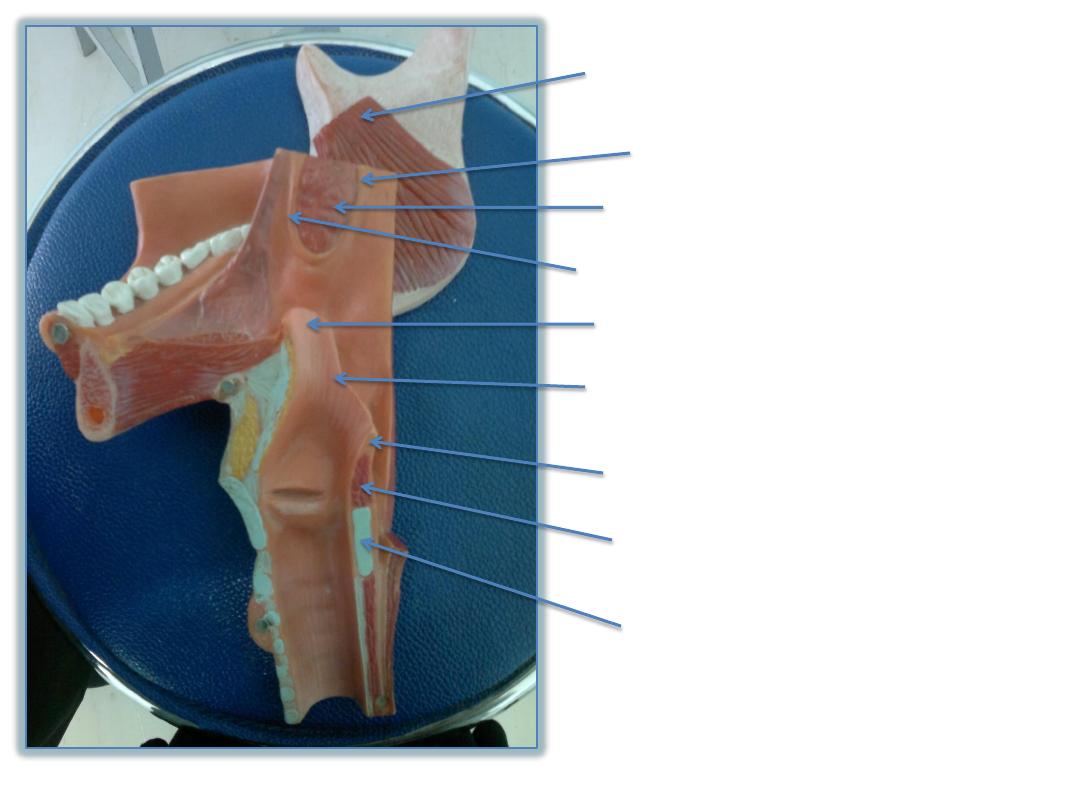
Palatopharyngeal fold
Palatine tonsils
Palatoglossal fold
epiglottis
Aryepiglottic fold
Corniculate cartilage
Aryteniod caetilage
Lamina of cricoid cartilage
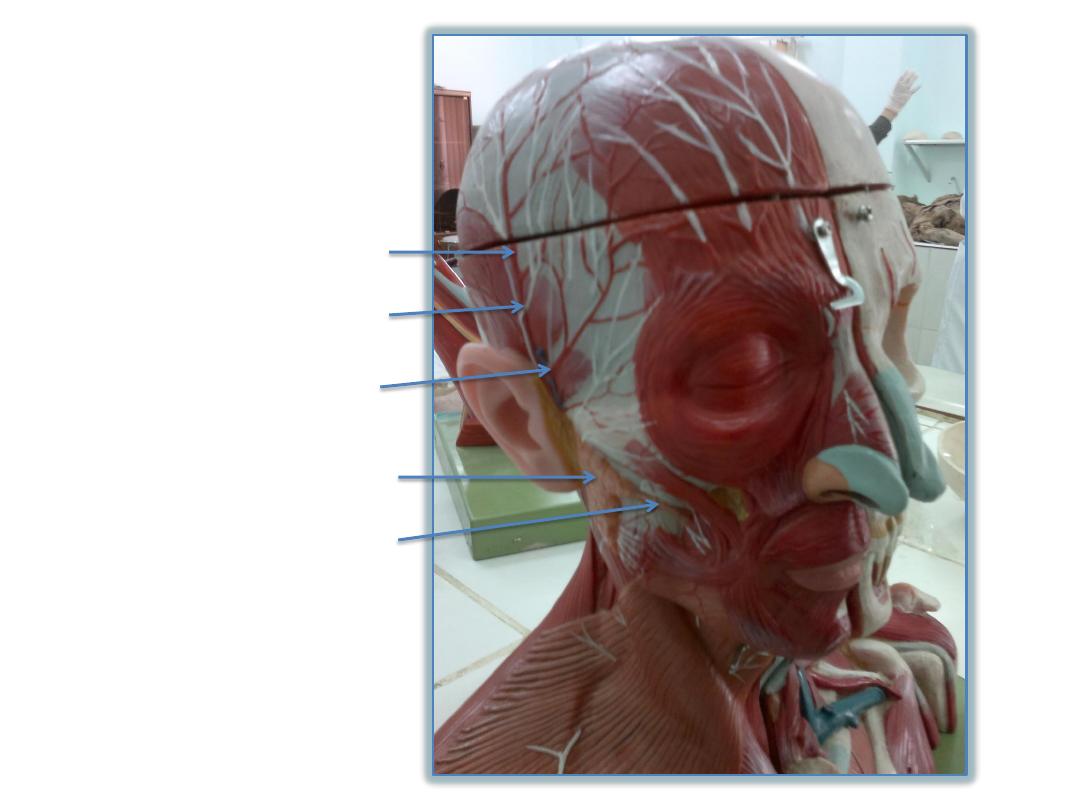
Superficial temporal artery
Auriculotemporal nerve
Superficial temporal vein
Parotid gland
Parotid duct
Maxillary artery
Posterior belly of
digastric
Stylohyoid
muscle
Superior belly of
omohyoid
Intermediate
tendon of omohyoid
Inferior belly of
omohyoid
Temporalis
muscle
Superficial
temporal artery
Spinal part of
accessory nerve
Levator
scapulae
External auditory
meatus
Scalenus anterior
Internal jugular
vein
Posterior
auricular artery
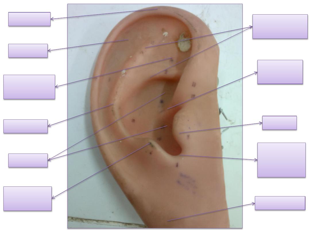
tragus
helix
Lobe of ear
Crus of
helix
Anti helix
Anti
tragus
concha
Inter
tragic
notch
Crur of anti
helix
scapha
Triangular
fossa
umbo
Anterior malleular fold
Posterior
malleular
fold
cone of light
pars flaccida
Pars tensa
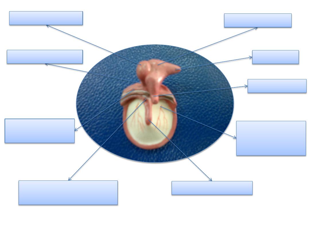
Handle of malleus
Head of malleus
Body of incus
Short limb
Long limb
Neck of malleus
Anterior process of
malleus
Tympanic
branch of
maxillary
Chorda tympani
(gray in color)
External
audiotory
meatus
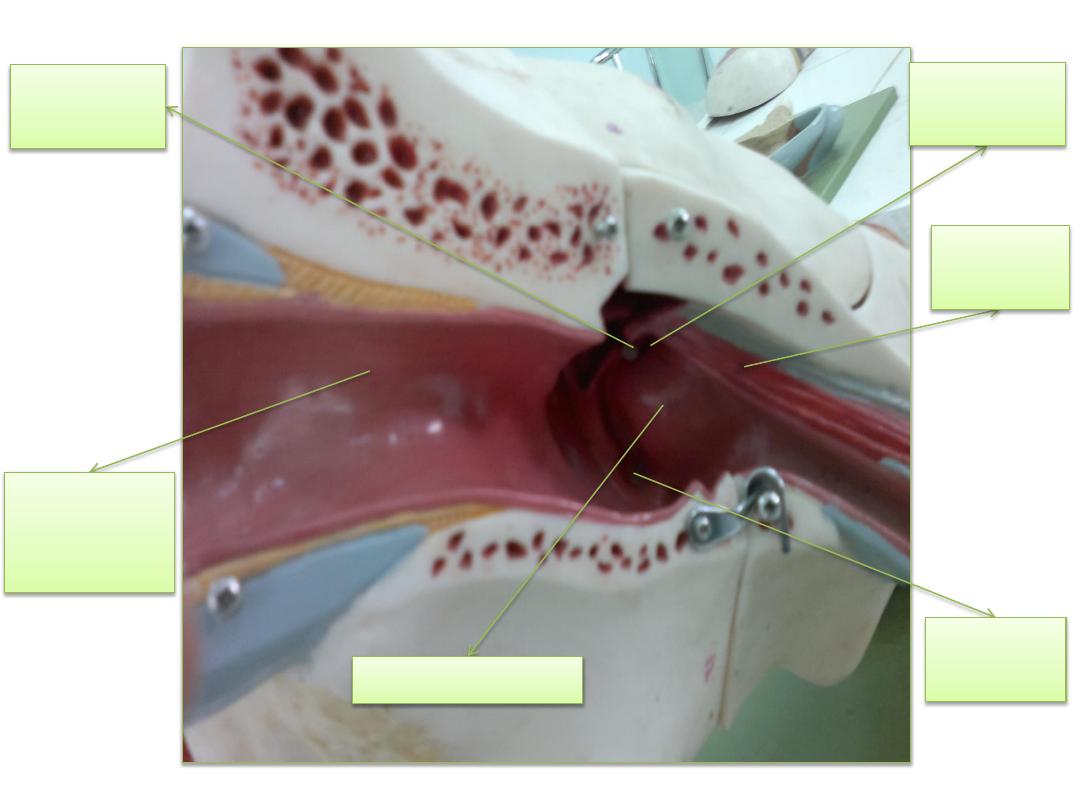
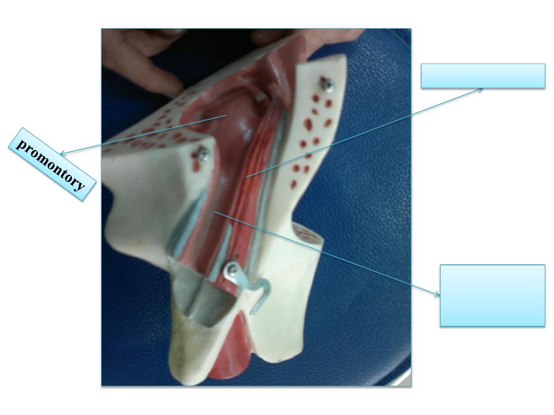
promontory
Oval
window
Round
window
Tensor
tympani
Head of
the stapes
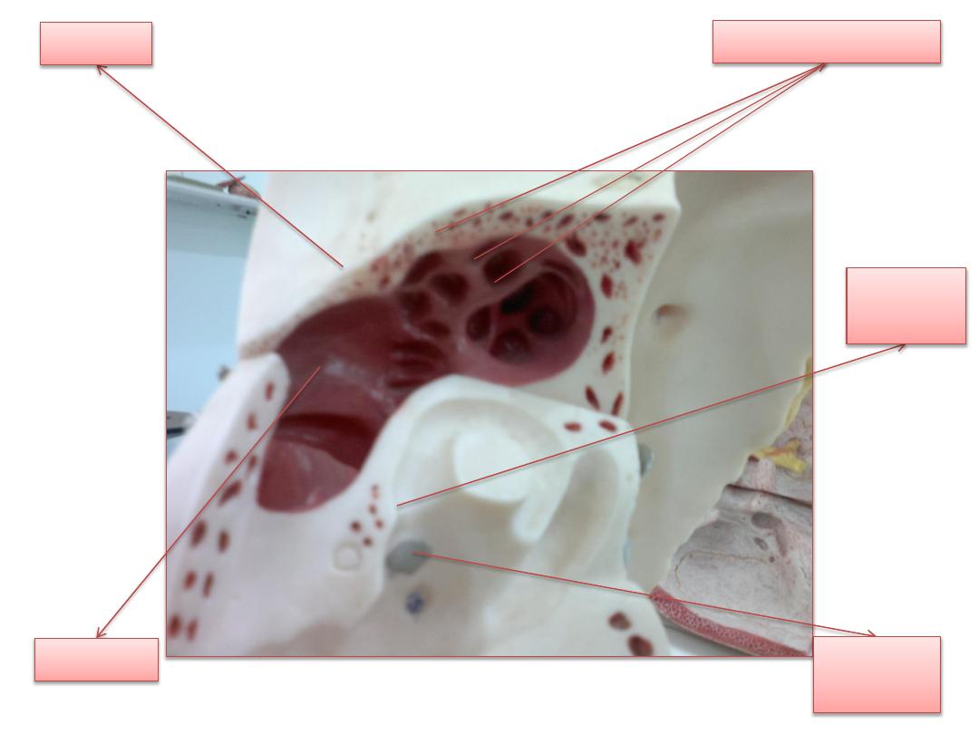
Mastoid air cells
aditus
Oval
window
Round
window
antrum
Pharyngo
tympanic
tube
Tensor tympani
cochlear nerve
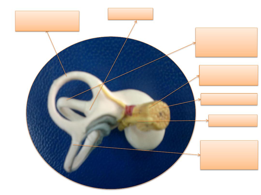
facial nerve
Vestibular nerve
superior
semicircular
canal
Lateral
semicircular
canal
Posterior
semicircular canal
ampulla
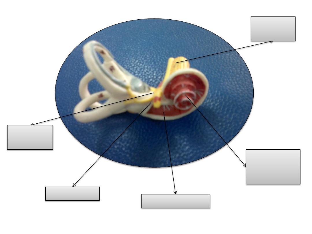
Cochlear
nerve
Vestibular
nerve
Osseus
spiral
lamina
Basal membrane
geniculate
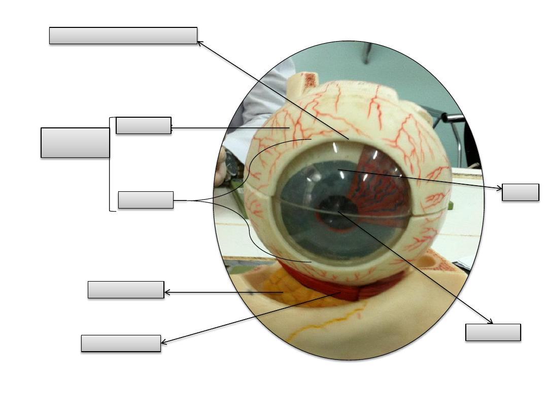
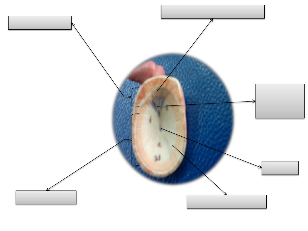
sclera
cornea
Orbital fat
iris
pupil
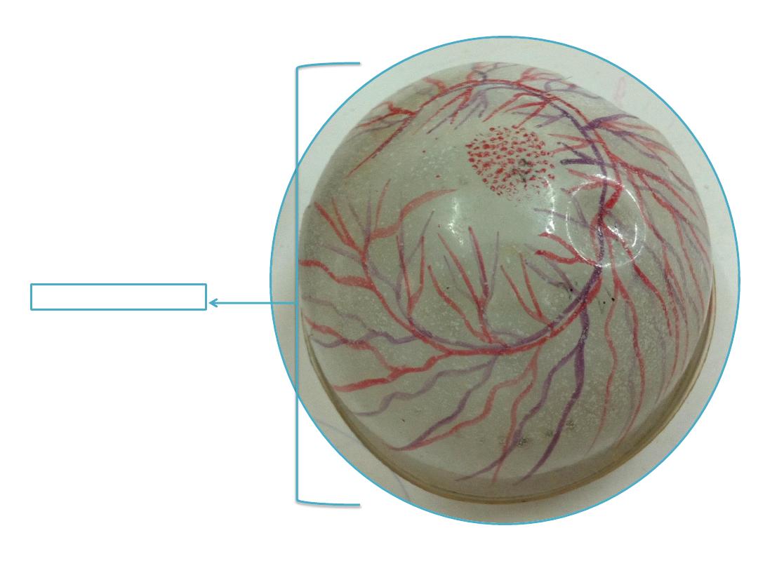
Fibrous coat
of the eye
Corneoscleral junction
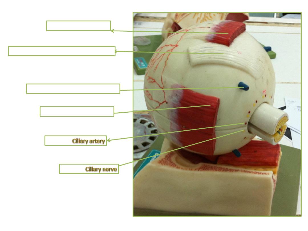
Inferior oblique
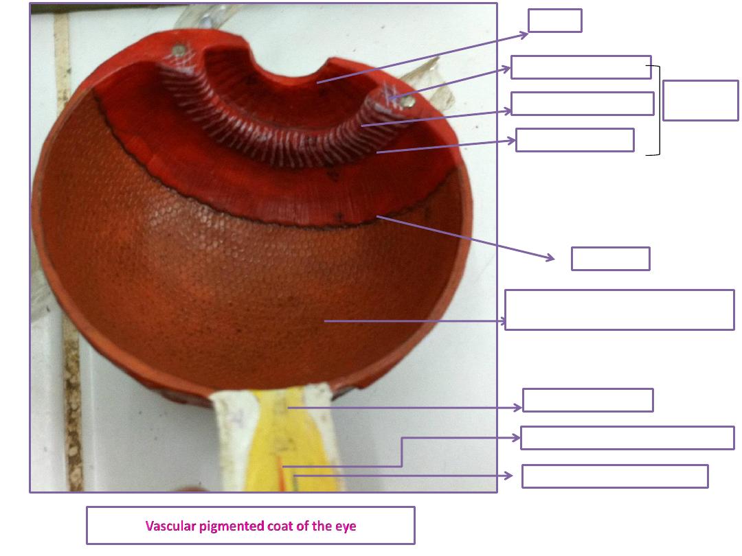
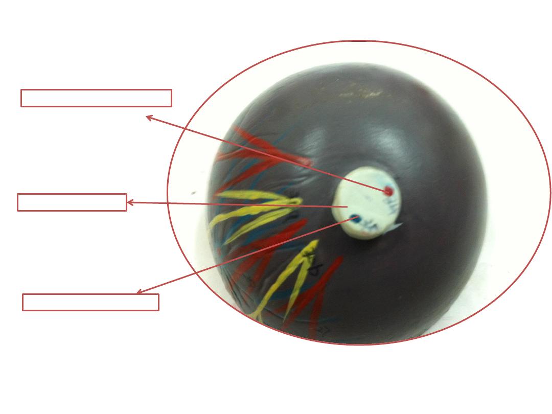
Optic nerve
Central retinal artery
Central retinal vein
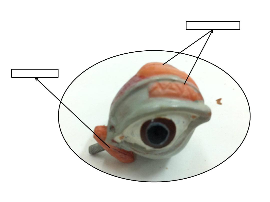
Ciliary muscle
Ciliary process
Ciliary ring
iris
Ciliary
body
Retina
(nervous coat of the eye)
Ora serrata
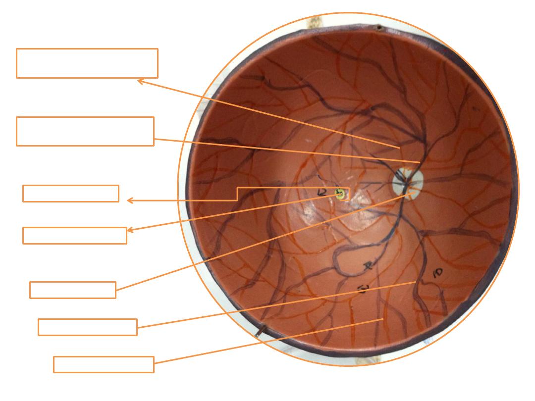
Optic disk
Macula lutea
Fovea centralis
Superior temporal retinal
arteriole
Superior temporal retinal
venule
Inferior t.r.v
Inferior t.r.a
Central retinal artery
Central retinal vein
Optic nerve
Vitreous body
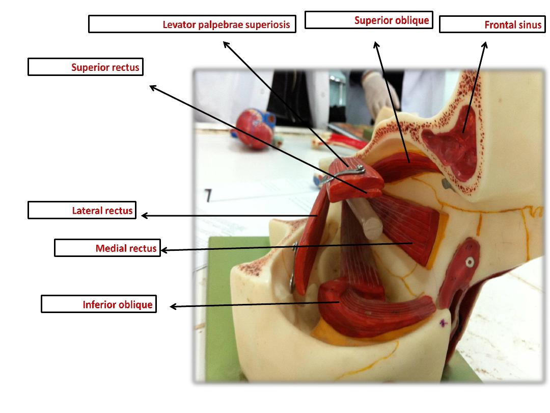
Lateral rectus
Vorticose vein
Tendon of superior oblique
Superior rectus
Lacrimal sac
Lacrimal gland
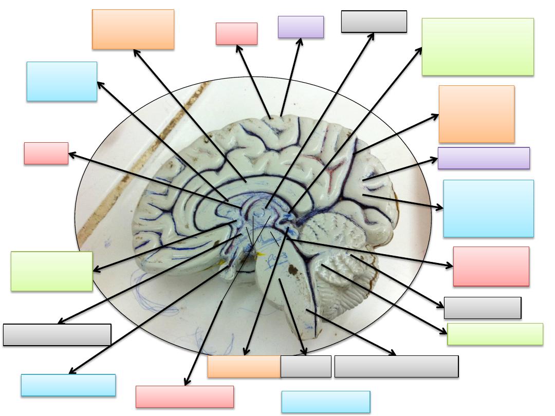
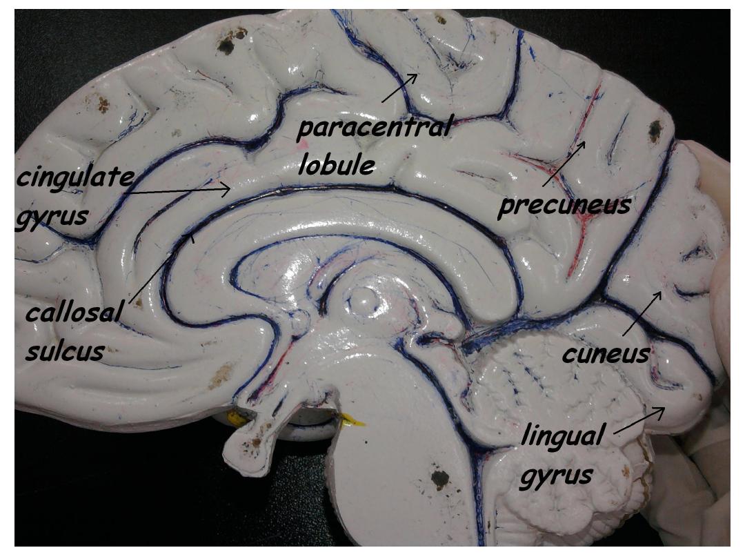
cerebellum
Occipital lobe
Parieto -
occipital
sulcus
Corpus
callosum
Calcarine
sulcus
(visual area)
4
th
ventricle
Pineal
body(gland)
“epithalamus”
Cerebral
aqueduct
3
rd
ventricle
hypo-thalamus
fornix
Septal
Pellucidus
thalamus
gyrus
sulcus
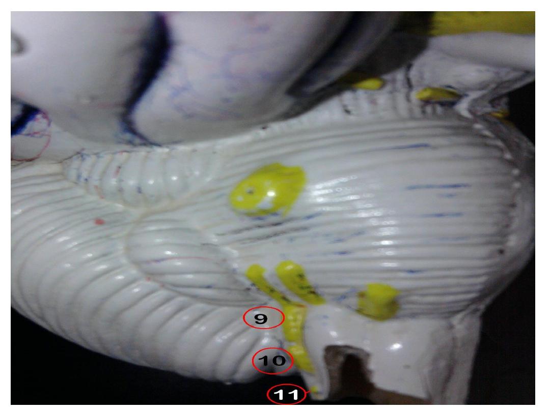
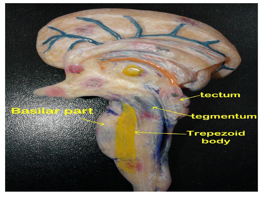
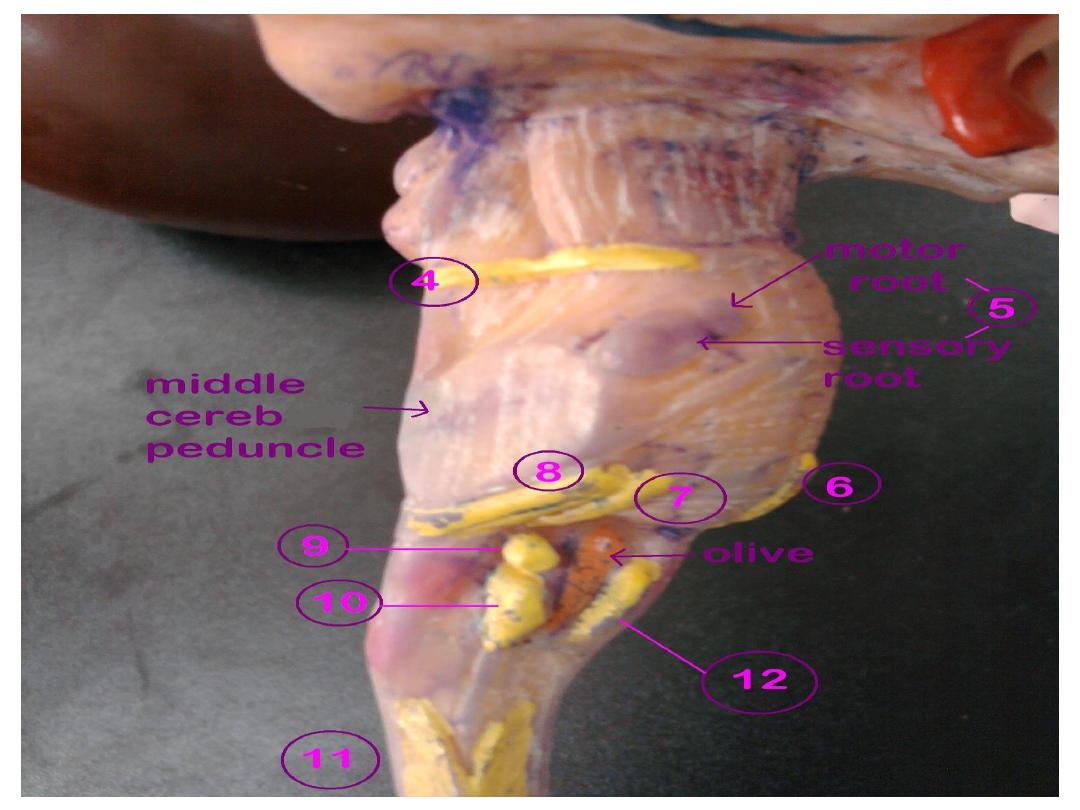
Midbrain
pons
Medulla oblongata
Brain stem
Anterior
commisure
Pitutary gland
Body of corpus
callosum
splenium of
corpus
callosum
genu of
corpus
callosum
diencephalen
septum pellucidum
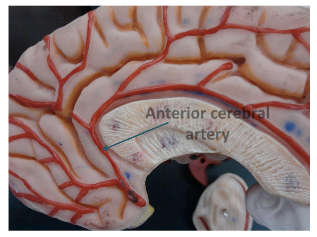
anterior
cerebral
artery
mid brain
pons
medulla
oblongata
br
ain
st
e
m
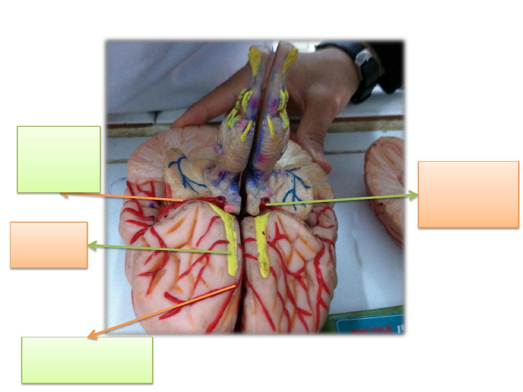
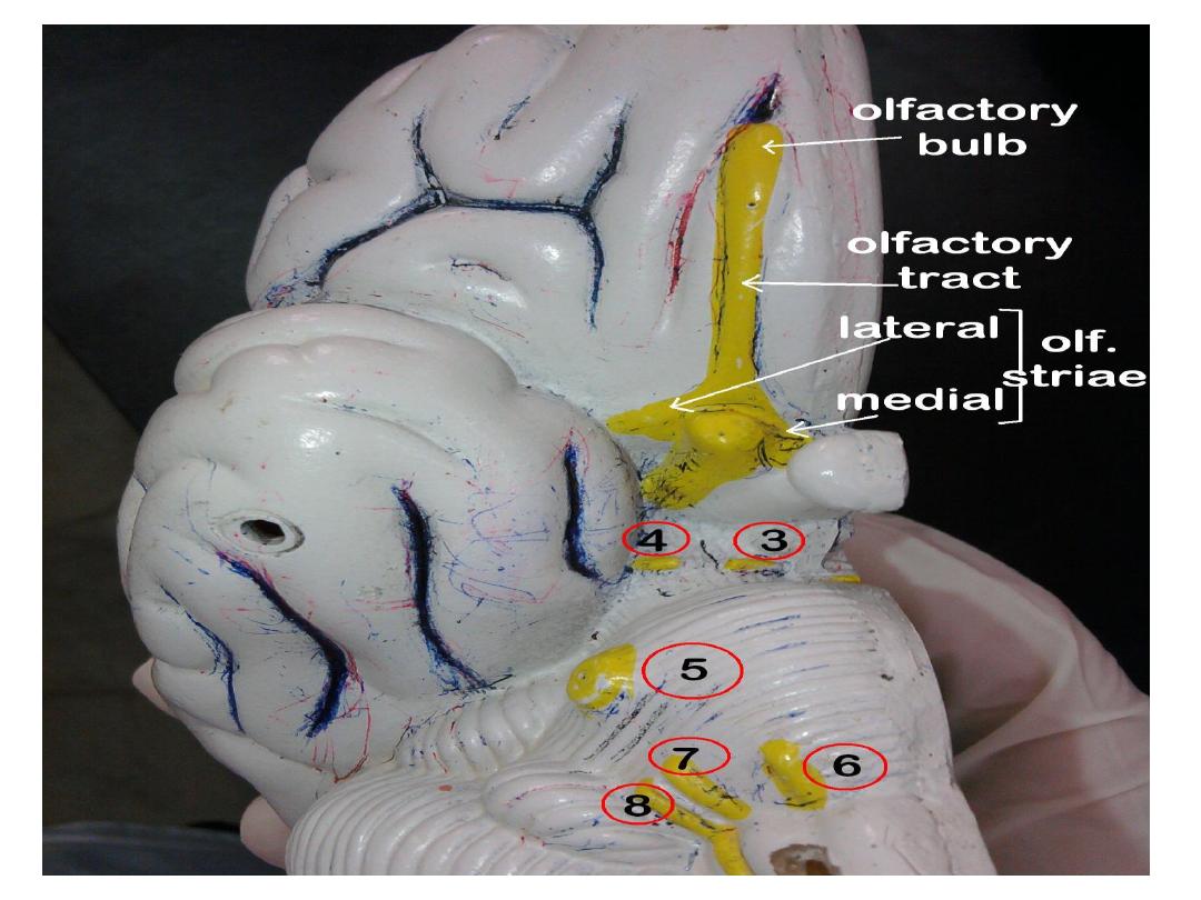
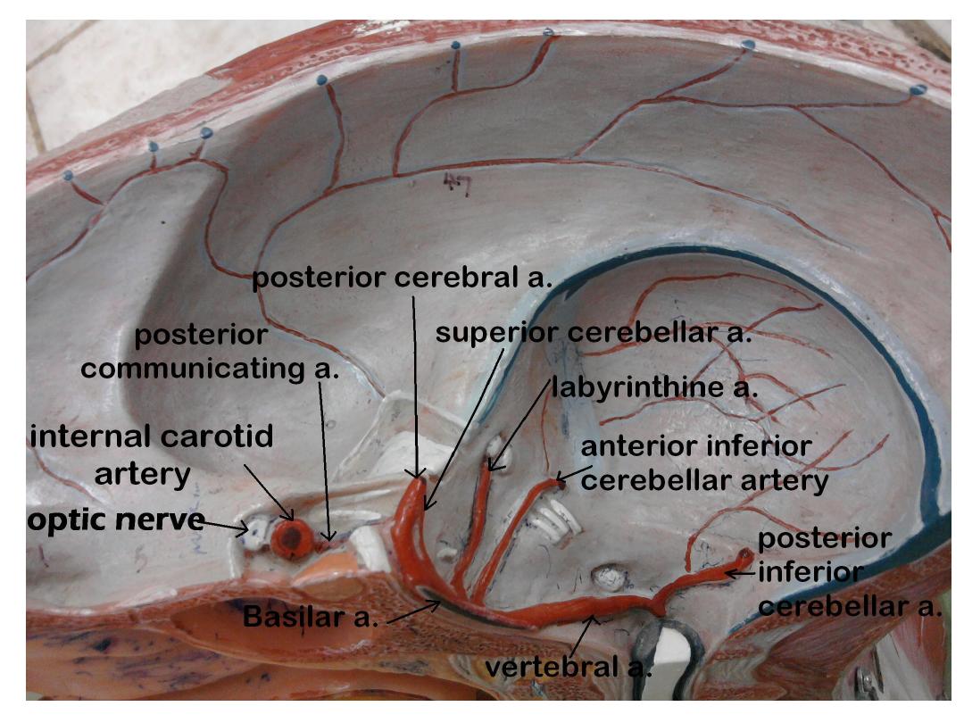
olfactory
tract
internal
carotid
artery
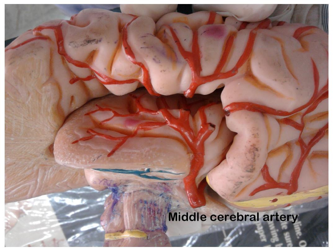
middle
cerebral
artery
anterior cerebral
artery
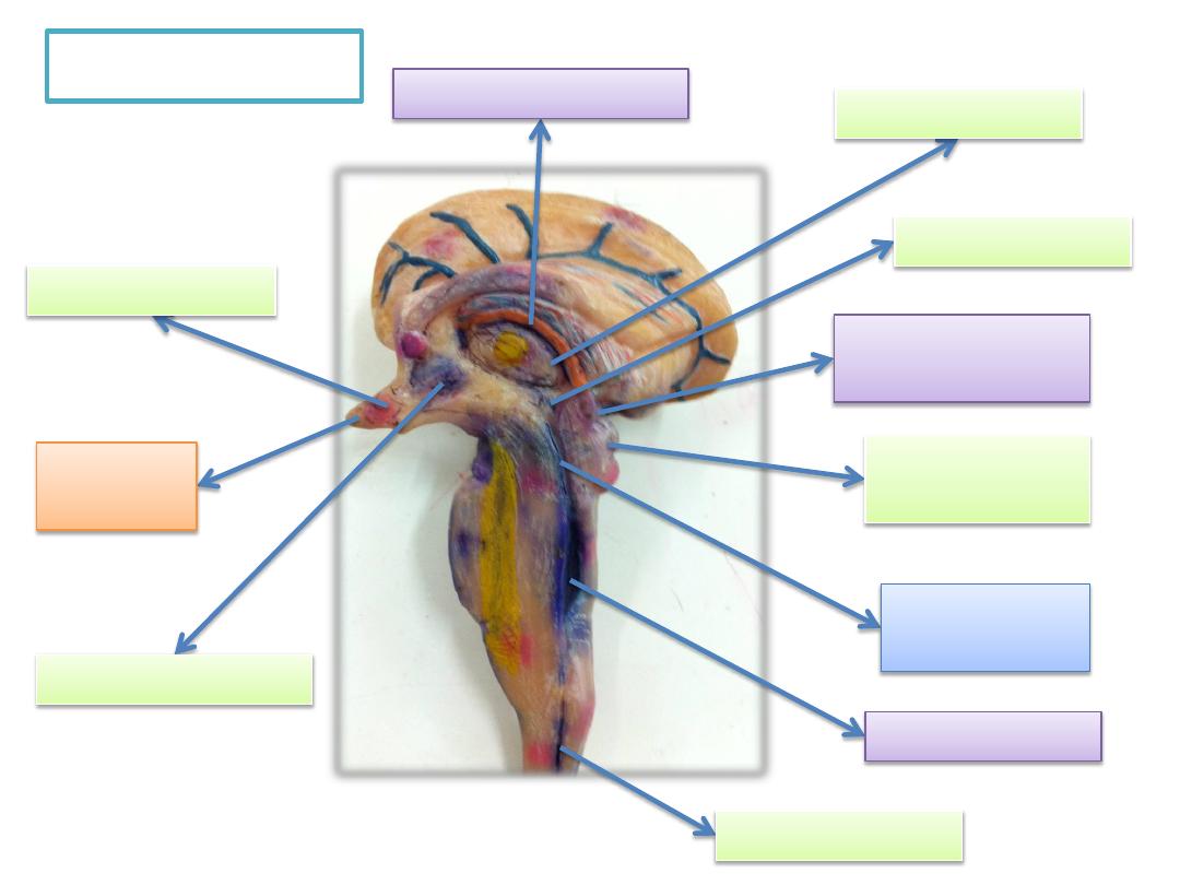
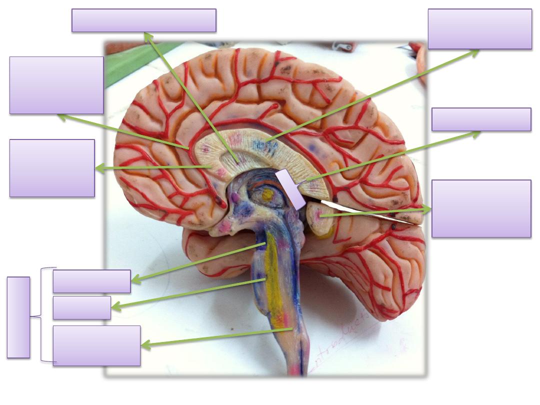
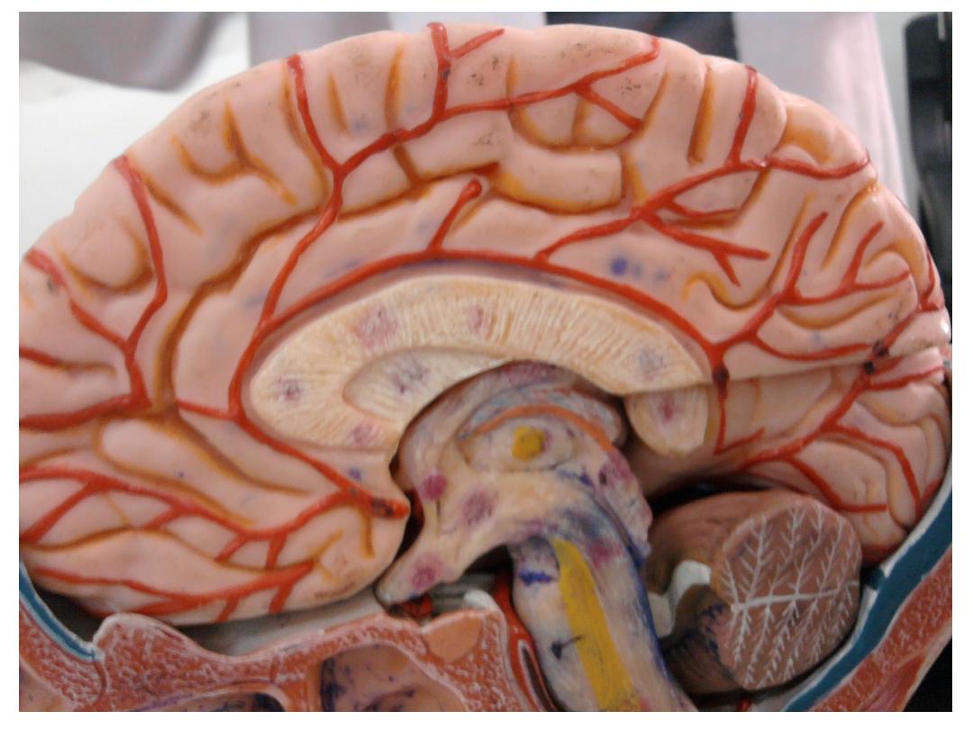
Medial view
central canal
4
th
ventricle
cerebral
equeduct
thalamus
hypothalamus
pineal body
(epithalamus)
3
rd
ventricle
choroid plexus
infandibulum
pitutary
gland
superior &
inferior colliculi
Middle cerebral
artery
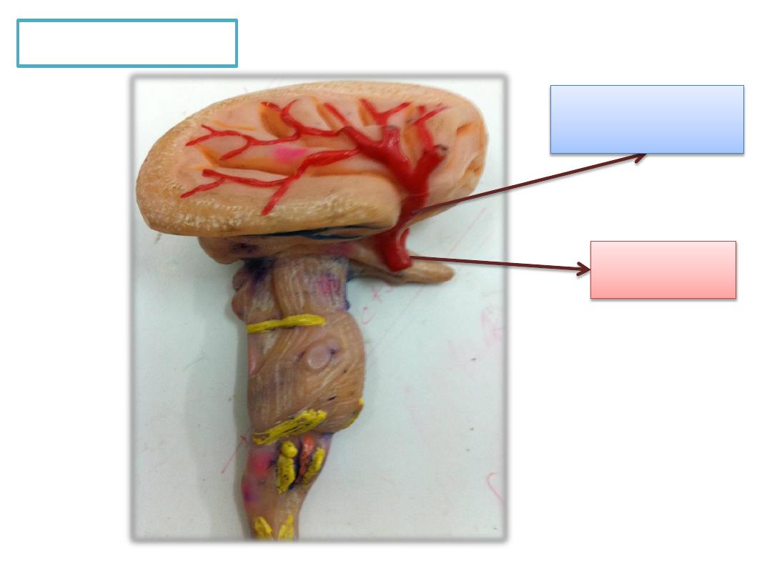
Lateral view
Internal
carotid artery
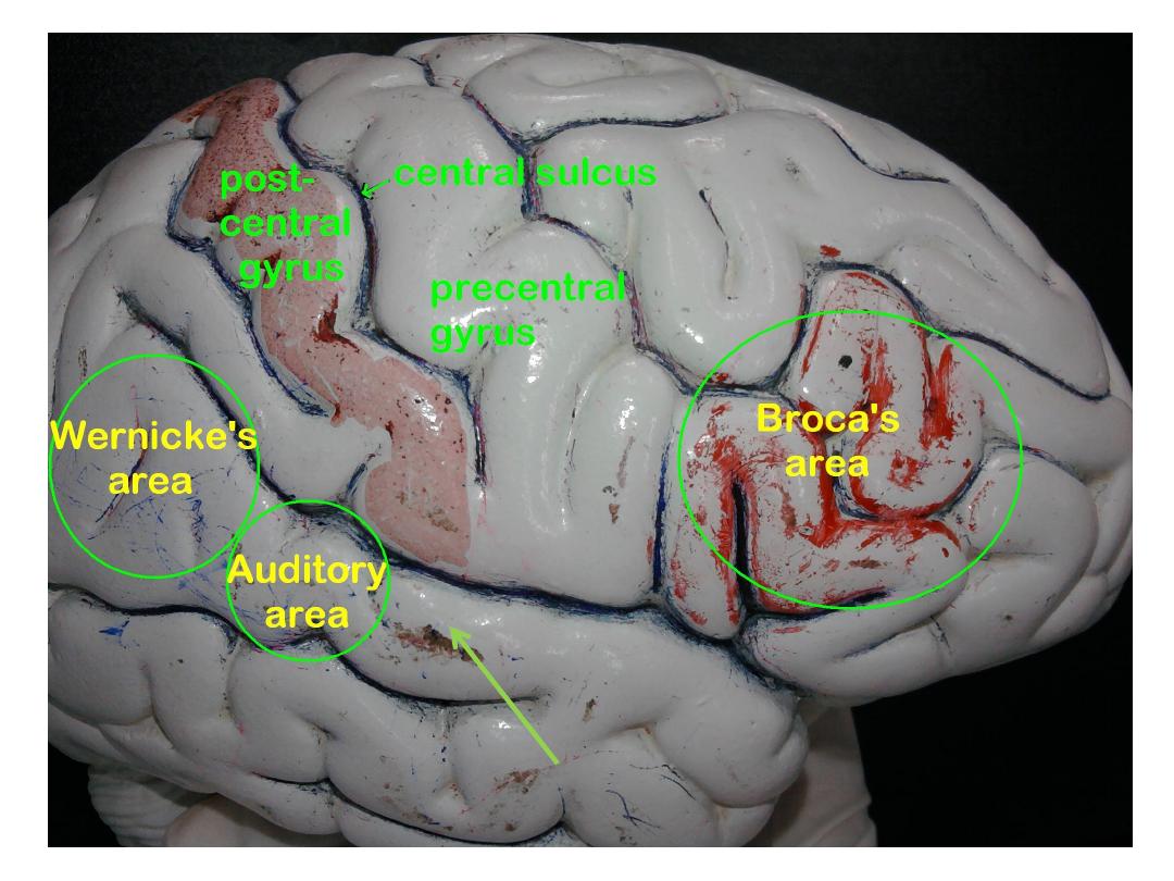
Lateral
sulcus
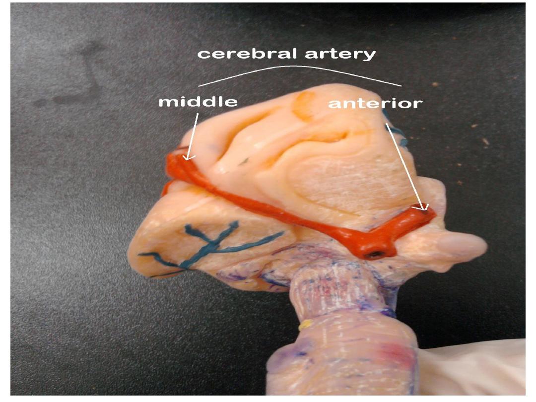
Anterior cerebral
artery
artery

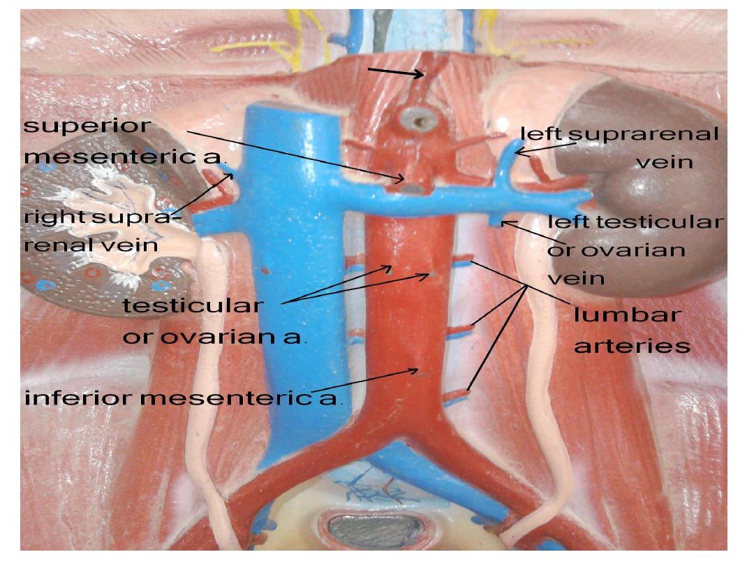
Inferior
phrenic artery
Superior mesenteric
vessels
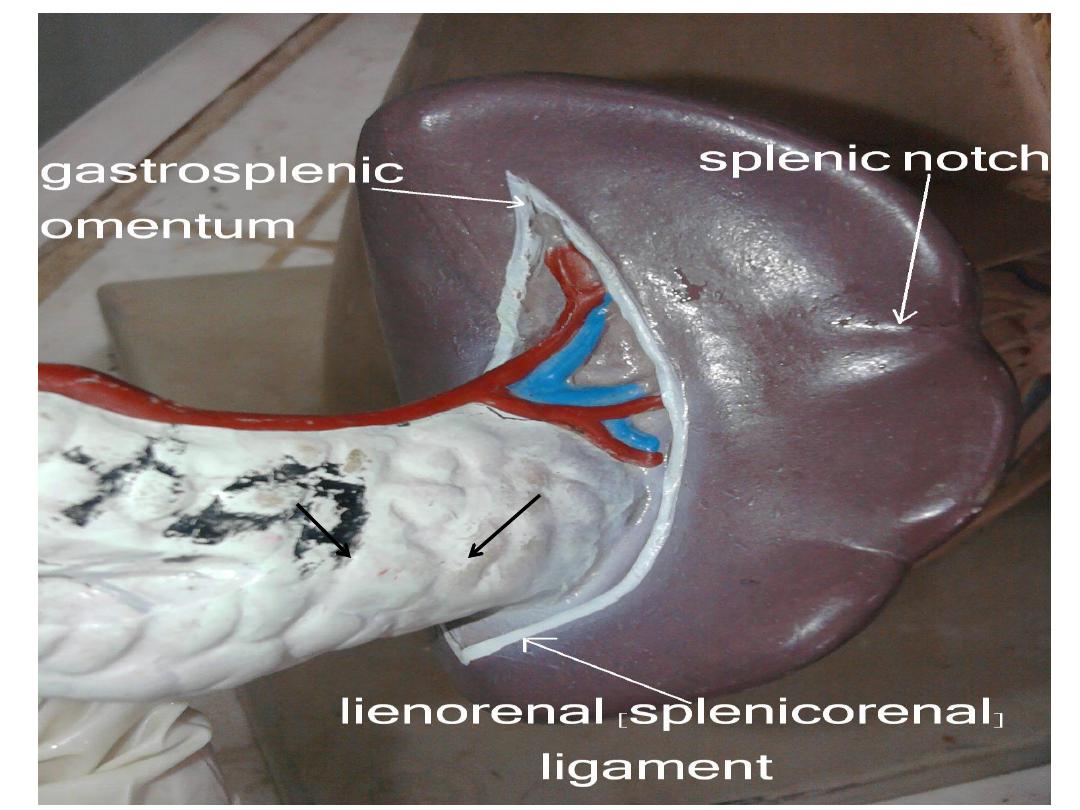
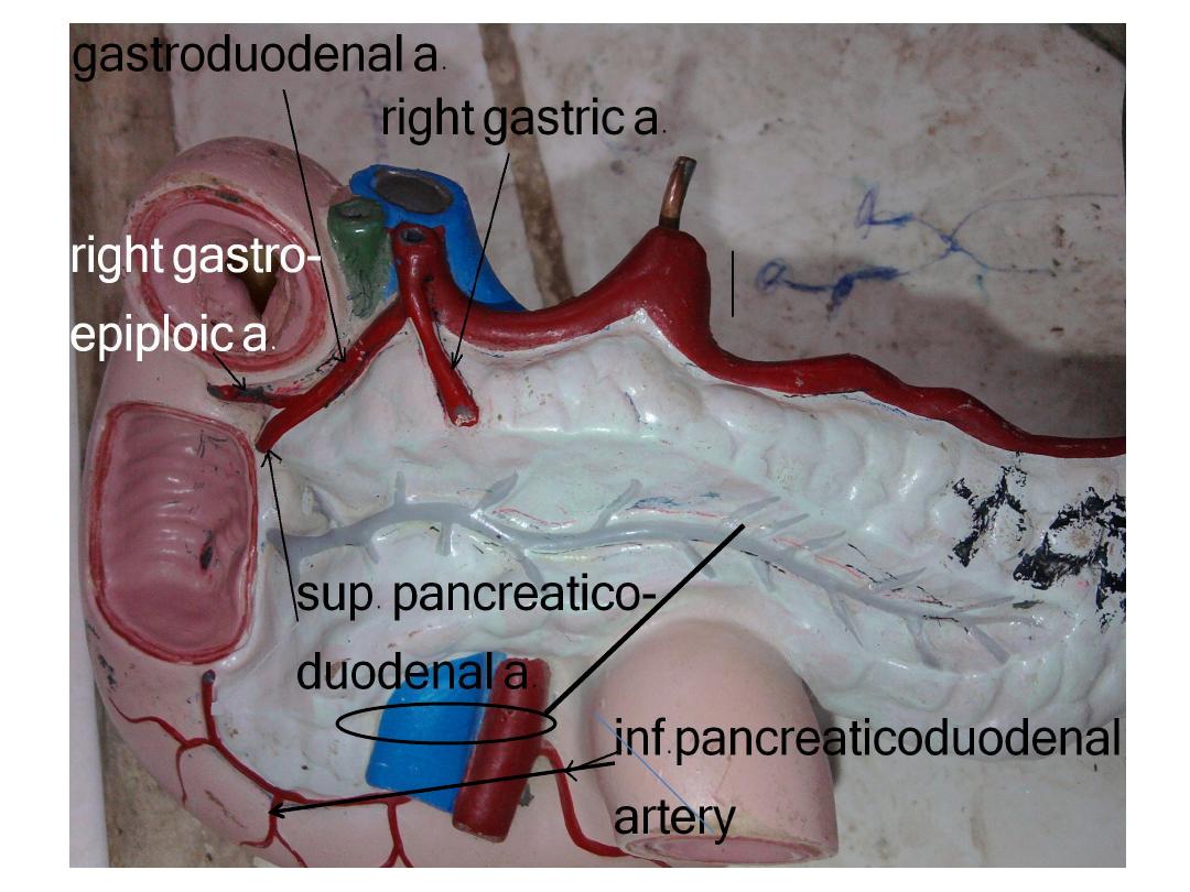
Tail
Head
Of Pancreas
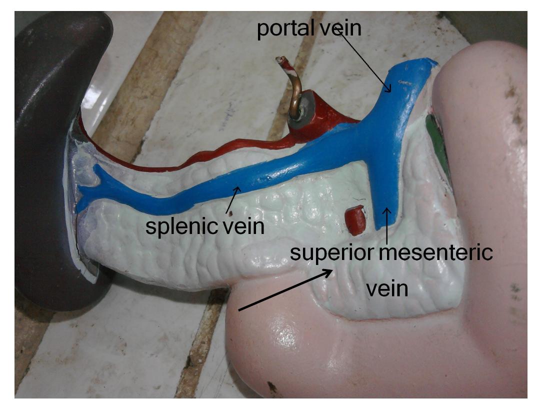
Uncinate process of
pancreas