Anatomy
For
Upper limbs
http://goo.gl/rjRf4F
I
LOKA
©
http://www.muhadharaty.com/anatomy-upper
I

Content
Topics:
Page:
The axilla
3
The breast
11
The arm
16
The forearm
24
The hand
29
Joints
32
Arteries
36
Make it easy!
38
Part1
: The Axilla
1- Is a pyramidal space between the upper part of
the arm & the lateral thoracic wall
2- It has :
4 walls (anterior , posterior , medial , lateral)
Apex
Base
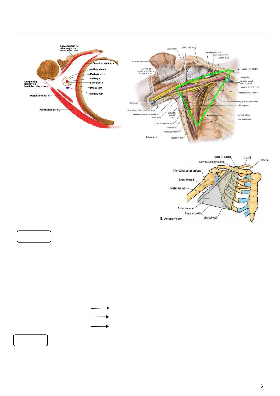
1- Is pointed upward in the direction of the root of the neck (to receive the brachial plexus)
2- It communicates with the superior aperture (inlet thorax) of thorax (to receive the
axillary artery –(continuity of subclavian artery)-)
3- Apex is known as "Cervico-axillary canal"
4- It allows the passage of the "Neuro-vascular bundle"( brachial plexus & Axillary artery)
to the upper limb
5- It has bony boundaries :
The clavicle anteriorly.
Outer border of the first rib medially
Upper part of the scapula posteriorl
1- Is formed by skin & superficial fascia of the axilla
2- Its bounded by ( anterior axillary fold anteriorly, posterior axillary fold posteriorly , the
chest wall medially )
The apex
The base
Note :
The lateral border of Pectoralis major M forms the anterior fold of the axilla.
The posterior fold forms by latissimus dorsi & teres major Ms
3- it is concave when the limb is beside the trunk
4- it becomes flat & straight on raising and abducting the limb due to suspensory ligament
of the axilla.
1- is formed by :
The clavicle
3 muscles ( Pectoralis major & minor Ms in addition to the subclavius M)
The Clavi-pectoral fascia
muscle
Origin (arises from) insertion
Nerve supply
(innervate)
Main actions
Pectoralis
major
1- clavicular head :
anterior surface
of medial half of
clavicle
2- sternocostal
head : anterior
surface of
sternum ,
superior six
costal cartilages
3- abdominal head
Outer lip of
the
intertubercul
ar
groove(crest
of Greater
tubercle )
Medial and
lateral
pectoral
nerves
Adducts and
medially rotates
humerus at
shoulder joint
Pectoralis
minor
From 3
rd
to 5
th
ribs
near their costal
cartilages
Superior
surface of
coracoid
process of
scapula
Medial
pectoral nerve
Produces the
depression of the
scapula
subclavius
From costal
cartilage of the first
rib
Subclavius
groove on
inferior
surface of
clavicle
Nerve to
subclavius
Anchors and
depresses clavicle
at steronclavicular
joint.
The anterior wall
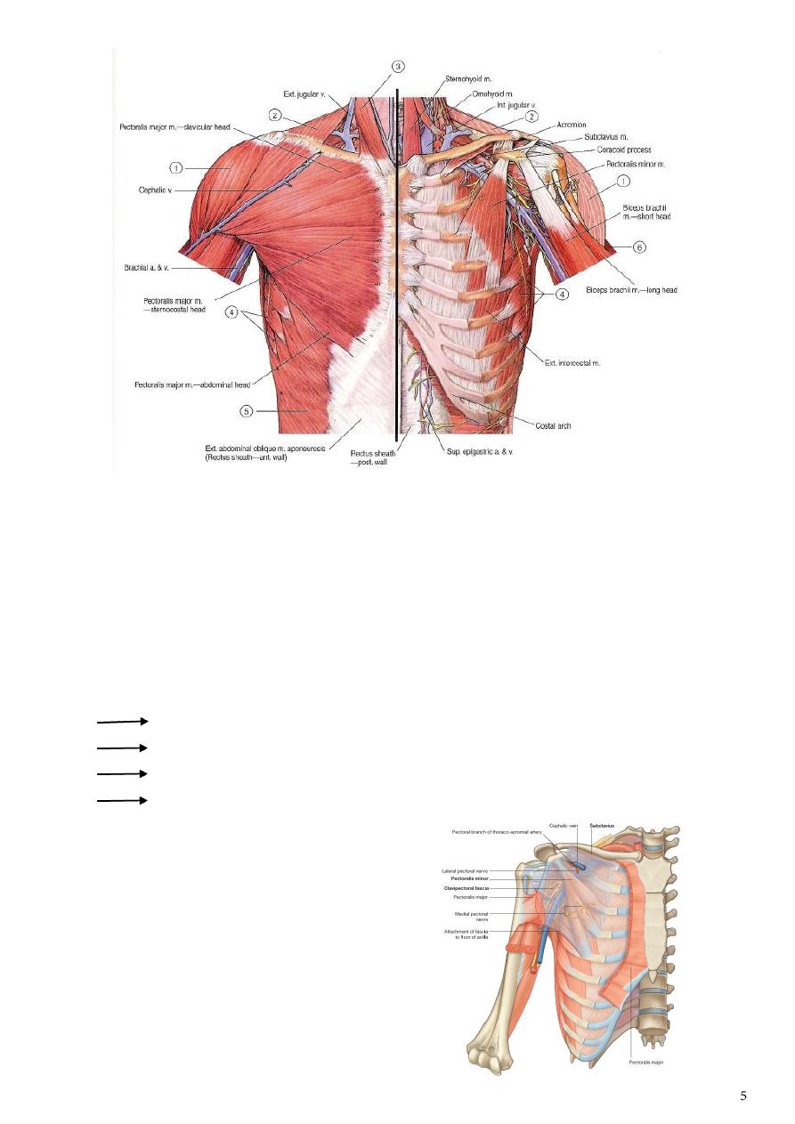
The clavipectoral fascia:
1- Is part of the deep fascia attached to the clavicle
2- it encloses the subclavius M
3- then descends down ward deep to Pectoralis major & enclosing Pectoralis minor M
4- ends as suspensory ligament of the axilla
5- Function : it protects the contents of the axilla by filling in the interval between the
clavicle and the Pectoralis minor M.
6- It is pierced by the following structures:
Lateral pectoral nerve.(as it passes to the Pectoralis major M.)
Cephalic vein
Pectoral branch of thoracoacromial artery.
Some lymphatic vessels

Is formed by 3 muscles, these are subscapularis, teres major & latissimus dorsi Ms.
muscle
Origin (arises from) insertion
Nerve supply
(innervated)
Main actions
subscapularis Subscapular fossa
of scapula
Lesser
tuberosity of
humerus
Upper and
lower
subscapular
nerve (from
posterior cord)
1- Medially
rotates
shoulder joint
and adducts it
2- Helps to hold
humeral head
in glenoid
cavity
Teres major
Posterior surface of
lateral border of
scapula near the
inferior angle
Medial lip of
intertubercular
groove (crest
of lesser
tubercle)
Lower
subscapular
nerve
Adducts and
medially rotates
shoulder joint
Latissimus
dorsi
A- Spines of T7_T12
vertebrae.
B- Thoracolumbar
fascia.
C- Iliac crest of the
Hip bone.
D- Inferior angle of
scapula
Into the floor
of
intertubercular
(Bicipital)
groove
The middle
subscapular
(thoracodorsal)
nerve
Adducts shoulder
joint
Elevates body
toward arms
during climbing
Is formed by:
The upper 4-5 ribs
And their intercostal spaces
And the upper part of serratus anterior muscle covering them
The posterior wall
The medial wall
Is formed by the intertubercular ( Bicipital ) groove containing the coracobrachialis M & short
head of Biceps
muscle
Origin
insertion
Nerve
supply
Main actions
serratus
anterior
External
(outer)
surfaces of
lateral parts of
1
st
to 8
th
-9
th
ribs
Anterior
surface of
medial border
of scapula
Long
thoracic
nerve
When it contract it pulls the scapula
forward around the rib cage / when its
upper or lower fibers contract
separately they help to produce
downward or upward rotation of the
scapula
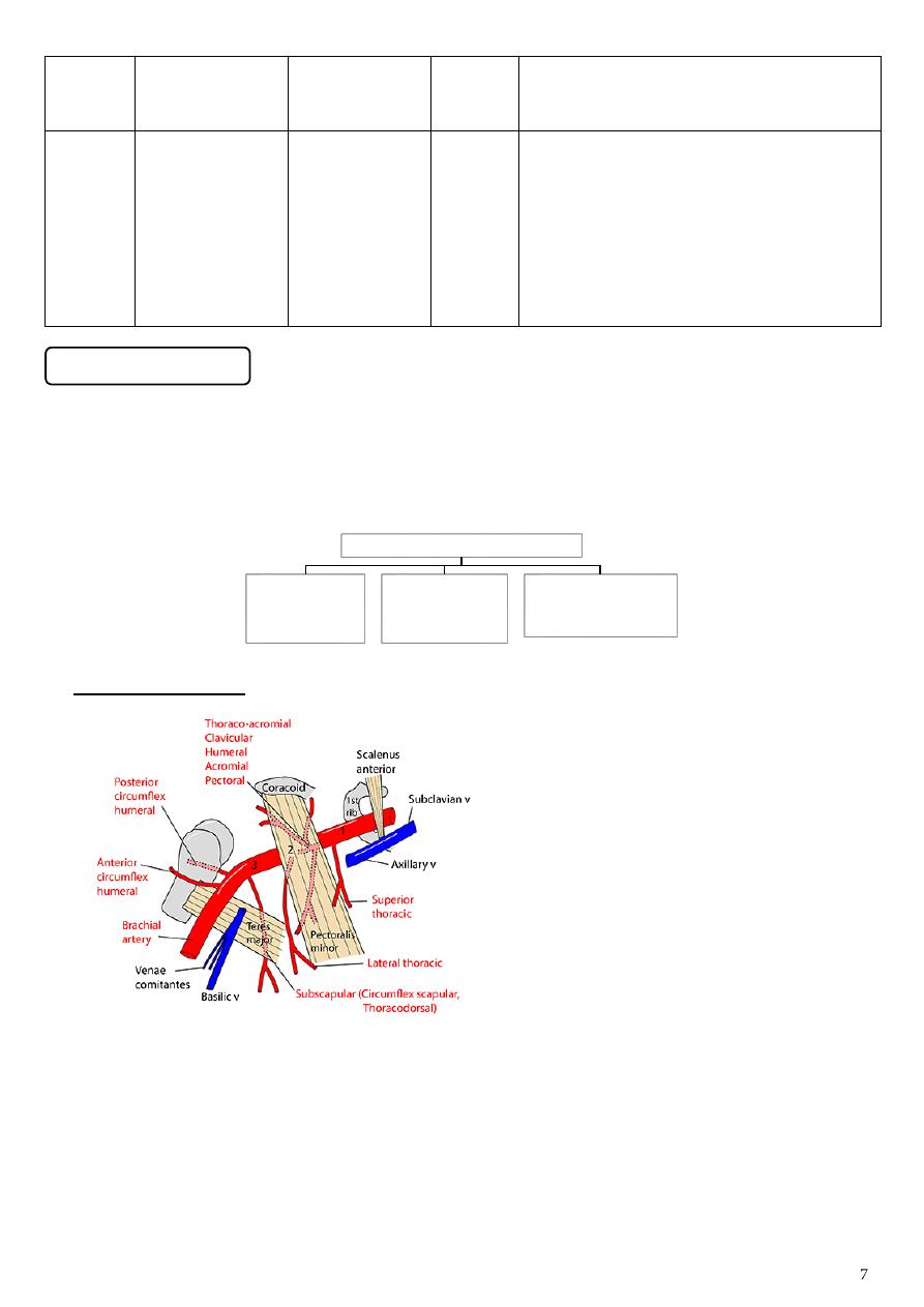
1-The axillary vessels:
1- The axillary artery, starts as the continuity of the subclavian artery at the outer border of the
first rib& ends at the lower border of teres major M( the lower limit of the axilla),where it
continue as the Brachial artery.
2- The axillary artery is closely related to the cords of the brachial plexus and their branches and it
is enclosed with them in a connective tissue sheath , called the "Axillary sheath"
3-
is crossed by the Pectoralis minor M ,which divides it into 3 parts
:
The lateral wall
The Contents of the Axilla
The axillary
vessels
The Brachial
plexus
The axillary lymph
nodes
The first part
The second part
The third part
1- Between outer
border of first rib
& the upper
border of
Pectoralis minor
M
2- it gives a single
branch known as
highest thoracic
or superior
thoracic A ( is
small and runs
along the upper
border of
Pectoralis minor)
3- Relations :
Anteriorly :
Pectoralis major
and the covering
fascia and skin .
the cephalic vein
crosses the
artery.
Posteriorly : long
thoracic nerve
(nerve to
serratus ant.)
Laterally : three
cords of the
brachial plexus
Medially : axillary
vein.
1- lies behind Pectoralis minor M
2- is related to the 3 cords of the
brachial plexus
laterally to lateral cord
medially to medial cord
posteriorly to the posterior cord
while anteriorly it is related to
Pectoralis minor M
3- Relations :
Anteriorly : Pectoralis minor ,
Pectoralis major , and the
covering fascia and skin
Posteriorly : posterior cord of
brachial plexus , subscapularis
muscle , and shoulder joint
Laterally : lateral cord of brachial
plexus
Medially : medial cord of the
brachial plexus and the axillary v
1- extends from lower border of
Pectoralis minor to lower border
of teres major muscle where it
continues as the Brachial artery
2- it is related to the derivatives of
the 3 cords of the brachial plexus
3- Relations :
Anteriorly : Pectoralis major for a
short distance , lower down the
artery is crossed by medial root of
median nerve.
Posteriorly : subscapularis ,
latissimus dorsi and teres major .
the axillary and radial nerve also
lie behind the artery
Laterally : coracobrachialis , biceps ,
and the humerus . the lateral root
of median nerve and
musculocutaneous nerve also lie
on the lateral side
Medially : ulnar nerve , axillary vein
, and medial cutaneous nerve of
the arm
Note :
1- Thoracoacromial artery : pierces the clavipectoral fascia and immediately divides into
terminal branches
2- Lateral thoracic artery : runs along the lower border of Pectoralis minor
3- Subscapular artery : runs along the lower border of subscapularis muscle
4- Anterior and posterior circumflex humeral arteries: run around the front and the
back of the surgical neck of the humerus.
It gives 2
branches
thoracoacromia
l (it gives 4
branches)
2 of them to
bones (acromial
& clavicular)
other 2 to
muscles (Deltoid
& pectoral
branches).
lateral thoracic
descends to the side
of the chest wall to
accompany the long
thoracic nerve
within the substance
of serratus anterior
muscle.
it gives 3 branches
subscapularis
thoracodorsal
branch
circumflex
scapular branch
anterior &
posterior
circumflex
humeral arteries
around the
surgical neck of
the humerus
5- Axillary vein :
it is formed in the region of the lower border of teres major muscle by the union of the
venae comitantes of the brachial artery and basilic vein
it runs upward on the medial side of the axillary artery
and ends at the lateral border of the first rib becoming the subclavian vein
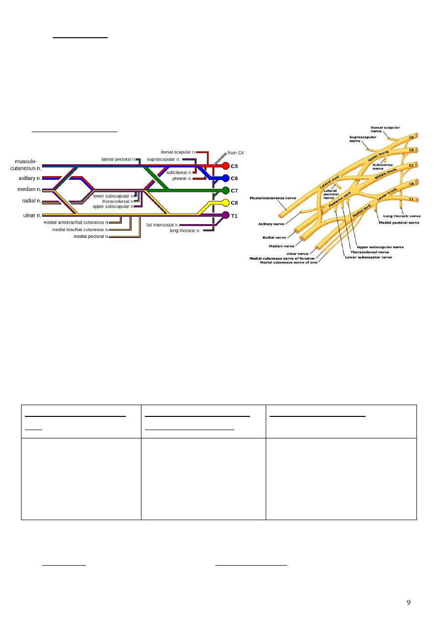
2-The Brachial plexus:
1- It is formed by :
the ventral rami of lower 4 cervical nerves
& the ventral ramus of the first thoracic nerve.
2- The first stage is roots arrangement to form trunks (C5& 6th form the upper trunk, C 7
alone forms the middle trunk while C 8 & T 1 form the lower ( inferior ) trunk .The
second stage is the splitting of each trunk to form anterior & posterior divisions. The
third stage is the formation of the 3 cords by the Re-union of these divisions .The
posterior divisions of the 3 trunks unite to form the posterior cord ,the anterior division
of the upper & middle trunks unite to form the lateral cord, while the anterior division of
inferior trunk Forms the medial cord of the brachial plexus.
3- The last stage is the derivatives of each cord as follows:
The posterior cord gives
off: (ULTRA)
The lateral cord gives the
following derivatives:(2ML)
The medial cord gives: (4MU)
1- Upper subscapular.
2- Middle subscapular
(Thoracodorsal).
3- Lower subscapular
4- Axillary nerve.
5- Radial nerve.
1- Lateral pectoral nerve.
2- Musculocutaneous
nerve.
3- Lateral root to median
nerve.
1- Medial pectoral nerve.
2- Medial cutaneous of Arm.
3- Medial cutaneous of forearm.
4- Ulnar nerve.
5- Medial root to median nerve.
4- In addition to these derivatives ,the upper trunk gives 2 branches suprascapular & nerve
to subclavius muscle while the roots gives dorsal scapular & long thoracic nerve (C5-7)
(Roots branch : 1- dorsal scapular 2- Long thoracic // Upper trunk branches : 1-Subclavian n. 2- Suprascapular n
)
5- At the end of the brachial plexus the four main nerves of the arm emerge
(musculocutaneous , median , ulnar, radial)
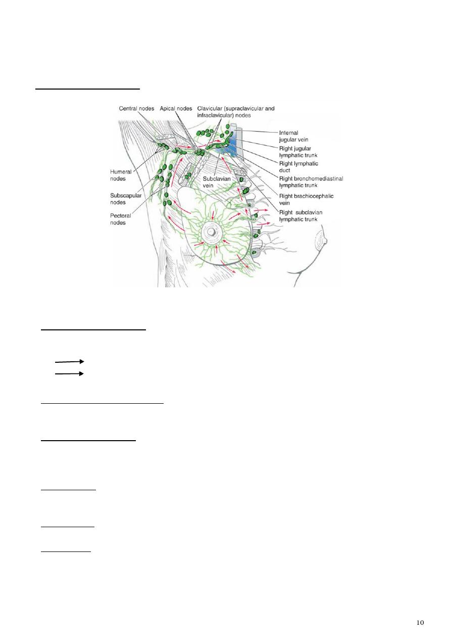
3-The axillary lymph nodes:
Which are arranges in the following groups:
1- Anterior ( pectoral ) group
Under anterior border of Pectoralis major M.
Receives lymph vessels from :
lateral quadrants of the breast
Superficial vessels from the anterolateral abdominal wall above the level of the
umbilicus
2- Posterior ( subscapular ) group
Along the course of subscapular vessels.
Receives superficial lymph vessels from the back , down as far as the level of the iliac crests
3- Lateral (humeral) group
Along the course of the axillary V near Bicipital groove.
Receives most of the lymph vessels of the upper limb (except those superficial vessels draining
the lateral side)
4- Central group
Within the loose areolar tissue of the base of the axilla.
Receives lymph from the above three groups
5- Medial group
Along the course of lateral thoracic vein.
6- Apical group
in the apex of the axilla
It receives lymphatic from the above groups & takes them (direct them) to the deep cervical
nodes in the root of the neck.
Part2
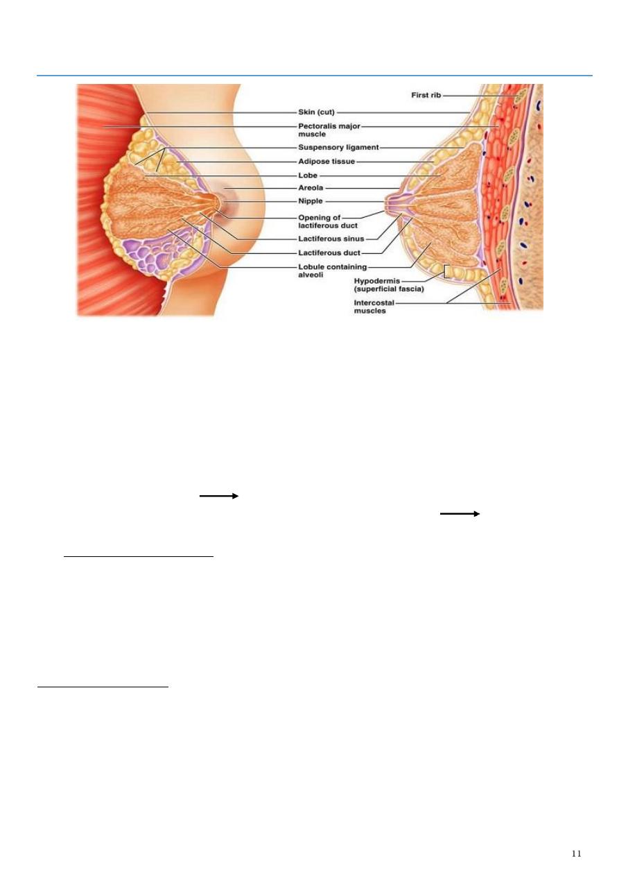
: The Breast
1- Is rudimentary in male & well developed in the female specially in lactating woman
2- It is a modified sweat gland
3- Located :
- under the superficial fascia covering the pectoral region
- and lying on the deep fascia covering Pectoralis major & part of the serratus anterior Ms.
4- It extends from the side of the sternum medially to the anterior axillary fold laterally
5- ( part of it extends into the axilla as axillary tail of the breast)
6- while supero-inferiorly it extends from the level of 2nd rib to the 6th rib.
7- The gland consists of 15-20 lobes extending from the periphery of the gland to the
area near the nipple
Each lobe has its own duct( lactiferous duct) which
opens externally in to the nipple( has about 15-20 openings)
The nipple is a
small conical projecting part surrounded by a lighter area ( Areola)
o The breast is supplied by:
1- Pectoral branch of thoracoacromial artery.
2- Mammary branches from the lateral thoracic artery.
3- Perforating branches from the internal thoracic artery (i.e internal mammary A ).
4- Branches from intercostal arteries for the spaces 3rd-5th.
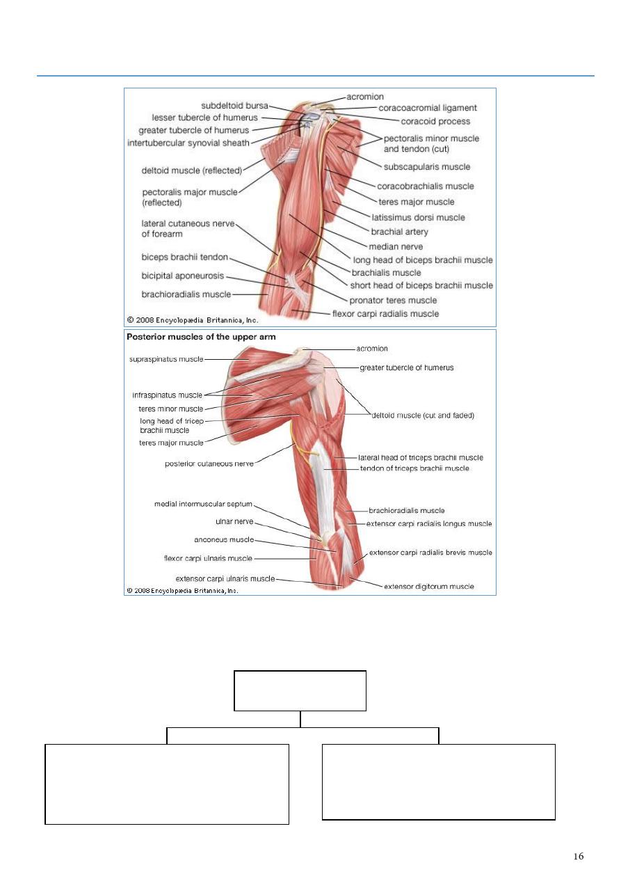
Rotator Cuff Muscles:
- are 4 in number surrounding the capsule of the shoulder joint to support &share in
stabilizing the shoulder joint
- One of these Ms. inserts into the lesser tuberosity ( Subscapularis )
- the other 3 are inserted into the greater tuberosity (Supraspinatus ,Infraspinatus & teres
minor muscles)
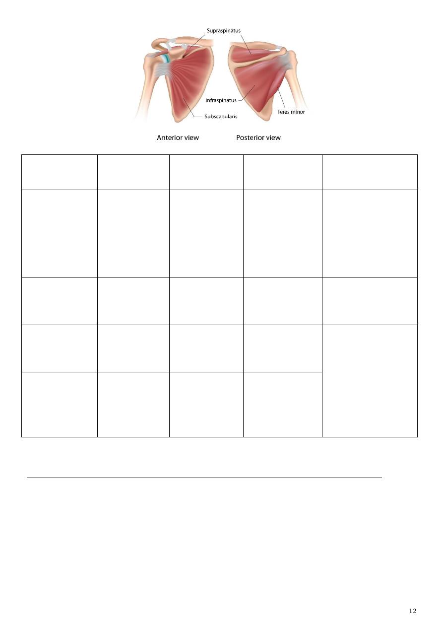
muscle
Origin (arises
from)
insertion
Nerve supply
(innervated)
Main actions
subscapularis
Subscapular
fossa of scapula
Lesser
tuberosity of
humerus
Upper and lower
subscapular
nerve (from
posterior cord)
1- Medially rotates
shoulder joint and
adducts it
2- Helps to hold
humeral head in
glenoid cavity
Supraspinatous
Supraspinous
fossa of scapula
superior facet of
greater
tuberosity
Suprascapular
nerve
Abduct the humerus
(shoulder joint)
Infraspinatous
Infraspinous
fossa of scapula
Middle facet of
greater
tuberosity
Suprascapular
nerve
1- Laterally rotates
shoulder joint
2- Helps to hold
humeral head in
glenoid cavity of
scapula
Teres minor
Superior part
of lateral
border of
scapula
Inferior facet
of greater
tuberosity
Axillary nerve
The muscles responsible for Abduction movement of the arm at shoulder joint are:
1- From 0 – 18 degree by Supraspinatous muscle.
2- 18—90 degree by Deltoid muscle(innervated by the Axillary nerve ).
3- Beyond 90 degree & above the head is by Trapezius & Serratus anterior muscles.
The muscles attaching the limb to the back
muscle
Origin (arises from) insertion
Nerve supply
(innervated)
Main actions
Trapezius
1- From medial
third of the
superior nuchal
line.
2- From
ligamentum
nuchae.
3- From the spine
of C 7 vertebra
4- From spines of
T1 –T 12
vertebrae
- into the front
of lateral third
of the clavicle
- acromion
process
- upper lip of the
spine of the
scapula.
the spinal root of
accessory
nerve(11th
cranial
nerve)which is
motor ,while
proprioception
sensations from
C4 & C5 nerves.
Elevates,
retracts, and
rotate the
scapula.
Depresses
scapula.
Superior rotation
of scapula.
Levator
scapulae
transverse
processes of upper
4 cervical vertebrae.
In the area
around the
superior angle
of scapula
dorsal scapular
nerve from the
ventral ramus of
C5.
Abduct the
humerus
(shoulder joint)
Rhomboid
minor
spines of C 7 & T 1
vertebrae
dorsal aspect of
vertebral border
of scapula at the
base of the
spine.
Dorsal scapular
nerve.
1- Retract
scapula and
rotate it to
depress
glenoid

Rhomboid
major
spines of T 2 – T 5
vertebrae
into dorsal
aspect of
vertebral border
below the base
of the spine till
inferior angle of
scapula
dorsal scapular
nerve
2- Fix scapula to
thoracic wall
Latissimus
dorsi
A- Spines of T 7 __T
12 vertebrae.
B- Thoracolumbar
fascia.
C- Iliac crest of the
Hip bone.
D- Inferior angle of
scapula
Into the floor of
intertubercular
(Bicipital)
groove
The middle
subscapular
(thoracodorsal)
nerve
Adducts
shoulder joint
Elevates body
toward arms
during climbing
Note :
Leveator scapulae , Rhomboid major and minor :
1- are known collectively as Elevators of the scapula( one of them is levaetor scapulae)
2- all the 3 has a common nerve supply( dorsal scapular nerve)
3- all the 3 has a common action ( all of them work in elevating the scapula)
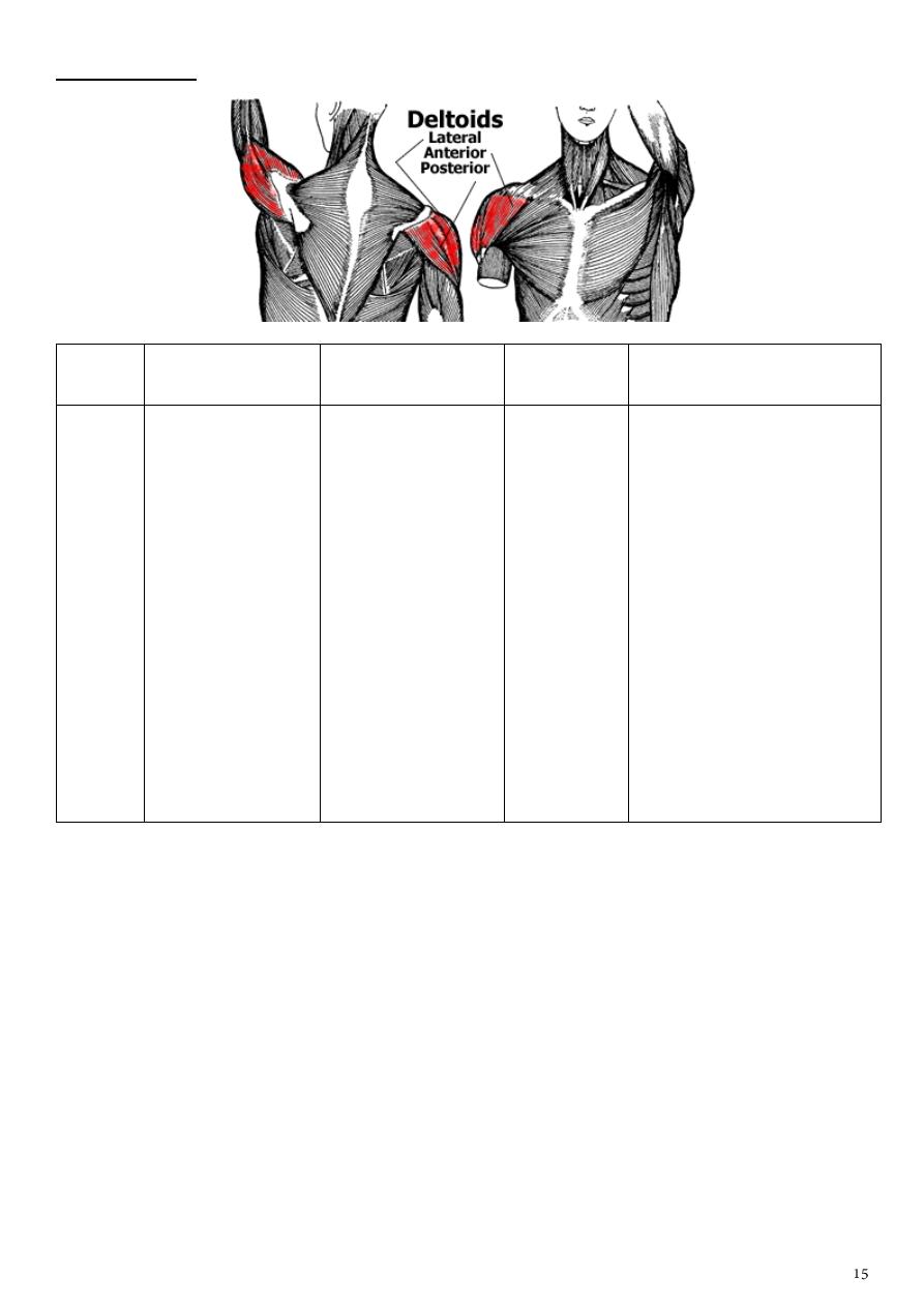
Deltoid muscle:
muscle
Origin (arises
from)
insertion
Nerve
supply
Main actions
Deltoid
the same areas of
the insertion of
the trapezius M
- the inferior
aspect of the
crest of spines of
scapula
- acromion
process
- lateral
third of the
clavicle
The M fibers from
the 3 sites of
origin converted
into a single
tendon of
insertion & being
inserted into the
Deltoid tuberosity(
on the lateral
aspect of the
middle part of the
humerus).
Axillary
nerve
1- flexion of arm at
shoulder joint(anterior
fibers)
2- extension of the arm
(posterior fibers)
3- abduction of the arm
at shoulder ( middle
fibers)
4- in fact it is considered
as powerfull & main
abductor M of the arm(
from 18-90 degree)
Note : if the Axillary nerve is injured or compressed by local haematoma due to fracture
at the surgical neck of the humerus ,abduction becomes impossible because of loss of
innervation of the deltoid M
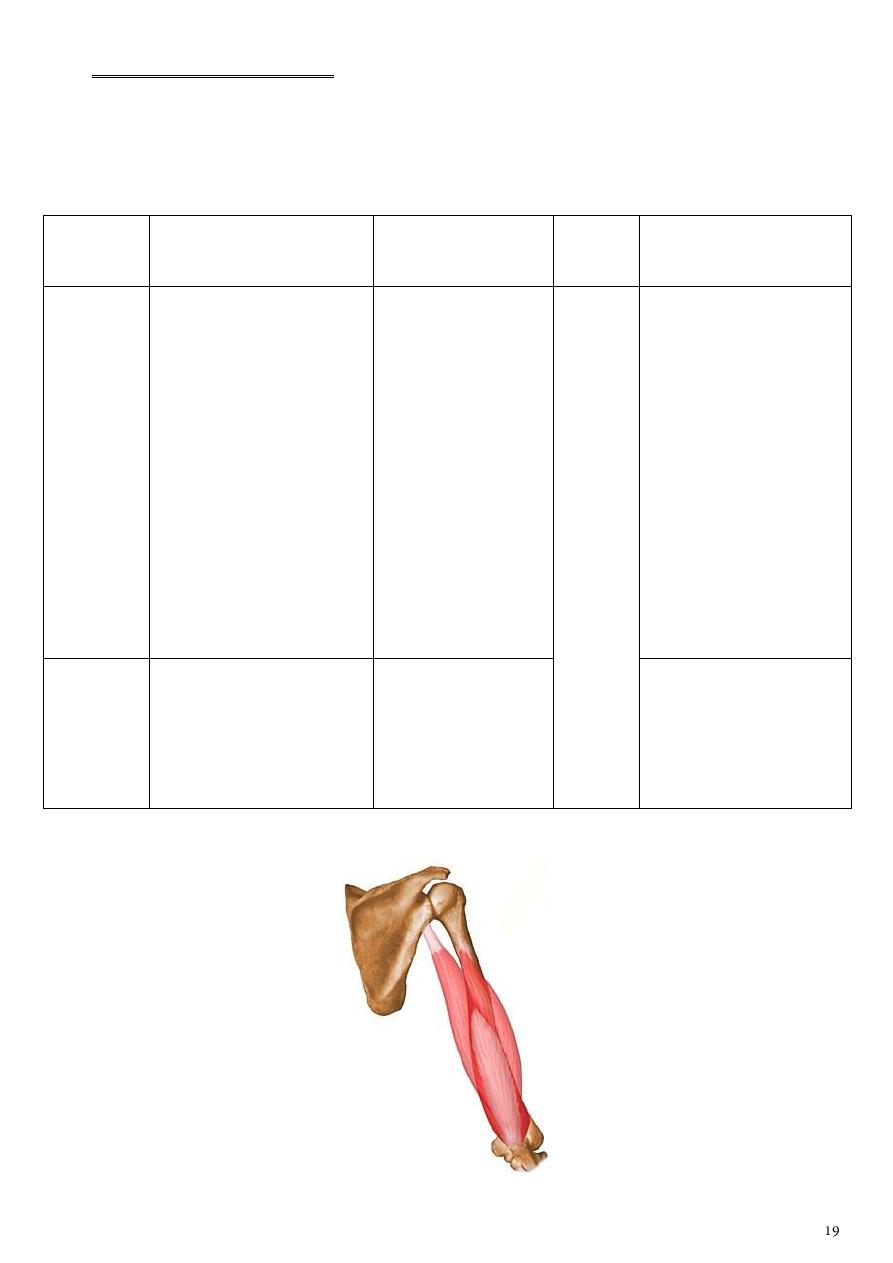
Part3
: The Arm
- The arm is surrounded by deep fascia ( brachial fascia )
- This fascia sends 2 septae attaching to medial & lateral compartments (muscles)
compartments
(muscles)
anterior ( B.B.C muscles)
which are flexors at the Elbow joint)
these Ms are Biceps brachi,Brachialis &
Coracobrachialis.
posterior compartment
is an Extensor compartment at the
Elbow joint mainly Triceps M & lower
down a small M (Anconeus M ).

A. The anterior compartment
contains the followings:
1- Muscles : The 3 Ms. ( B.B.C .)
2- blood : The Brachial Artery.
3- Nerve : Three nerves ( the Musculocutaneous ,Median & Ulnar nerves)
4- The stracture passing through :
1- musclucotanous nerve 2- median nerve 3- ulnar nerve
4- brachial artery 5- basilic vein 6- radial nerve (lower compartment)
muscle
Origin
insertion
Nerve
supply
Main actions
Biceps brachii
-
long head: from
supraglenoid
tubercle of the
scapula,
-
short head: from
coracoid process
of scapula in
common origin
with the
Coracobrachialis
muscle
the 2 head leads
to a common
belly of the
muscle before
going to insert (
by a single
tendon) into the
radial tubercle of
the radius bone
Biceps is a
powerful supinator
of the forearm in
addition to flexion
at the Elbow joint.
Brachialis
anterior surface of
the lower part of
the shaft of the
humerus
its insertion into
the front(anterior
surface) of
Coronoid process
of the Ulna bone
It is a flexor at
Elbow joint along
with the Biceps M.
Coracobrachialis Coracoid process
of the Scapula in
common with the
short head of
Biceps
on the medial
aspect of upper
part of the shaft
of the humerus.
Assists in flexion
and adduction of
shoulder joint.
Notes:
1- The three B.B.C muscles are supplied by musculocutaneous nerve ( from lateral cord ).It
is motor to the 3 Ms. & sensory to the skin on the lateral side of the F.A (How?) , where
after supplying motor to the 3 Ms. of anterior compartment of Arm ,it will continue as
lateral cutaneous nerve to the forearm.
2- The Musculocutaneous nerve motor component is only to the 3 Ms. of the anterior
compartment , while both the Median & Ulnar are not supplying any structure in the
Mu
sculocut
an
eou
s N
( fr
om
lat
er
al
cor
d
).
Arm ,these 2 nerves just passing through the anterior compartment to reach their areas
of destination( supply) in the F.A & the Palm of the Hand.
Bicipital Aponeurosis
As the tendon of Biceps passes through the front of Elbow (Cubital fossa) in its way to its
insertion site
it sends a flat ribbon like Aponeurosis medially & superficially to
cover the terminal part of the brachial A+ the Median nerve known as Bicipital Aponeurosis.
The Sensory Nerve supply of the Arm:
1- Medial cutaneous nerve of Arm from the Medial cord of the Brachial Plexus.
2- Upper lateral cutaneous of Arm from the Axillary nerve.
3- Lower lateral cutaneous nerve of Arm from the Radial nerve while running in the
spiral ( Radial ) groove on the back of the shaft of the Humerus.
4- Posterior cutaneous nerve of Arm from Radial nerve as it descends through the Axilla.
The Ulnar nerve
runs in the anterior
compartment
then inters the medial intermuscular septa
(leave the arm ) to reach the F.A by passing
behind the medial Epicondyle of the
humerus( by passing the cubital fossa)
then it passes through the
2 heads of origin of flexor
carpi ulnaris M of the F.A.
The Median nerve
is formed on the
anterolateral aspect
of the beginning of
the brachial A
then it becomes on
the lateral side of
the upper third of
the Brachial A
then crossing obliquely in front of the
middle third of the A ( from lateral to
medial ) to becomes on the medial side
of the lower third of the Brachial A
then both the terminal part
of the Brachial A + the
Median nerve are sheltered
by Bicipital aponeurosis
within the Cubital fossa
B. The posterior compartment
It contains:
1- mainly the Triceps M
2- the Radial nerve
3- Profunda Brachi A
4- and just inferiorly near the back of the Elbow joint, there is a small superficially placed slightly
triangular muscle known as "Anconeus"
muscle
Origin (arises from)
insertion
Nerve
supply
Main actions
Triceps
brachii
-
The long head: from
infraglenoid tubercle of
the scapula
-
The lateral head: from
posterior aspect of the
shaft of the Humerus
above the spiral groove
-
the Medial head: from
the back of the humerus
below the spiral groove
& slightly from the
medial side of the
Humerus
The muscle fibers of
the 3 heads of origin
converge inferiorly
into a single tendon
which is inserted
into the top of the
Olecranon process
of the Ulna bone
it is a powerful Extensor
muscle at the Elbow
joint.
Anconeus
Lateral epicondyle of
humerus
Lateral surface of
Olecranon and
superior part of
posterior surface of
ulna
assists triceps in
extending elbow joint,
stabilizes elbow joint ,
adducts ulna during
pronation
Radi
al
Ner
ve
Notes:
1- The spiral groove separates the lateral head from the medial head of the Triceps.
2- The Triceps is supplied by many branches from the Radial nerve at different levels.
The Intermuscular Spaces
Note :
-
The Axillary nerve as it passes through the Quadriangular space ,it divides into:
anterior division : supplies the major part of the Deltoid M,
posterior division :supplies teres minor ,the remaining part of the Deltoid and then
continues as upper lateral cutaneous nerve of the Arm ( supplies the skin on the upper
lateral part of Deltoid)
There are 2 main
Triangular spaces
a superior horizontal one
- between:
1- teres minor ( above )
2- teres major ( below )
3- the Surgical neck of the Humerus laterally.
- This space is divided by the long head of
Triceps into:
a smaller triangular space
medially
- between
1- teres minor
2- teres major
3- long head of Triceps
- transmits the Circumflex scapular
branch of the Subscapular artery)
a lateral Quadriangular space -
bounded by :
1- teres minor above
2- teres major below
3- long head of Triceps mediall
4- the Surgical neck of the
Humerus laterally
- transmits the Axillart nerve &
posterior circumflex humeral A.
The inferior vertical Triangular
space
- between:
1- teres major ( above )
2- long head of Triceps (medially )
3- the side of the Humerus.
It transmits the Radial
nerve & Profunda Brachi
A (a branch from the
Brachial A ).
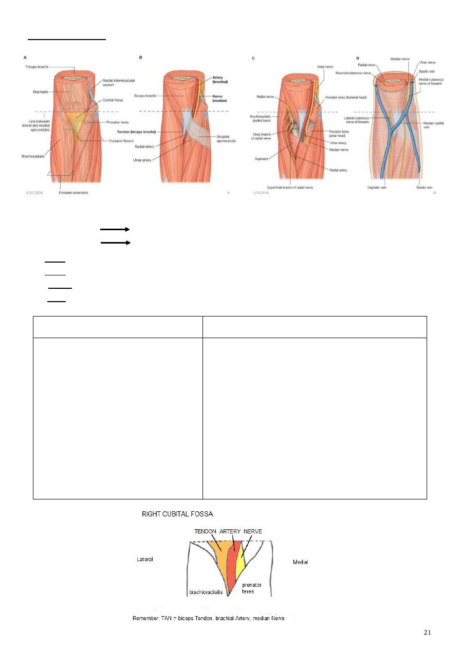
The Cubital Fossa
-
Is a triangular depressed space in front of the Elbow joint
-
It is bounded
by : the Pronator teres medially
Brachioradialis laterally
-
its base is formed by an imaginary line joining the 2 epicondyles of the humerus
-
its apex is formed as brachioradialis M crosses over the pronator teres
-
The floor is formed by the insertion of Brachialis M & the supinator M below it
-
its roof is formed by the skin & fascia
-
The contents includes 2 groups as follows:
A-The superficial contents are
B-Deep group of structures includes
1-Median Cubital vein joining the
Cephalic & Basilic veins.
2-Lateral cutaneous nerve of the F.A
laterally.
3-Medial cutaneous nerve of F.A
medially.
4-Bicepital Aponeurosis .
5-Some superficial lymphatic vessels
& lymph nodes
1-The termination of the Brachial A & its
bifurcation into Radial and Ulnar As.
2-The Median nerve just medial to the terminal
part of the Brachial A.
3-Tendon of Biceps Brachii in its way to reach its
insertion site.
4-Radial nerve laterally emerging in the groove
between Brachialis & Brachioradialis.
THE BRACHIAL ARTERY
-
Is the direct continuity of the Axillary artery at the lower border of Teres major M
-
it runs in the anterior compartment of the Arm & ends opposite the Neck of the Radius
bone ( in the Cubital fossa ) by dividing into Ulnar & Radial As
-
It gives the following branches:
1- Profunda brachi ( Deep brachial , deep artery of arm)
which goes to the Radial( spiral ) groove on the back of the shaft of the humerus
accompanies by the Radial nerve ( after the Axilla)
2- Superior ulnar collateral A which accompanies the Ulnar nerve into the medial
intermuscular septum.
3- Nutrient branch to the Humerus bone .
4- Muscular branches to supply B.B.C muscles of anterior compartment of Arm.
5- Inferior ulnar collateral which arises just above the Cubital fossa& goes to the medial
intermuscular septum to join the Ulnar N.
The A divides into 3 smaller branches while running in the spiral
groove
ascending branch to reach
surgical neck of humerus &
share in the anastomosis there
with both anterior & posterior
circumflex humeral A from 3rd
part of Axillary A
the second one is middle
collateral descends on the
back of the Arm to reach
the back of Elbow joint
the 3rd branch is the Radial collateral
(considered the continuity of the
profunda brachi A.This branch
accompanies the Radial nerve into the
front of lateral epicondyle
.
THE RADIAL NERVE
Is the direct
continuity of the
posterior cord after
giving off its
branches.
In the Axilla
it gives branches to :
- long head
- medial head of
Triceps M - posterior
cutaneous nerve of
Arm.
In the spiral groove it gives 4
branches :
- 2 muscular ( many branches to
medial & lateral heads of triceps +
Nerve to Anconeus M)
- other 2 branches as sensory
these are :
1-lower lateral cutaneous of Arm
2- posterior cutaneous of F.A.
Then the Radial N
leaves the spiral groove
& enters the lateral
intermuscular septum
with the Radial
collateral A
then it leaves the
septum to appear in
the Cubital fossa
between Brachialis &
Brachioradialis Ms (
gives branches to
both )
then it divides within the Cubital
fossa into:
- Superficial (sensory)
- deep branch (motor).
The deep branch will pierce
supinator & becomes posterior
interosseous N.
THE MEDIAN
NERVE
Is formed by a
contribution
from both lateral
& medial cords
of the Brachial
plexus
then descends
within the
anterior
compartment of
the Armin direct
relation with the
Brachial A
It reaches the
Cubital fossa just
medial to the
terminal part of
the Brachial A&
becomes
sheltered with
the A by the
Bicepital
aponeurosis
then the N leaves
the Cubital fossa
by passing
between the 2
heads of the
Pronater teres M
- It supplies 4 Ms of the superficial group
of the flexor Ms of the F.A directly
1- Pronater teres
2- Flexor carpi radialis
3-Palmaris longus
4-Flexor digitorum superficialis)
- it supplies two & a half Ms of the deep
Flexor group indirectly (via the anterior
interosseous branch of the Median N )
1-to the lateral half of Flexor digitorum
profundus
2-Flexor pollicis longus
3- Pronater quadratus Ms
Finally it leaves
the F.A by passing
deep to Flexor
Retinaculum to
reach the Palm of
the Hand.
Part4
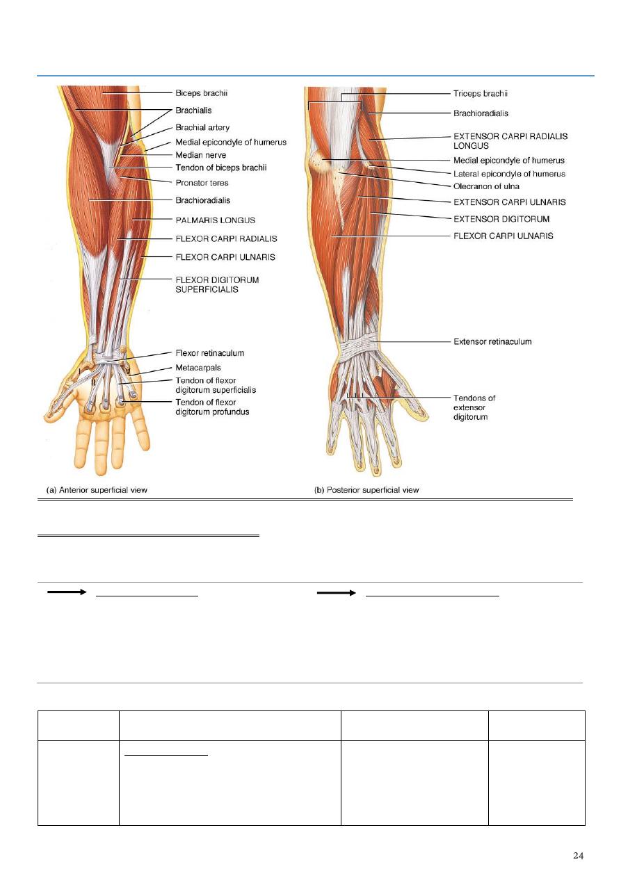
: The Forearm
The Flexor Compartment of Forearm
-
Is the anteromedial compartment of the F.A
-
It includes 8 Ms. ( 5 of them are superficial & 3 deep)
The superficial are :
-
Pronater teres
-
Flexor carpi radialis
-
Palmaris longus
-
Flexor digitorum superficialis
-
Flexor carpi Ulnaris
The other 3 deep Ms are :
-
Flexor digitorum profundus
-
Flexor pollicis longus
-
Pronater quadratus Ms
muscle
Origin (arises from)
insertion
Nerve supply
Pronater
teres
humeral head : arises from medial
epicondyle& medial supracondylar
line
into the lateral surface
of the middle part of the
shaft of the Radius
bone.
-
Four of them
are
innervated
directly by

ulnar head: from medial side of
Coronoid process of the ulna.
median N
,two & half
indirectly
(via the
anterior
interosseous
branch of
the median
N ).
-
Flexor carpi
ulnaris &
medial half
of Flexor
digitorum
profundus
are supplied
by the ulnar
nerve.
Flexor carpi
radialis
medial epicondyle of humerus
into the base of the 2nd
metacarpal bone.
Palmaris
longus
medial epicondyle
palmer aponeurosis.
Flexor carpi
ulnaris
humeral head : from medial
epicondyle
ulnar head: from medial side of
olecranon process of ulna bone
into the Pisiform bone
(one of the carpal
bones).
Flexor
digitorm
superficialis
,humeral head from medial
epicondyle, ulnar collateral ligament
& medial margin of coronoid process
of ulna bone. While the radial head
arises from oblique line on the
anterior surface of the radius bone
The M gives 4 tendons
to the medial 4 fingers(
each tendon inserts into
the sides of the middle
phalange of the
corresponding finger).
Flexor
digitorum
profundus
from the upper 3/4(three fourth)of
anterior, medial &posterior surfaces
of the ulna bone and from anterior
surface of interosseous membrane.
The M gives 4 tendons
to the 4 medial fingers (
inserts into the base of
the distal phalanges).
Flexor
pollicis
longus
takes origin from middle 2/4 of
anterior surface of Radius bone
&interosseous membrane
into the base of distal
phalanx of the thumb
Pronater
quadratus
oblique line on the lower 1/4 of
anterior surface of ulna bone
into the lower 1/4 of
anterior surface of the
radius bone
THE FLEXOR RETINACULUM
-
It is the thickened part of the deep fascia of the F.A
-
located anteriorly at the junction between the F.A & palm of the hand in front of some
carpal bones
-
It is attached to :
pisiform & hook of hamate medially
scaphoid & trapezium laterally
-
this fibrous retinaculum bridges over some carpal bones forming a fibro—osseous
tunnel known as the "carpal tunnel"
-
the carpal tunnel through which pass the following structures:
1- Four tendons of Flexor digitorum superficialis.
2- Four tendons of Flexor digitorum profundus.
3- Tendon of Flexor pollicis longus .
4- The Median nerve.Thus the median N is liable for compression in certain
circumstances leading to what is called Carpal Tunnel Syndrome.
-
The following structures cross superficial to Flexor Retinaculum:
1-Tendon of palmaris longus M.
2-The Ulnar Nerve.
3-The Ulnar Artery.
4-Palmer cutaneous branch of the Median nerve.
5-Palmer cutaneous branch of the Ulnar nerve
Posterior compartment of Forearm
It includes 7 superficial & 5 deep Ms :
The superficial are :
-
Brachioradialis
-
Extensor carpi radialis longus
-
Extensor carpi radialis brevis
-
Extensor digitorum
-
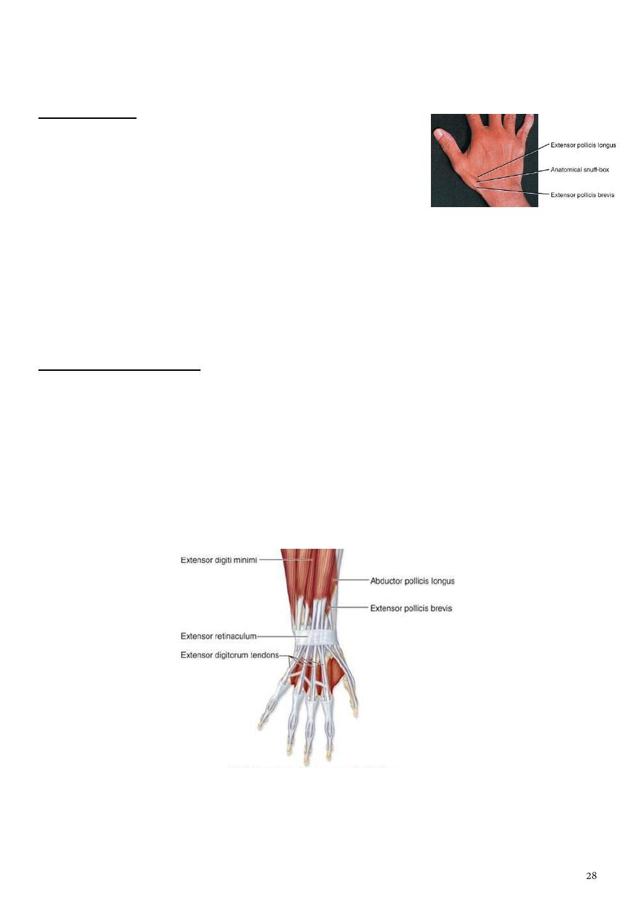
Extensor digiti minimi
-
Extensor carpi ulnaris
-
Anconeus
The deep Ms are :
-
Abductor pollicis longus
-
Extensor pollicis brevis
-
Extensor pollicis longus
-
Extensor indices
-
Supinator
muscle
Origin (arises from)
insertion
1- Brachioradialis
-
upper two thirds of lateral
supracondylar ridge
-
lateral intermuscular septum
into the lateral side of Radius bone.
2- Extensor carpi
radialis longus
-
lower third of lateral suracondylar
ridge
into the base of 2nd metacarpal bone
The Ulnar Nerve
Is one of the branches
of the medial cord of
the Brachial plexus
it doesn’t supply any structure
in the arm
just above the middle part of
the Arm it pierces the medial
intermuscular septum of the
Arm (accompanied by both
superior & inferior Ulnar
collateral As ).
It leaves the Arm by passing
behind the medial
epicondyle of the humerus
to reach the F.A
It passes between the 2
heads of Flexor carpi ulnaris
to reach F.A.
In the F.A it descends under
cover the Flexor carpi ulnaris
M accompanied by the Ulnar
A .
It supplies :
1- Flexor carpi ulnaris
2- medial half of Flexor
digitorum profundus M
then at the lower part
of the F.A it gives
dorsal ulnar cutaneous
branch which goes to
the back of the hand to
supply sensations
Just before it leaves the
F.A it gives palmer
cutaneous branch which
passes superficial to the
Flexor retinaculum to
reach the Palm of the
hand.

-
lateral intermuscular septum
3- Extensor carpi
radialis brevis
-
lateral epicondyle
-
radial collateral ligament
into the base of the 3rd metacarpal
bone
4- Extensor
digitorum
lateral epicondyle
.It gives 4 tendons to the medial four
fingers on the back& insert via
extensor expansion to middle & distal
phalanges.
5- Extensor digiti
minimi
lateral epicondyle too
Its tendon join the tendon of extensor
digitorum for the little finger
6- Extensor carpi
ulnaris
-
lateral epicondyle
-
posterior border of Ulna bone
the medial side of the base of 5th
metacarpal bone.
7- Anconeus
lateral epicondyle
-
into the lateral side of Olecranon
process
-
upper one fourth of posterior
surface of Ulna bone.
8- Abductor
pollicis longus
-
posterior surface of Ulna
-
Radius below the Anconeus
into the base of 1st metacarpal bone
9- Extensor
pollicis brevis
-
posterior surface of Radius-
-
from interosseous membrane
To the base of the proximal phalanx
of the Thumb.
10- Extensor
pollicis longus
-
posterior surface of Ulna
-
interosseous membrane
to the base of the distal phalanx of
the Thumb
11- Extensor
indices
-
from posterior surface of Ulna
-
interosseous membrane
Its tendon goes with the tendon from
extensor digitorum for the index
finger & joins its extensor expansion
12- Supinator
from many sites as:
-
lateral epicondyle
-
Radial collateral ligament
-
annular ligament of superior Radio-
ulnar joint
-
from Supinator crest of Ulna bone.
It wraps round the upper third of
Radius to get insertion to the
posterior surface ,lateral & anterior
surfaces of the upper third of Radius
bone
Note: the following for innervations:
1-Brachioradialis & Extensor carpi radialis longus from Radial nerve before its division.
2-The Supinator by deep branch of Radial N as it pierces its substance.
3-Anconeus by the nerve to Anconeus given off by Radial N as it runs in Spiral groove.
4-All the rest of the Extensor Ms are supplied by the posterior interosseous N which is the
direct continuity of the deep branch of Radial N as it leaves the substance of Supinator M to
run on the posterior surface of the interosseous membrane.
THE SNUFF BOX
1- Is seen at the base of the Thumb posteriorly
2- it is bounded :
laterally ( or anteriorly )by the tendons of Abductor
pollicis longus & that of Extensor pollicis brevis
medially ( or posteriorly) by the tendon of Extensor pollicis longus
3- Its roof is formed by the skin & fascia being crossed superficially by the terminal
branches of superficial branch of Radial N & the beginning of Cephalic vein
4- where as its floor is formed by Scaphoid bone + Styloid process of the Radius bone lying
on them the Radial artery ( feel pulsation here).
EXTENSOR RETINACULUM
1- Exactly like the Flexor Retinaculum on the back of the Wrist region at the junction
between back of F.A & Dorsal aspect of the Hand
2- It is attached to :
the lower end of anterior border of Radius ( laterally )
to the Pisiform + Triquetral bones with the Styloid process of Ulna ( medially )
3- Deep to it the extensor tendons pass via 6 compartments.
Part5
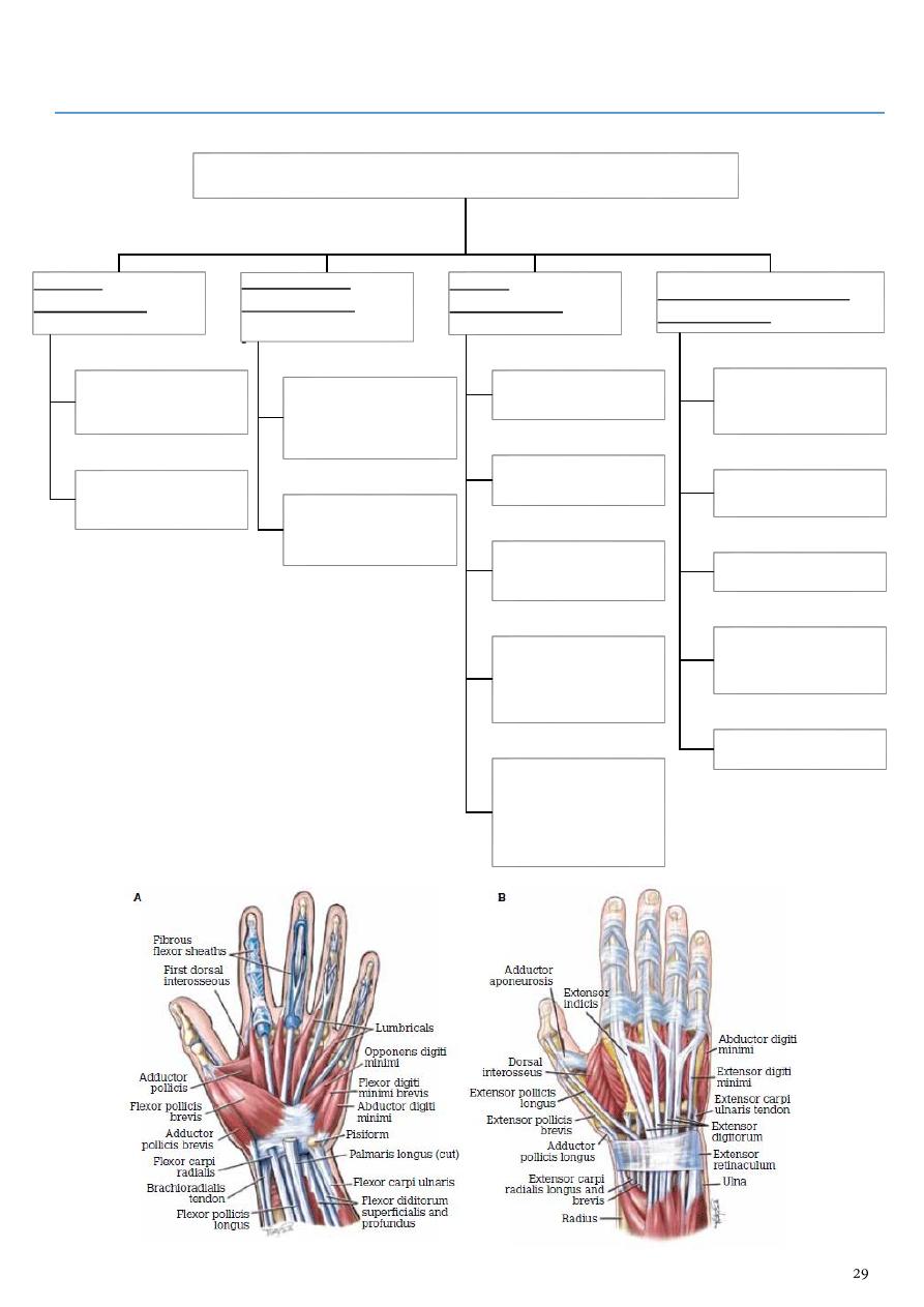
: The Hand
The palm of the hand consists of 4 compartment:
Thennar
compartment
(3muscles)
abductor ,flexor and
opponence pollicis
brevis
supplies by
recurrent branch of
median nerve.
Hypothennar
compartment
has (3ms)
abductor and flexor
digiti minimi and
opponence digiti
minimi
supplies by the deep
branch of ulnar
nerve.
Central
compartment
contains:
palmer aponeurosis
superficial palmer
arch
cutaneous branches
of median nerve
8 tendons belong to
flexor dugitorum
superficialis and
profundus
four lumbrical
muscles associated
with the 4 tendons
of flexor digitorum
profundus
Adductor -interosseous
compartment
has four palmer and
4 dorsal interossei
muscles
adductor pollicis
muscle
deep palmer arch
deep branch of
ulnar nerve
and metacarpal
bones

Sensory nerve supply of hand are:
A- Palm by
1- superficial branch of ulnar to medial one
and half fingers
2- median nerve to supply lateral 3 and half
finger
3- palmer cutaneous branches of median and
ulnar nerve
B- Dorsum of hand by
1- superficial branch of radial nerve
2- dorsal ulnar cutaneous branch
Motor innervation of hand by
A- Recurrent branch of median nerve to :
1- thennar muscles
2- the first 2 lumbrical also by median
nerve.
B- Deep branch of ulnar supplies :
1- adductor pollicis
2- 4palmer and 4 dorsal interossei
3- 3rd and 4th lumbrical muscles
4- the hypothennar muscles
Blood supply of palm of hand
A- Deep palmer arch
-
which is formed mainly by deep palmer
branch of radial artery
-
and to lesses extent by superficial palmer
branch of ulnar artery
B- Superficial palmer arch
-
mainly by superficial palmer branch of
ulnar artery
-
and to lesser extent by superficial palmer
bdanch of radial artery
-
In addition to princeps pollicis Nd radialis
indices from the radial artery
The palmer interossei : are unipennate each one arises from the metacarpal bone of the
same finger and they act in adduction of the fingers toward the middle finger.
The dorsal interossei : are bipennate each one arises from contigous sides of adjacent
metacarpal bone and acts in abduction of fingers or fanning out of fingers
- The interossei are inserted into the extensor expansion on the back of the corresponding
finger and all interossei are supplied by deep branch of ulnar nerve...
Note :
-
The extensor digitorum muscle has 4 tendons one for each finger running on the back
of the finger on reaching proximal phalanx
-
the tendon expand on each side of the proximal phalanx to meet together and inserts
into the distal phalanx
-
while the central part of the tendon inserts into the middle phalanx of corresponding
finger.
Each extensor expansion receives the insertion of 3 small muscles these are one
lumbrical, one dorsal and one palmer interossei

Spaces in the hand are:
1- The pulp space on the palmer aspect of the finger
2- The thenner space at the base of the thumb.
3- Mid-palmar space just medial to thenner space.
4- Ulnar bursa surround the tendons of flexor digitorum superficialis and profundus and
follows deep to flexor retinaculum and follows the flexor tendon to little finger.
5- Radial bursa follow the flexor tendons of flexor pollicis longus and that to index
finger.
- These spaces play a role in transmition of infection in the hand and with the forearm
just proximal to flexor retinaculum
The venous drainage of hand starts as dorsal venous arch which drains the fingers .From
lateral side of the arch the cephalic vein starts and from ulnar side the basilic vein starts
Part6
: Joints
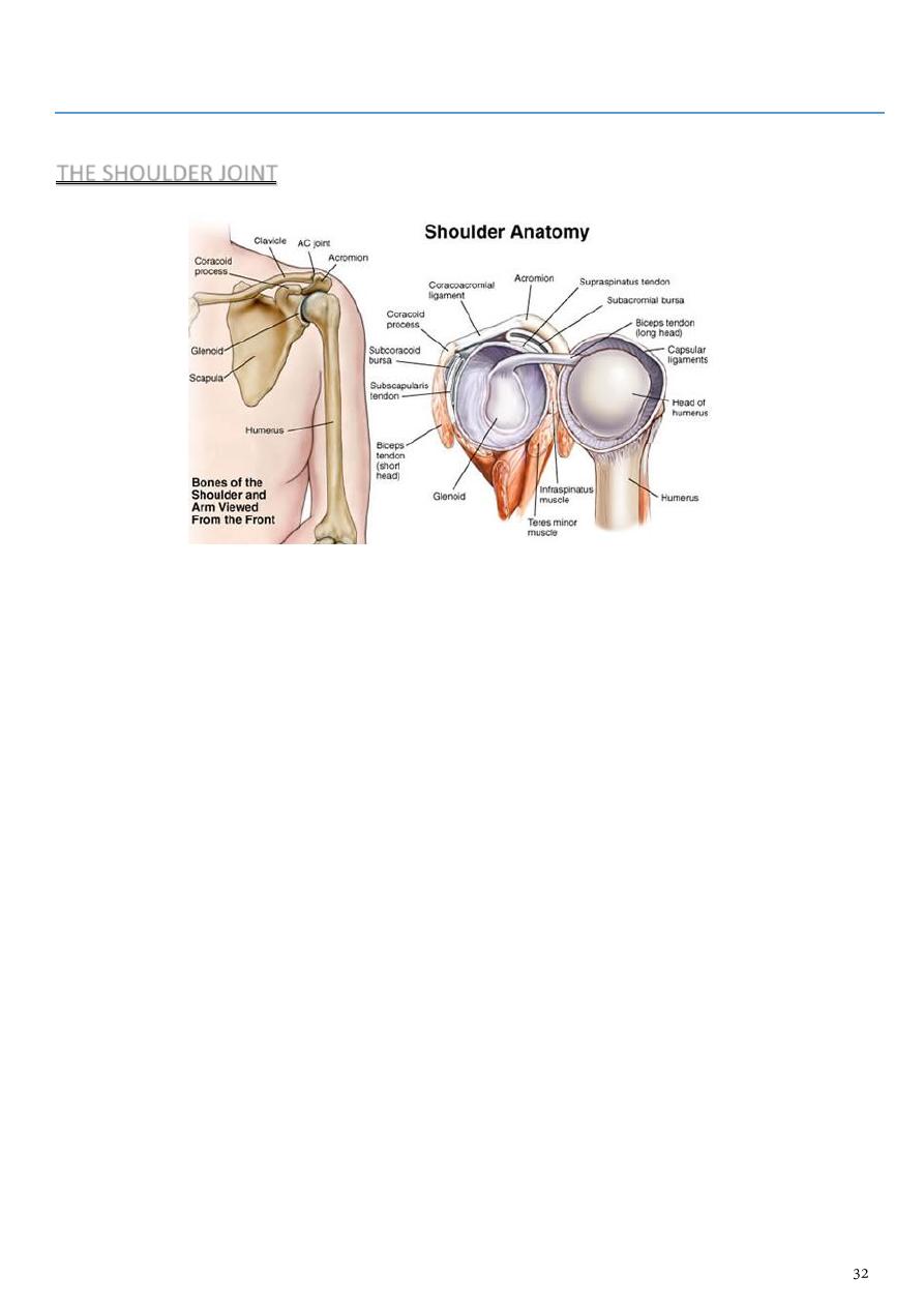
THE SHOULDER JOINT
-
It is a synovial joint of ball & socket variety between the shallow glenoid cavity of
Scapula & hemispheroidal head of Humerus ( both articular surfaces are covered by
hyaline cartilage)
-
The concavity of Glenoid fossa is deepened by a fibro-cartilagenous rim known as
Glenoid labrum.
-
The thin & lax capsule allows freedom of movements at the joint ,it is re-inforced by the
tendons of the Rotator cuff Ms.
-
The capsule is lined by from inside by synovial membrane which forms a cavity (synovial
cavity).
-
The ligaments of the joint are:
1- Glenohumeral ligaments which are 3 in number, superior ,middle & inferior (known
as intrinsic ligaments).
2- Coraco-humeral ligament( Extrinsic ligament ).It is a thick band from the root of
coracoid process to the upper part of the front of greater tuberosity.
3- Transverse humeral ligament ( Extrinsic ) ,it stretches between the lips of Bicepital
groove of the humerus converting it into a canal for the passage of the tendon of
long head of biceps.
4- Coraco-acromial ligament( accessory),its apex attached to the acromion & its base
attaches to the lateral border of the Coracoid process.
5- There are 4 bursae related to the joint ,these are the subscapular, infraspinatous,
Subacromial & subcoracoid bursae.
-
The joint receives articular ( sensory ) nerve supply from the Axillary & Suprascapular
nerves.
-
It receives blood supply from anterior & posterior circumflex humeral ,and also from
circumflex scapular & Suprascapular arteries.
-
The movements are as follows:
1- Flexion is performed by anterior fibers of Deltoid, clavicular head of Pectoralis
major, Biceps & Coracobrachialis muscles.
2- Extension by posterior fibers of Deltoid& teres major muscles.
3- Abduction by Supraspinatous up to 18 degree, then by the lateral fibers of Deltoid
from 18—90 degree. Beyond 80 degree occurs at the shoulder girdle due to
rotation of scapula by Trapezius & Serratus anterior muscles.
4- Adduction by Pectoralis major & Latissimus dorsi muscles.
5- Medial rotation by Pectoralis major, anterior fibers of Deltoid& Subscapularis .
6- Lateral rotation by posterior fibers of Deltoid, Infraspinatous & Teres minor Ms.
7- Circumduction is a combination of all the above movements.
Applied Anatomy:
1-
Due to instability of the joint & laxity of the capsule, it is frequently lible to inferior
dislocation( the capsule here is least protected by muscles.This dislocation may cause
injury or pressure on the Axillary nerve.
2-
Osteoarthritis & Rheumatoid arthritis which may needs artificial joint replacement,
3-
Supraspinatous tendinitis is usually secondary to subacromial bursitis,thus results in
the inability to initiate abduction.
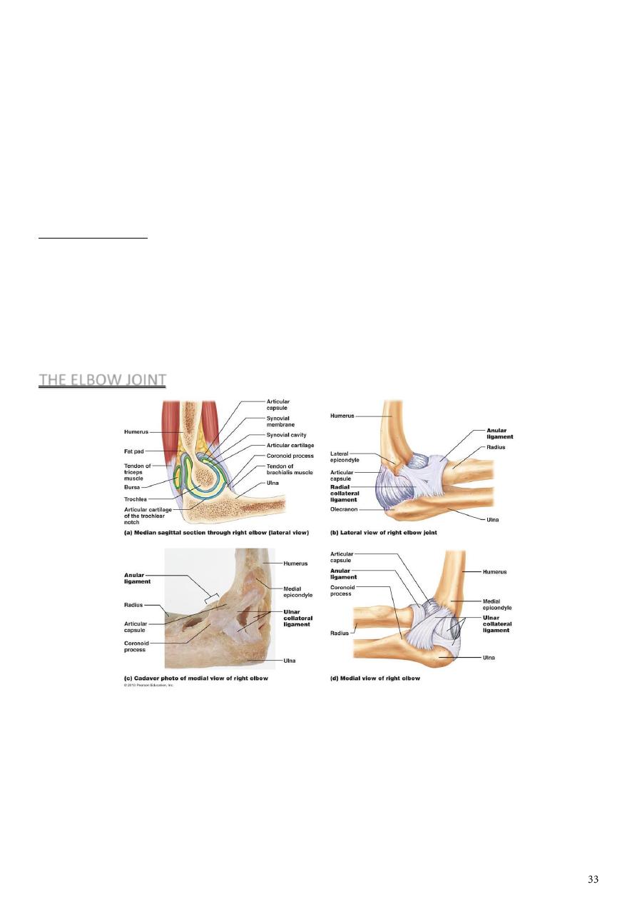
THE ELBOW JOINT
-
Is a compound synovial joint of hinge variety, includes 2 articulations a Humeroulnar
& Humeroradial.
-
The Trochlea of the humerus articulates with the Trochlear notch of Ulna.
-
The Capitulum of Humerus articulates with head of Radius, they are covered by
hyaline cartilage.
-
The capsule is attached
1- (above ) in front to the medial epicondyle and upper margins of Coronoid &
Radial fossa,

2- but from behind along the trochlear margin, margin of Olecranon fossa & over the
Capitulum.
3- Below along the margins of the Coronoid & Olecranon processes and to the
Annular ligament around the head of the Radius.
4- The inner surface of the Capsule & the 3 fossae are lined by synovial membrane.
Ligaments are the followings:
1- Ulnar collateral (Medial) ligament. It is triangular band with anterior, posterior &
inferior thick bands and middle thin part.
2- Radial collateral (lateral) ligament .It extends from lateral epicondyle of the Humerus
to the Annular collateral ligament.
3- Anterior & posterior ligaments which strengthen the capsule in front &behind.
The main relations are as follows:
1- Anteriorly by Brachialis, tendon of Biceps, Median N & Brachial artery.
2- Posteriorly by Anconeus & insertion of Triceps.
3- Medially by common Flexor origin & the Ulnar nerve.
4- Laterally by common Extensor origin & Supinator M.
-
The joint are supplied by articular (sensory) branches from Radial &
Musculocutaneous nerves.
-
The blood supply by branches from the anastomosis around the Elbow joint.
The main movements at the joint are the followings:
1- Flexion movement is performed by Brachialis & Biceps .
2- Extension is performed by Triceps & Anconeus.
Applied Anatomy includes the following cases:
1- Dislocation which is usually a posterior one &is often associated with fracture of the
Coronoid process. Here the Anatomical Triangular relation ship between the Olecranon
& the 2 Epicondyles is lost.
2- Subluxation of the head of Radius(pulled elbow) occurs in children when the F.A is
suddenly pulled in Pronation movement.The head of the Radius slips away from the
Annular ligament.The Elbow is kept fixed in slight Flexion & Pronation,while Supination
is limited and is painfull.
3- Tennis Elbow.Any abrupt Pronation may lead to pain & tenderness over the lateral
epicondyle.This is possibly due to the following factors:
A- The sprain of Radial collateral ligament.
B- Tearing of the fibers of Extensor carpi Radialis Brevis.
C- Inflamation of the Bursa related to M tendon.
4- Student's Elbow: Repeated excessive friction may cause inflammation of subcutaneous
Olecranon Bursa.Gout may cause subcutaneous Bursitis.
5- Effusion of the joint,leads to distension which occurs posteriorly ,because here the
Capsule is weak&the covering deep fascia is thin.Aspiration is done on any side of the
Olecranon to remove the fluids
Anastomosis
Around scapula:
1- Suprascapular a. (from subclavian a.).
2- Transverse cervical a. (from thyrocervical
trunk).
3- Circumflex scapular a. (from the subscapular of
3
rd
part of axillary a.).
4- Thoracodorsal a. (from the subscapular of 3
rd
part of axillary a.).
5- Posterior intercostal.
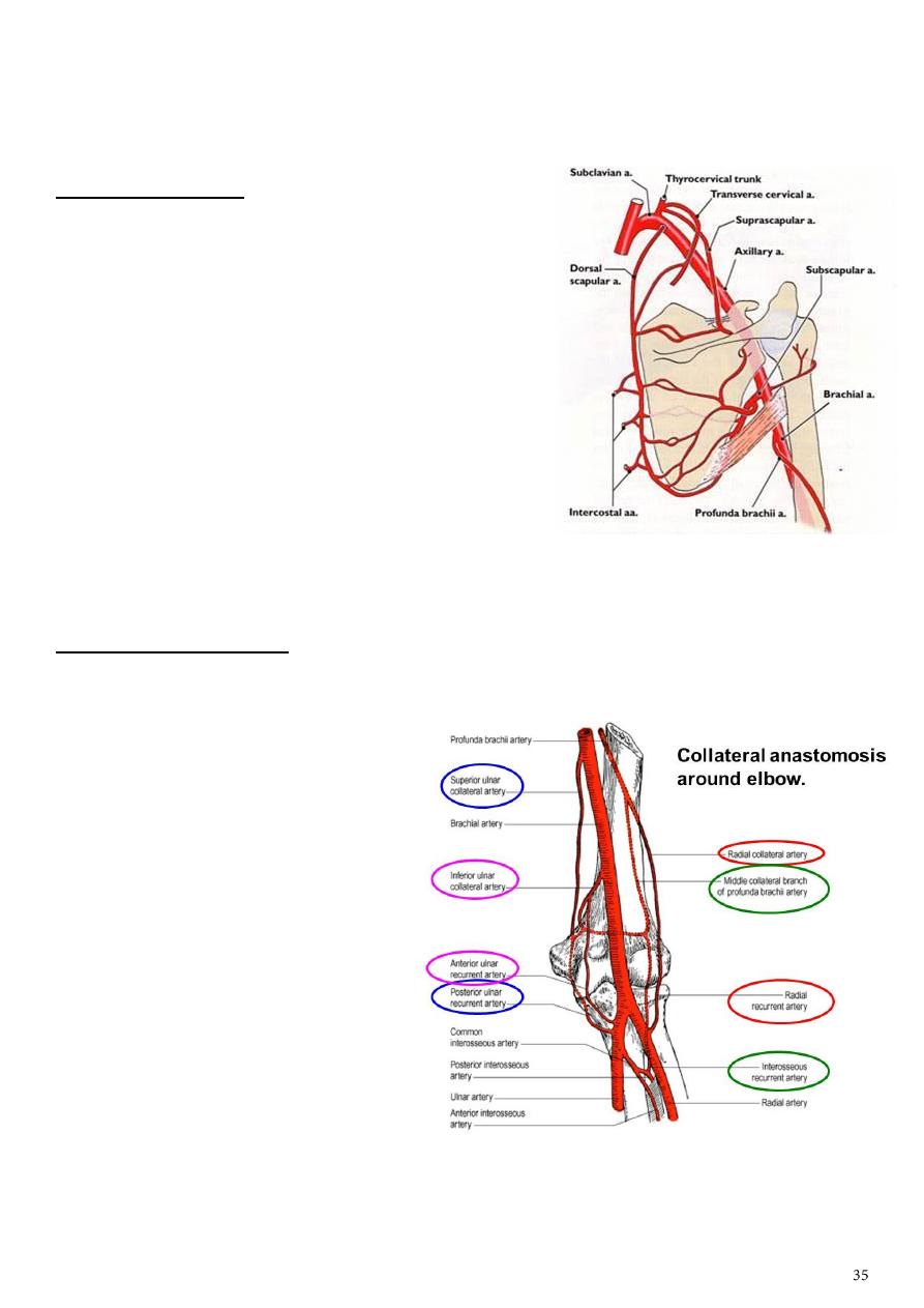
Around elbow joint:
1- Around the medial epicondyle:
From above:
Superior ulnar collateral a.
Inferior ulnar collateral a.
From below:
Anterior ulnar recurrent a.
Posterior ulnar recurrent a.
Interosseous a.
2- Around lateral epicondyle:
From above:
Radial collateral a.
Middle collateral a.
From below:
Radial recurrent a.

Part7
: Arteries
Subclavian artery axillary a. brachial Radial a. (laterally) and ulnar a. (medially)
deep palmar arch and superficial palmar arch.
Axillary artery:
1- First part:
Supreme thoracic (highest thoracic, superior thoracic).
Supply
first and second intercostal spaces and superior part of serrotus anterior.
2- Second part:
Thoraco-acromial (medial) clavicular, acromial, pectoral, deltoid.
Lateral thoracic (lateral) supplies lateral aspect of breast.
3- Third part:
Circumflex humeral (anterior and posterior).
Subscapular circumflex scapular, thoracodorsal.
Note: thoracodorsal a. + thoracodorsal nerve both enter latisimuss dorsi.
Brachial artery:
1- Profunda brachii ascending branch (deltoid), middle collateral, radial collateral.
2- Superior Ulnar collateral.
3- Inferior Ulnar collateral.
4- Common unterosseous artery (superior of interosseous membrane) anterior and
posterior.
Radial artery:
Start in the elbow rejoin at the level of head of radius.
Course:
1- Runs inferolaterally under cover of brachioradials.
2- Then it lies lateral to flexor carpi radialis tendon distal forearm.
3- Finally it winds around lateral aspect of radius and crosses the floor of snuff box to
pass between the 2 heads of first dorsal interosseous muscle.
4- Take part in forming the deep palmar arch.
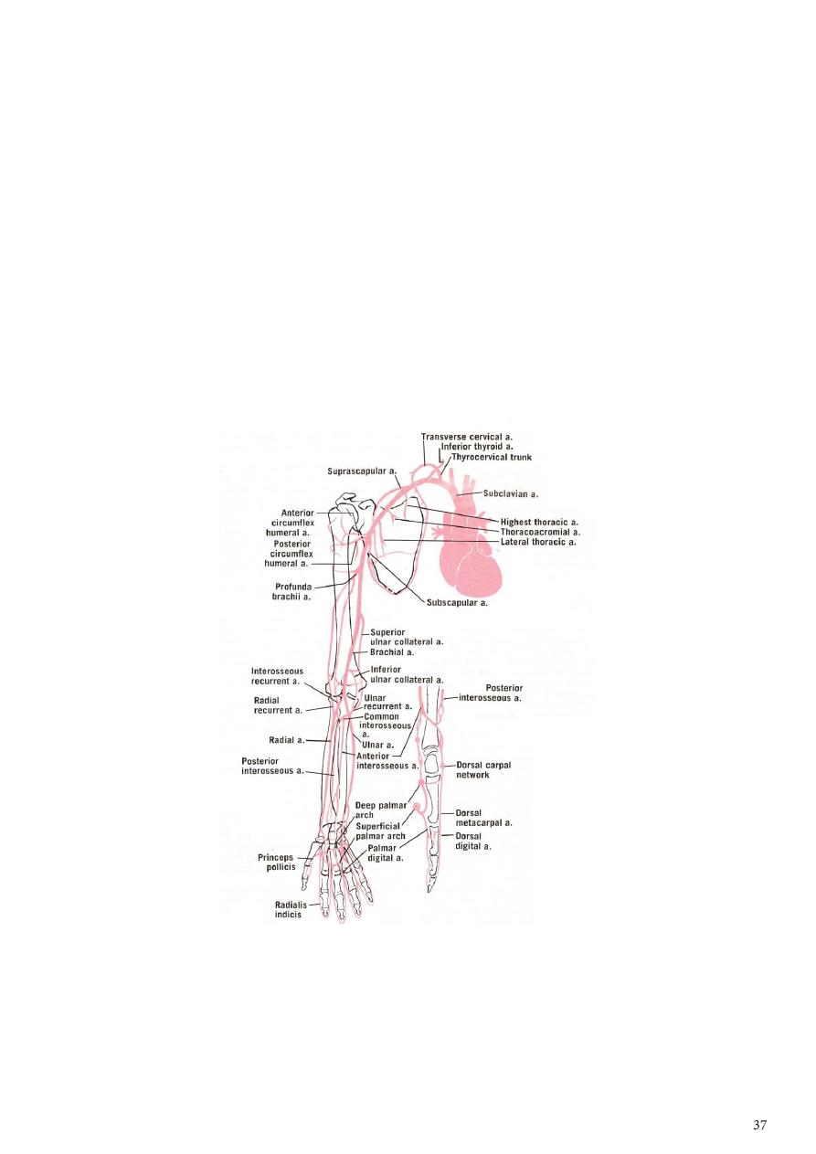
Branches of radial artery:
1- In the forearm radial recurrent a. – palmer carpal branch – superficial palmer
branch.
2- At the wrist dorsal carpal branch – first dorsal metacarpal artery.
3- In the hand princeps pollicis – radialis indicis – deep palmer arch.
Ulnar artery:
Disappears from the cubital fossa by passing deep to the deep head of pronator teres
and beneth flexor digitorum superficialis near the median nerve.
Leaves the median nerve and lies on flexor digitorum profunds with the ulnar nerve to
its medial side and passes down over the front wrist into the palm where it continues
as the superficial palmer arch.
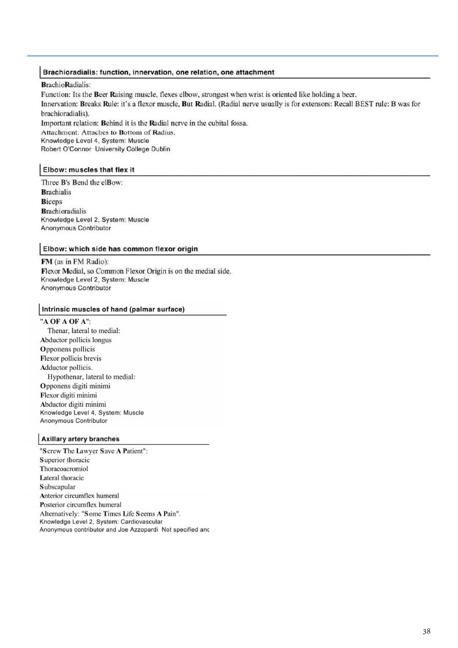
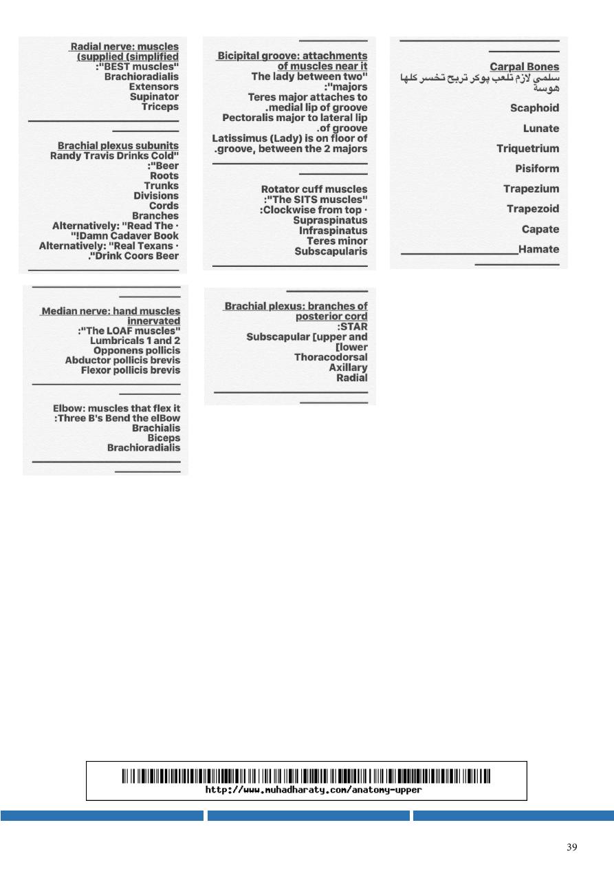
Part8
: Make it easy!