Germ cell tumor
Dr.AHMED JASIM
Origin : cells derived form oocytes
Incidence: 15- 20% of all ovarian tumors, 5% malignantAge: young age
A germ cell tumor (GCT) is a neoplasm derived from germ cells. Germ cells normally occur inside the gonads (ovary and testis). Germ cell tumors that originate outside the gonads may be birth defects resulting from errors during development of development of the embryo.
Etiology
Some investigators suggest that this distribution arises as a consequence of abnormal migration of germ cells during embryogenesis. Others hypothesize a widespread distribution of germ cells to multiple sites during normal embryogenesis, with these cells conveying genetic information or providing regulatory functions at somatic sites.
Classification
Germ cell tumors are classified by their histology,regardless of location in the body.
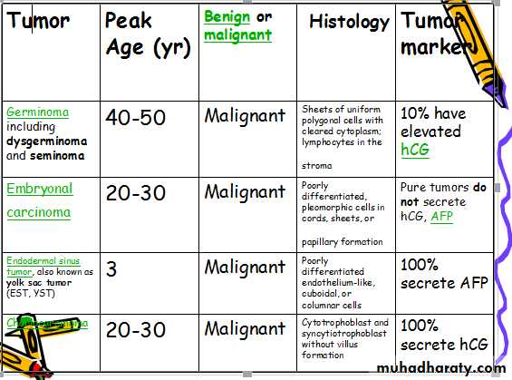
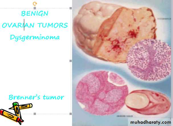
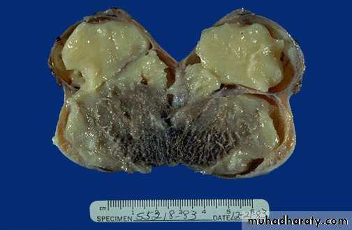
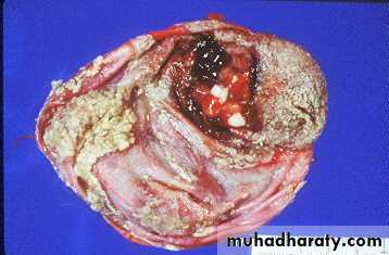
Dysgerminoma
Incidence : very commonAge : 20 – 20 yrs
Bilateral : 10 – 15 %
Macroscopic features :
Solid tumors, elastic rubbery consistency having smooth, firm capsule
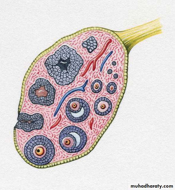
2. Teratoma
Derived from cells of all three germ layers
Types:
Mature or benign type (e.g. Dermoid cysts)
Immature or malignant type (e.g. Solid Teratoma)
Monodermal or highly specialized (e.g. Struma ovarii)
3. Choriocarcinoma and Embryonal Cell Carcinoma
Choriocarcinoma mostly of placental origin occurs in prepubertal girls. Highly malignantContains syncytiotrophoblasts and cytotrophoblasts
Secretes large quantities of the tumor marker - HCG
Embryonal cell carcinoma
Incidence : rare
Highly malignant
4. Ovarian Fibroma:
Meig’s syndromeAscites
Right sided effusion

Germ cell tumors are broadly divided in two classes:
The germinomatous or seminomatous germ cell tumors (GGCT, SGCT) include only germinoma and its synonyms dysgerminoma and seminoma.The nongerminomatous or nonseminomatous germ cell tumors (NGGCT, NSGCT) include all other germ cell tumors, pure and mixed.
Cystic Teratoma
Dermoid Cyst:
The two classes reflect an important clinical difference. Compared to germinomatous tumors, nongerminomatous tumors tend to grow faster, have an earlier mean age at time of diagnosis (~25 years versus ~35 years, in the case of testicular cancers), and have a lower 5 year survival rate. The survival rate for germinomatous tumors is higher in part because these tumors are exquisitely sensitive to radiation, and they also respond well to chemotherapy. The prognosis for nongerminomatous has improved dramatically, however, due to the use of platinum-based chemotherapy regimens.
Mixed
Mixed germ cell tumors occur in many forms. Among these, a common form is teratoma with endodermal sinus tumor.
Teratocarcinoma refers to a germ cell tumor that is a mixture of teratoma with embryonal carcinoma, or with choriocarcinoma, or with both.This kind of mixed germ cell tumor may be known simply as a teratoma with elements of embryonal carcinoma or choriocarcinoma, or simply by ignoring the teratoma component and referring only to its malignant component: embryonal carcinoma and/or choriocarcinoma.
Location
Despite their name, germ cell tumors occur both within and outside the ovary and testis.
In females, germ cell tumors account for 30% of ovarian tumors, but only 1 to 3% of ovarian cancers in North America. In younger women germ cell tumors are more common, thus in patients under the age of 21, 60% of ovarian tumors are of the germ cell type, and up to one-third are malignant. In males, germ cell tumors of the testis occur typically after puberty and are malignant (testicular cancer). In neonates, infants, and children younger than 4 years, the majority of germ cell tumors are sacrococcygeal teratomas.
Males with Klinefelter's syndrome have a 50 times greater risk of germ cell tumors (GSTs).In these persons, GSTs usually contain nonseminomatous elements, present at an earlier age, and seldom are gonadal in location.
Prognosis
The 1997 International Germ Cell Consensus Classification is a tool for estimating the risk of relapse after treatment of malignant germ cell tumor.
A small study of ovarian tumors in girls reports a correlation between cystic and benign tumors and, conversely, solid and malignant tumors. Because the cystic extent of a tumor can be estimated by ultrasound, MRI, or CT scan before surgery, this permits selection of the most appropriate surgical plan to minimize risk of spillage of a malignant tumor.