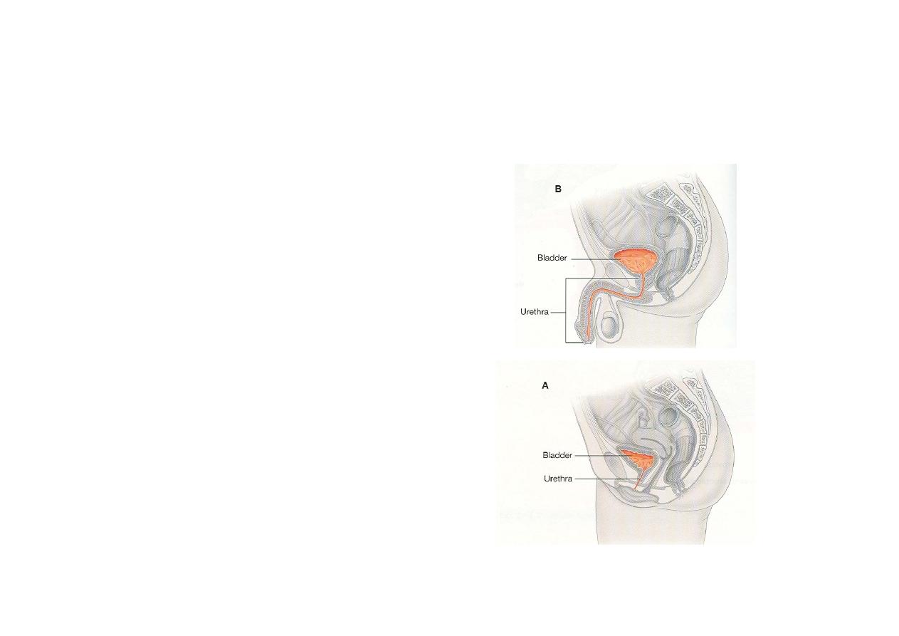
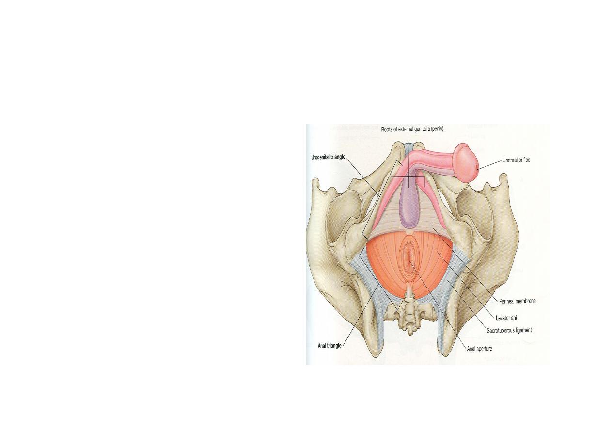
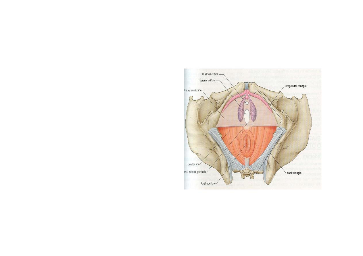
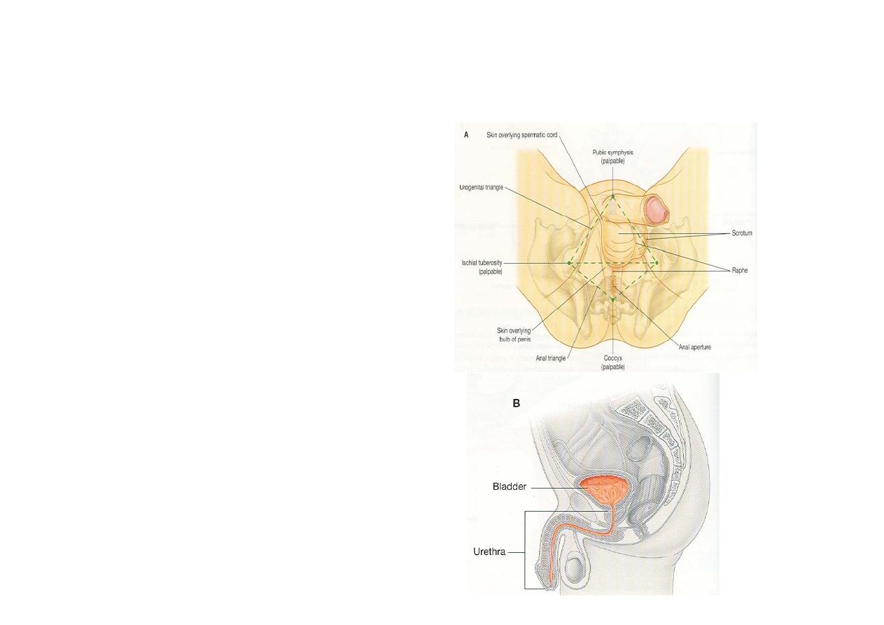
Perineum
Rhomboid space at the lower end of
abdomen which lies between two thigh
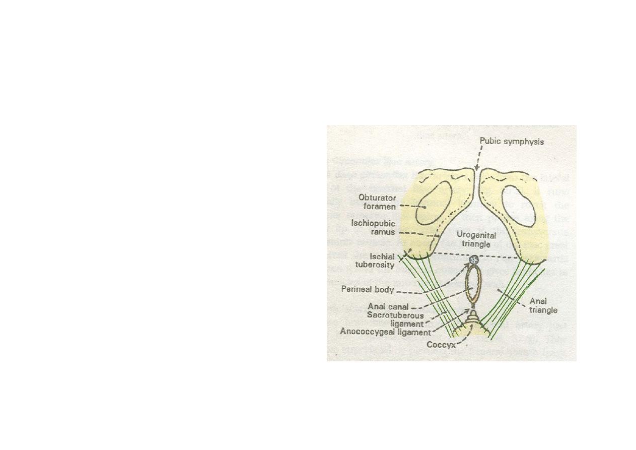
Boundaries
• Anteriorly bounded by
pubic arch and
Arcuate pubic
ligament
• Posteriorly the tip of
coccyx
• On each side
ischiopubic rami,
ischial tuberosity &
sacrotuberous
ligament
Division
• Divided into two
regions by a line
joining the anterior
part of ischial
tuberosity
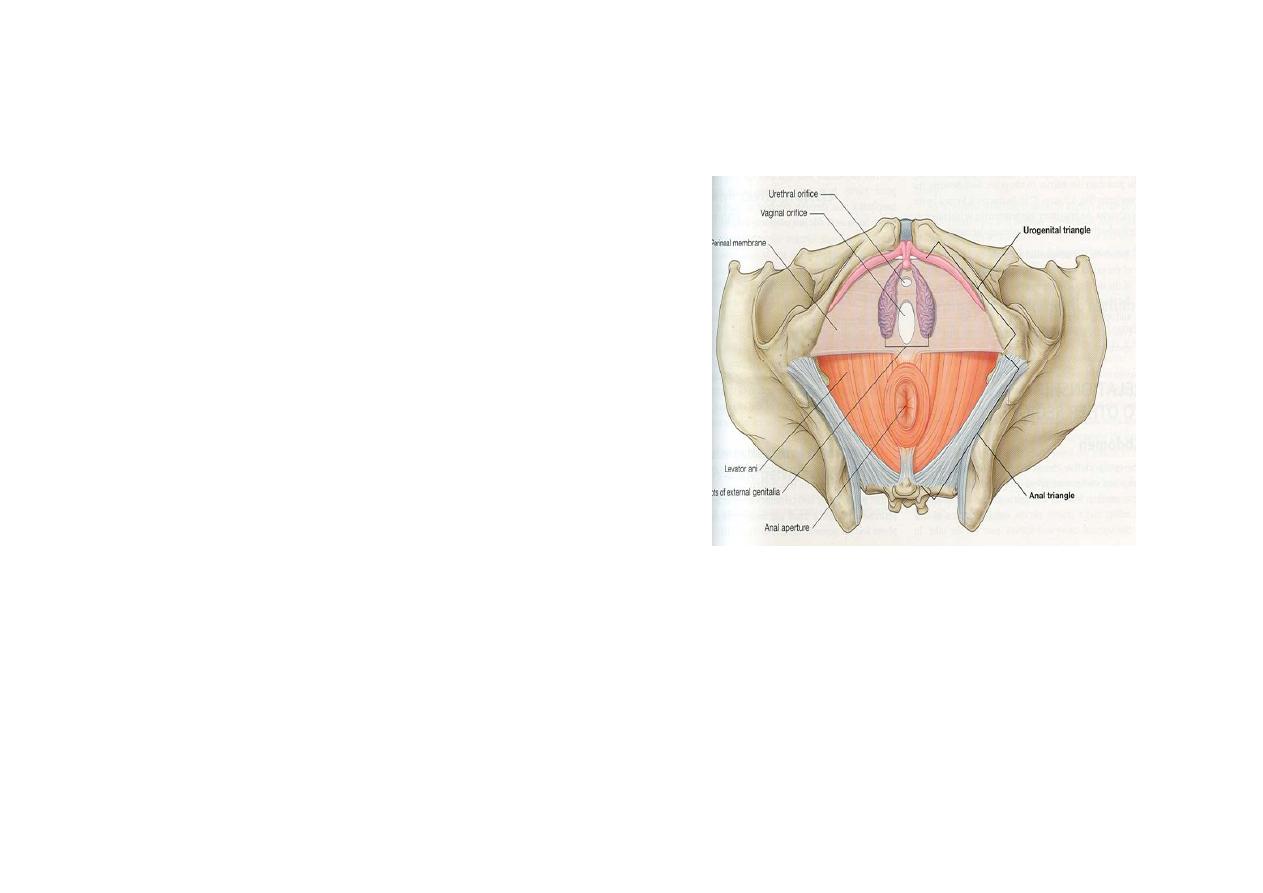
• Urogenital region
• Anal region
Urogenital region
• Placed between two
ischiopubic rami
•
In male contains urethra
enclosed by root of penis,
scrotum
• In females contains
urethral and vaginal
orifice & female external
genitalia
• Three membranes
• Two spaces
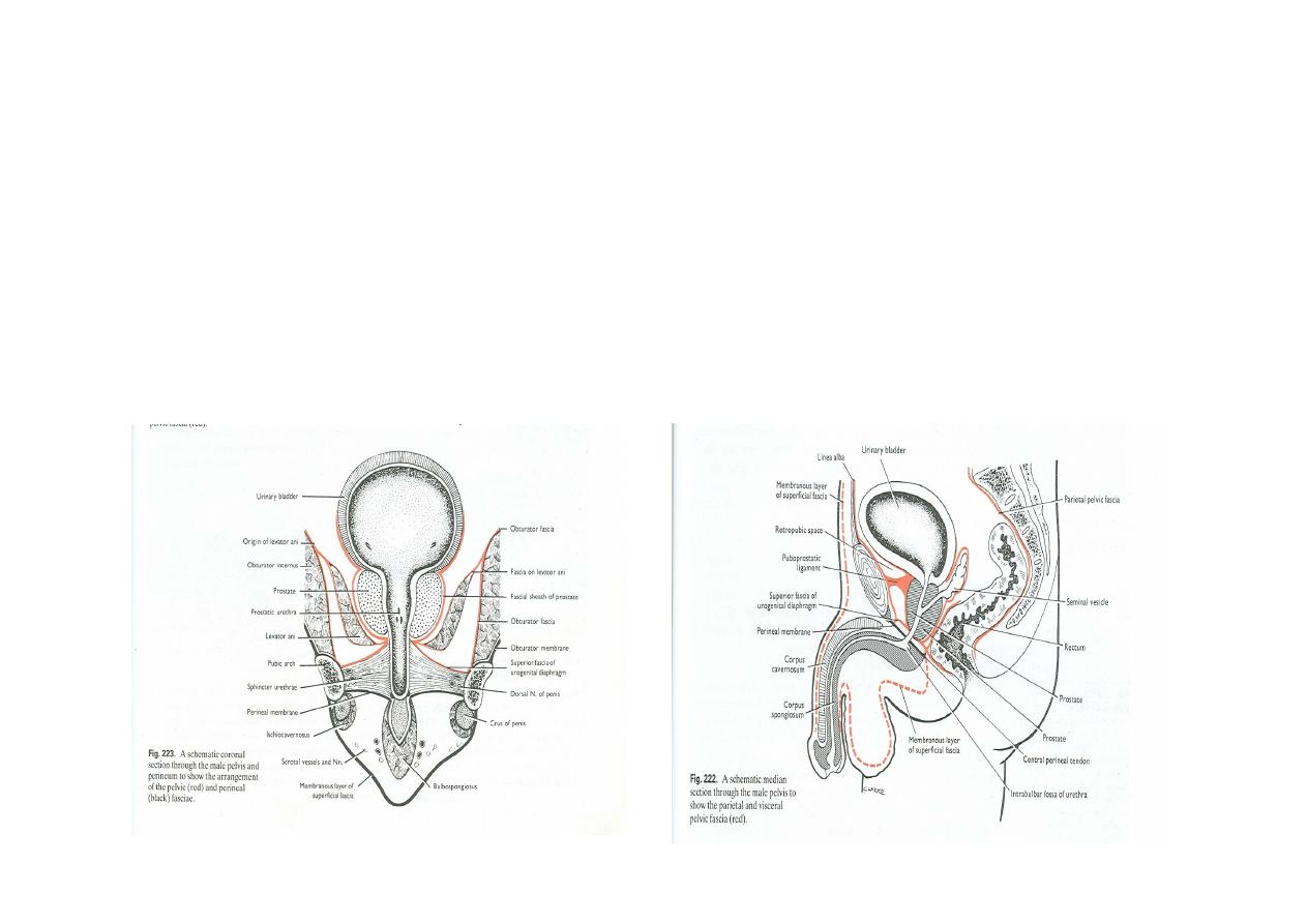
Three membranes
Two spaces
• Part of pelvic fascia
continuous laterally
with the fascia over
obturator internus &
constitutes superior
fascia of urogenital
diaphragm
• Second membrane is
inferior fascia of the
urogenital diaphragm
(Perineum)
• Most superficial
membrane is
membranous layer of
superficial fascia
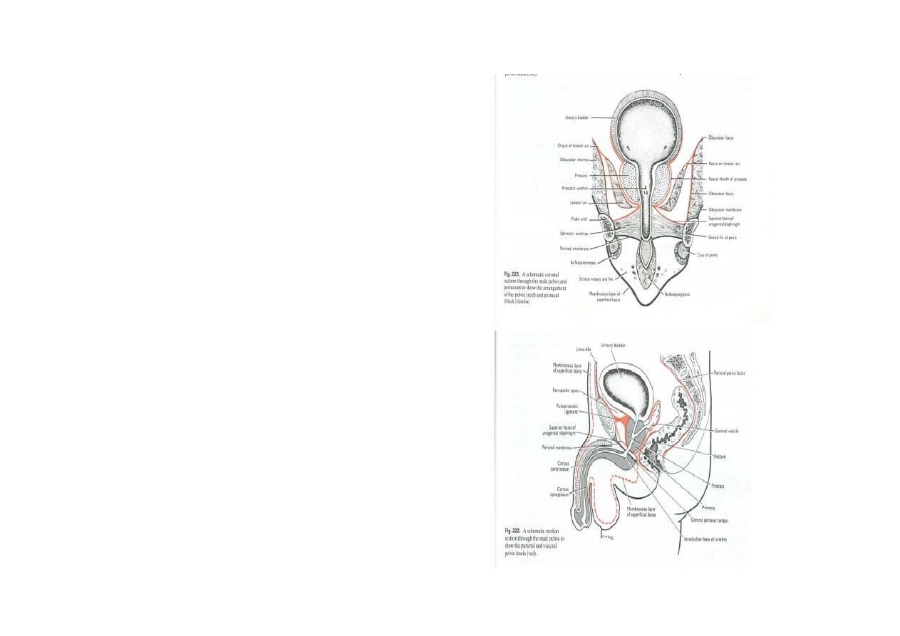
• Between upper and
middle layer is deep
perineal space
• Between the middle
and membranous
layer is superficial
perineal space
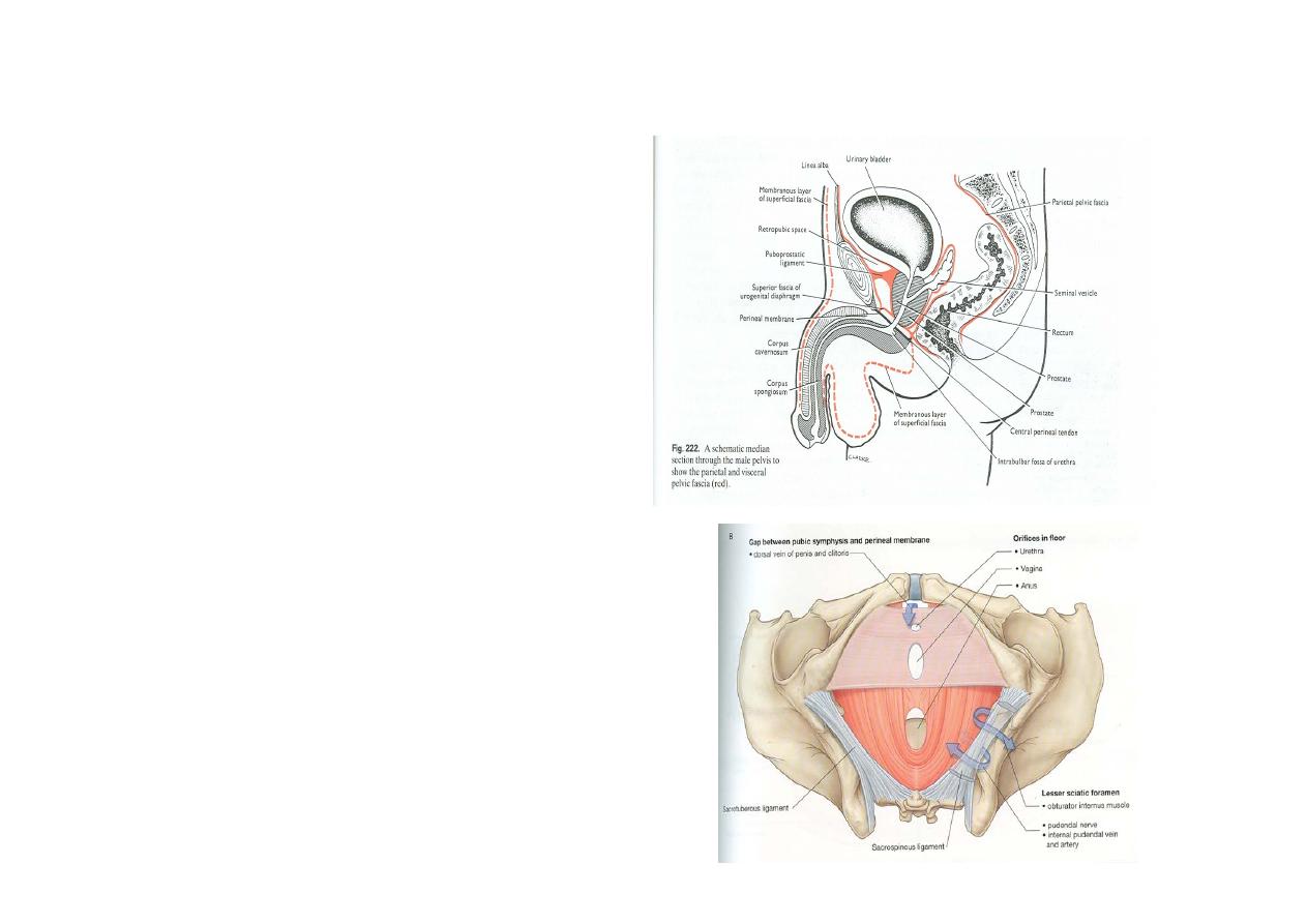
• Posteriorly all three
membranes are attached
to perineal body & to
each other thus closing
the perineal spaces
behind
•
Anteriorly the upper &
middle membrane fuse a
little behind the pubic
symphysis & form
transverse ligament of
the pubis
• Traced Anteriorly the
membranous layer is
continues with the
anterior abdominal wall

Structures piercing the perineal
membrane in males
• Urethra
• Duct of bulbourethral gland
• Artery & nerve to bulb, urethral artery,
deep artery & dorsal artery of penis
• Posterior scrotal nerves & vessels
• Branches of perineal nerve to superficial
perineal muscles

Structures piercing the perineal
membrane in females
• Urethra
• Vagina
• Artery & nerve to the bulb of the vestibule
• Deep & dorsal artery of clitoris
• Posterior labial arteries & nerves
• Branches of perineal nerve to superficial
perineal muscles
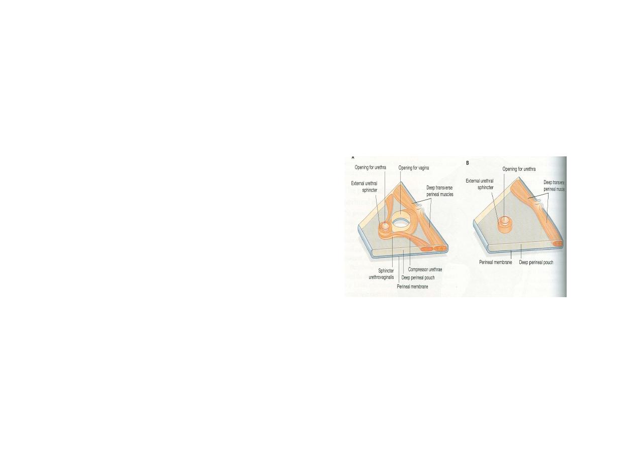
Urogenital diaphragm
Formed by
• Superficial fascia of
urogenital diaphragm
• Deep perineal muscles
Sphincter urethrae,
Deep transverse perinei
• Inferior fascia of
urogenital diaphragm
(perineal membrane)
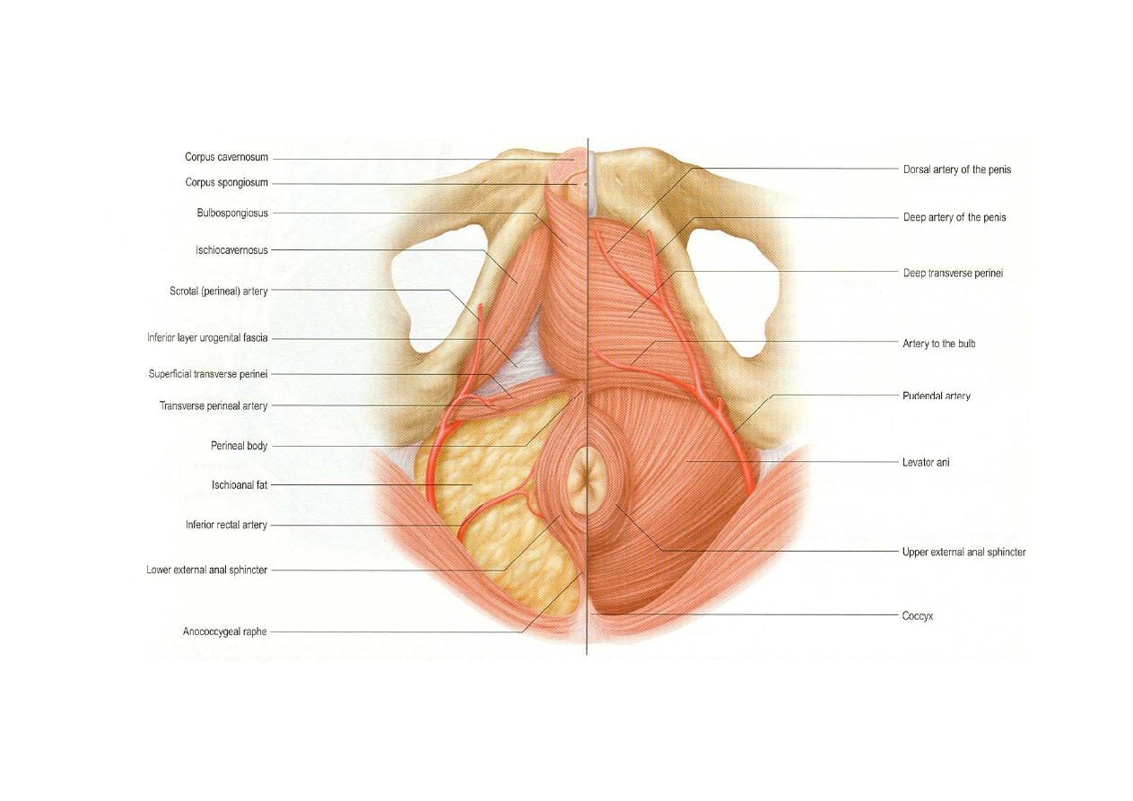
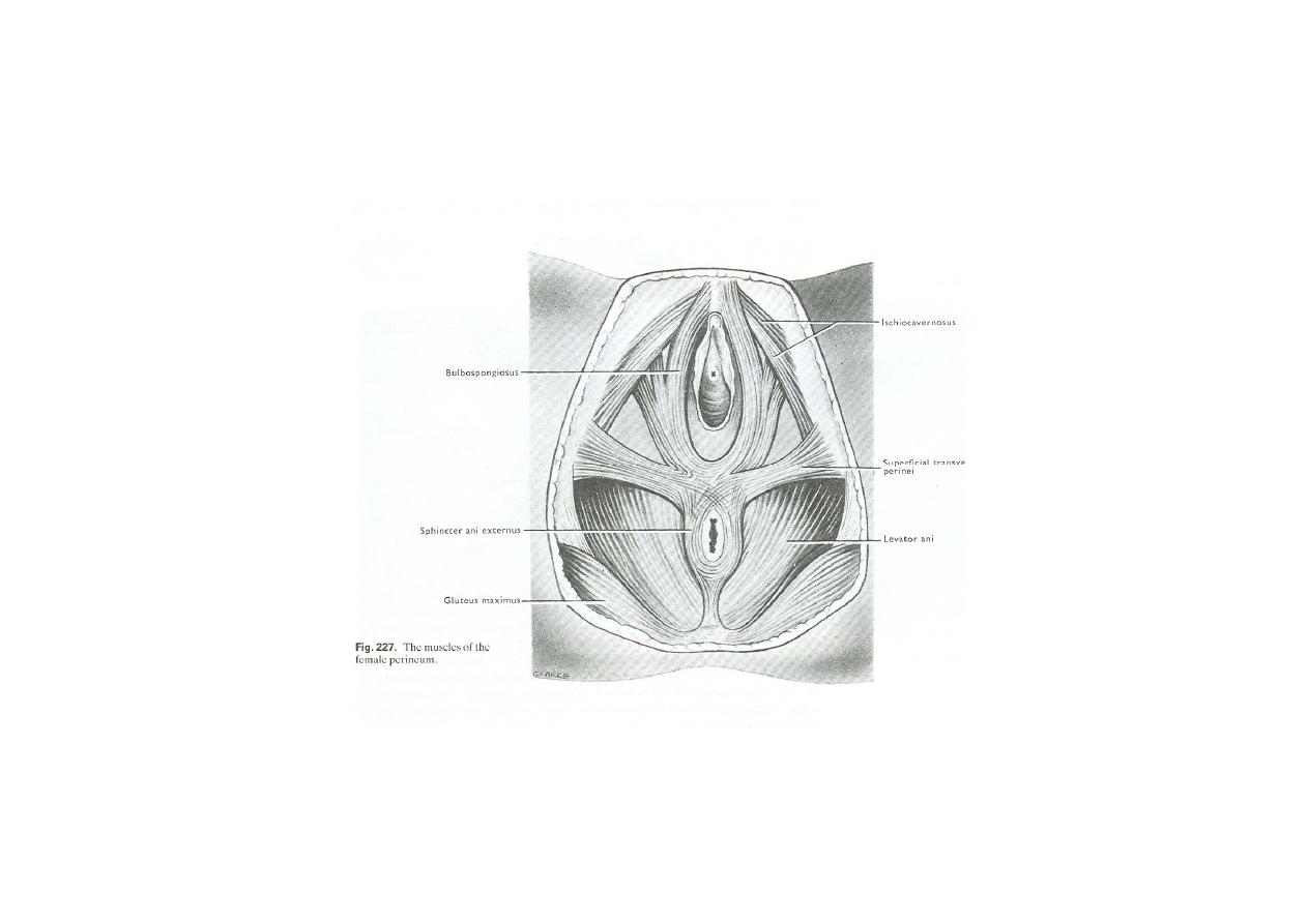
Superficial perineal space in the
male
•
Contents
•
Root Of Penis
Bulb
Right & left crura
•
Muscles
Bulbospongiosus
Ischiocavernosus
Superficial transversus
perinei
All muscles are supplied by
perineal branch of pudendal
nerve
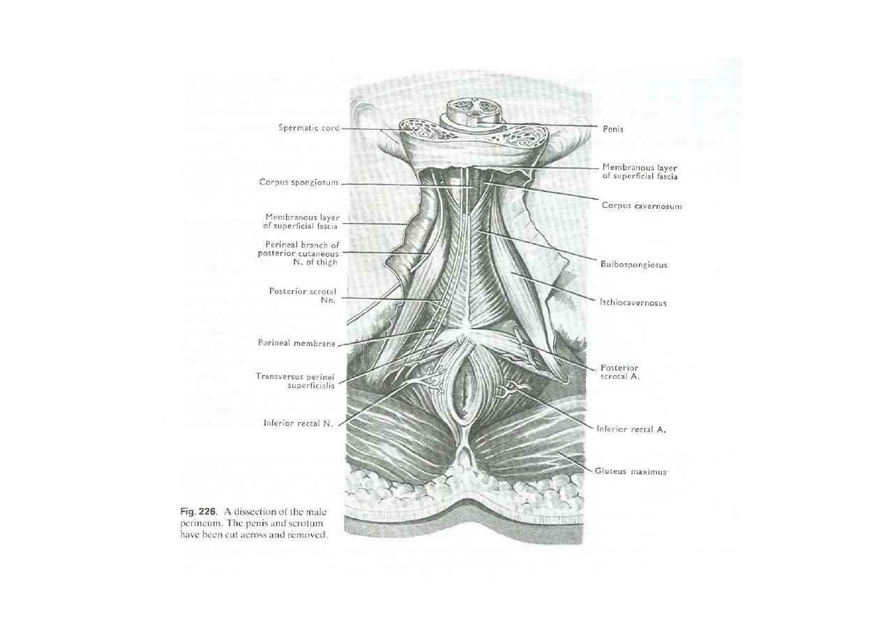

Nerves
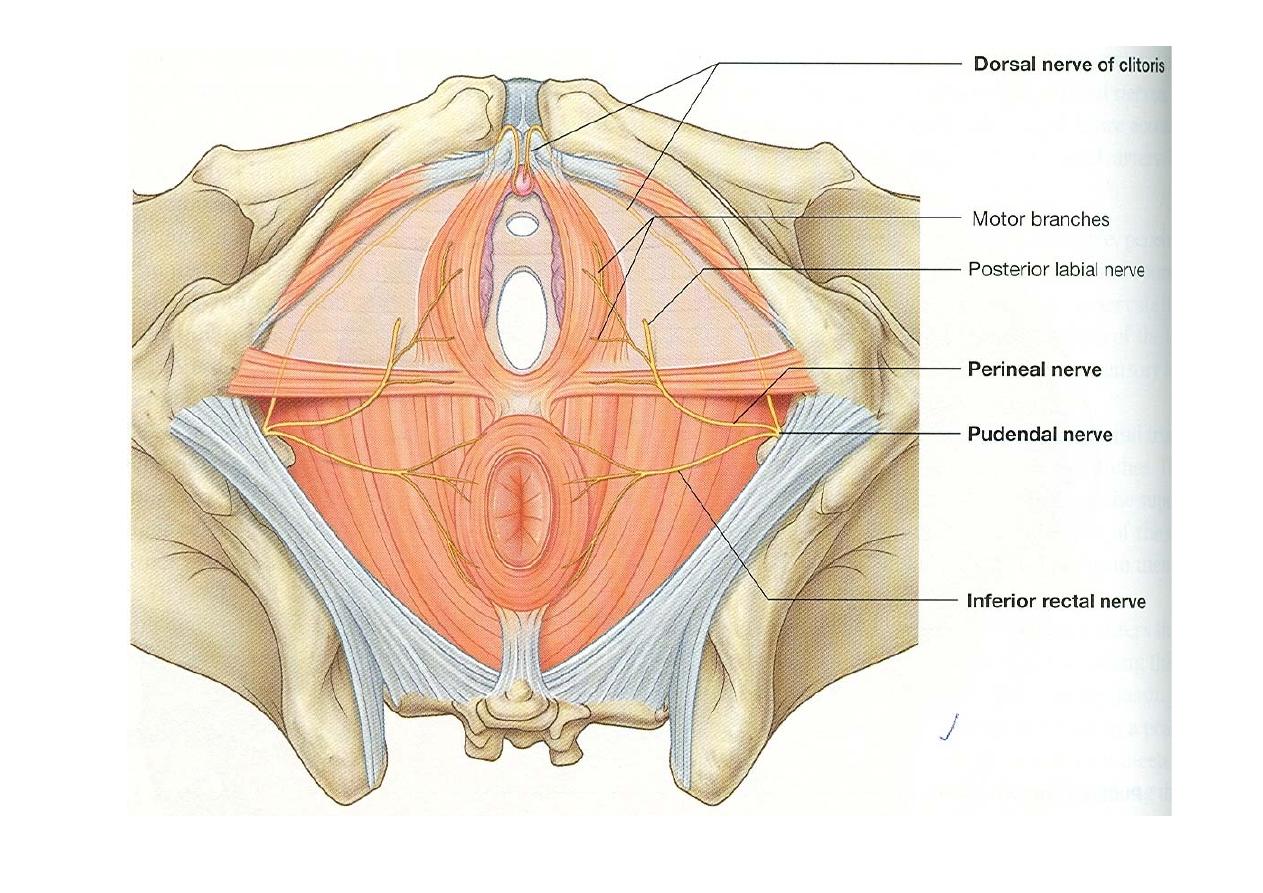
•Branches of perineal nerve- posterior
scrotal, nerve to bulb & muscular
•Long perineal nerve from posterior
cutaneous nerve of thigh
•Vessels- branches of perineal artery
namely posterior scrotal and transverse
perineal
•Branches of artery of penis namely
artery to bulb, urethral artery and deep
&dorsal artery of penis
Superficial perineal space in the
female
• Female external genitalia
• Muscles
Bulbospongiosus
Ischiocavernosus
Superficial
transversus perinei
Nerve supply of muscles is
by perineal branch of
pudendal nerve
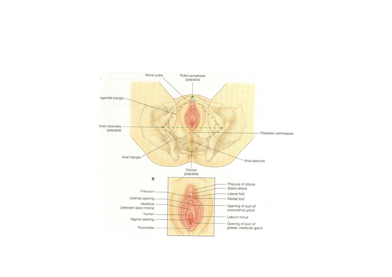
Female external genital organs
Muscles

Nerves
• Branches of perineal nerve- posterior labial,
nerve to bulb & muscular
• Long perineal nerve from posterior
cutaneous nerve of thigh
Vessels- branches of perineal artery namely
labial and transverse perineal
• Branches of artery of clitoris namely artery to
bulb,deep &dorsal artery of clitoris
• greater vestibular gland in females
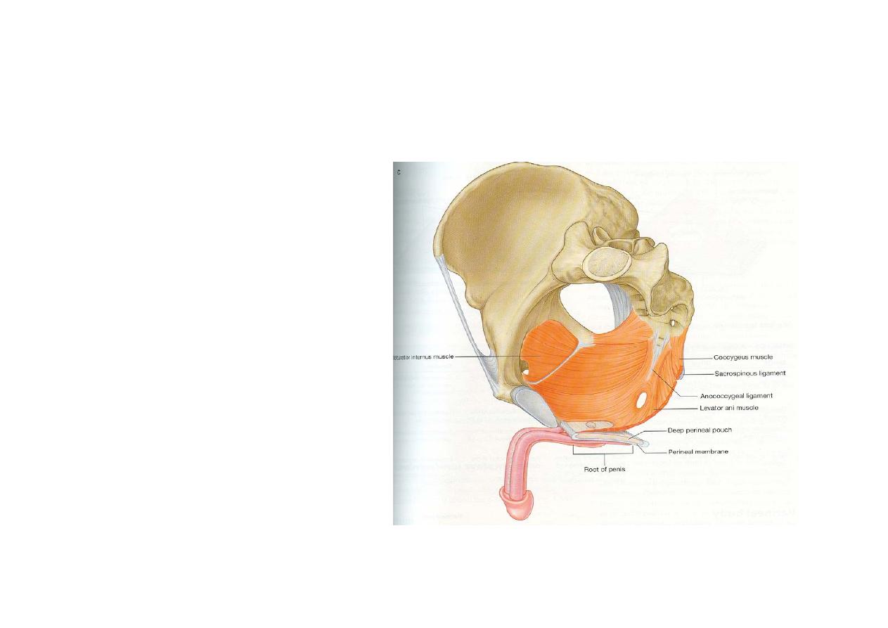
Deep perineal space
• Contents
• Membranous
urethra
• Muscles- sphincter
urethrae, deep
transverse perinei
Nerves-
• Dorsal nerve of
penis and muscular
branches from
perineal nerve
Vessels-
• Artery of penis
• Bulbourethral
Gland In Males

Applied
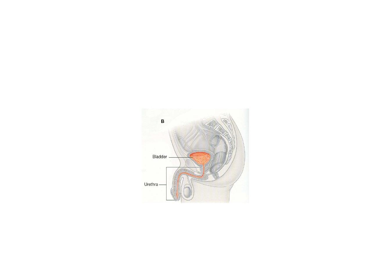
• Membranous part of urethra is narrowest
& least dilatable
• Extravasation of urine
• Proalpse of pelvic viscera due to perineal
body rupture
ANAL REGION
• Triangular area
between the
posterior margin of
the urogenital
diaphragm & the
coccyx
• Transmits the anal
canal
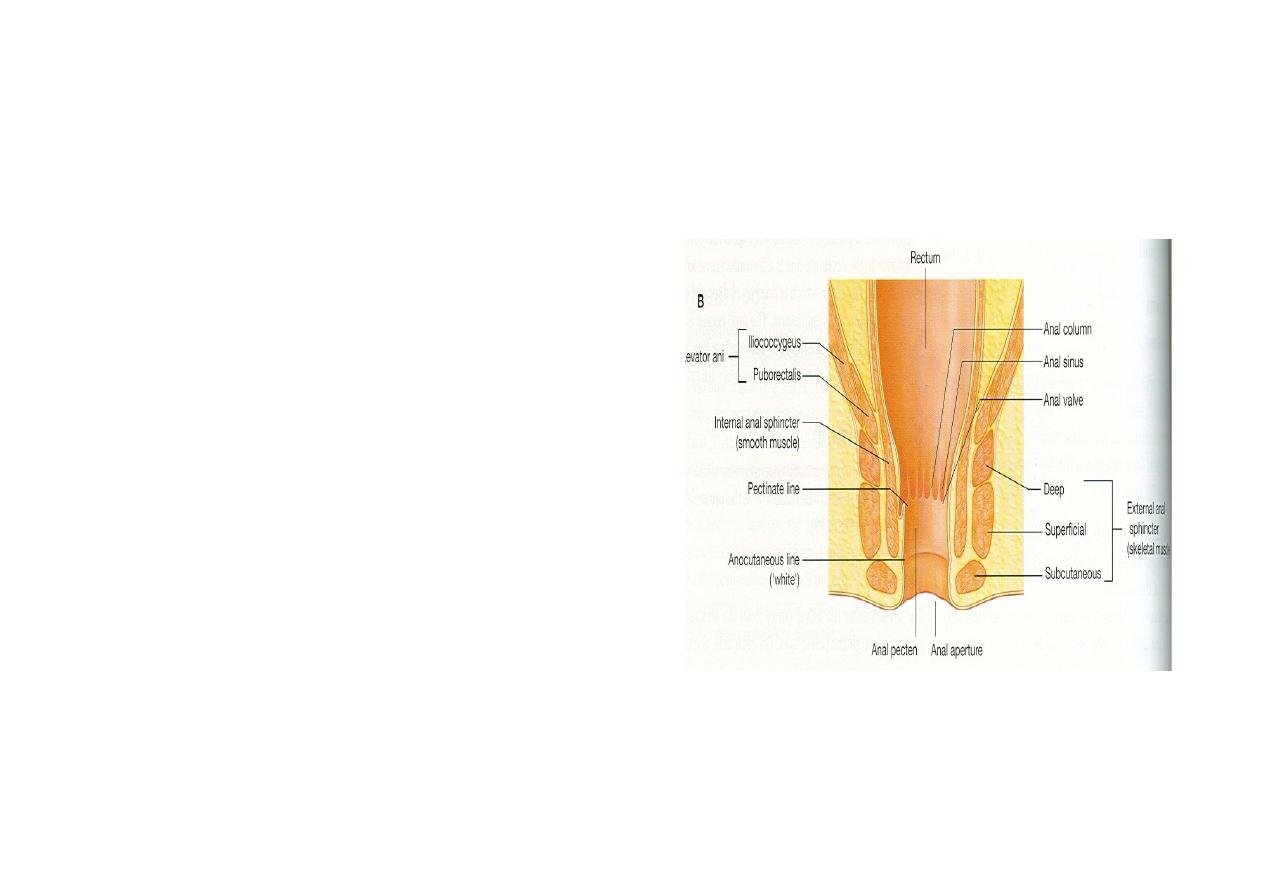
• Anal canal is
surrounded by external
anal sphincter
• Anal canal is connected
to the coccyx by
anococcygeal ligament
• On either side of canal
is a triangular space
called ischiorectal fossa
• Perineal body (central
tendon of perineum) is
situated 1.25 cm
anterior to anal canal
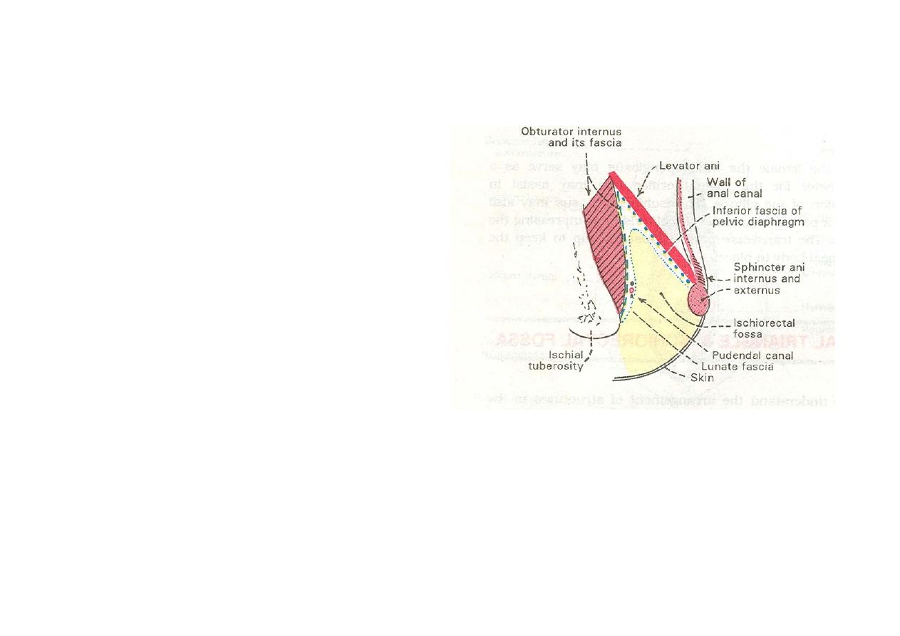
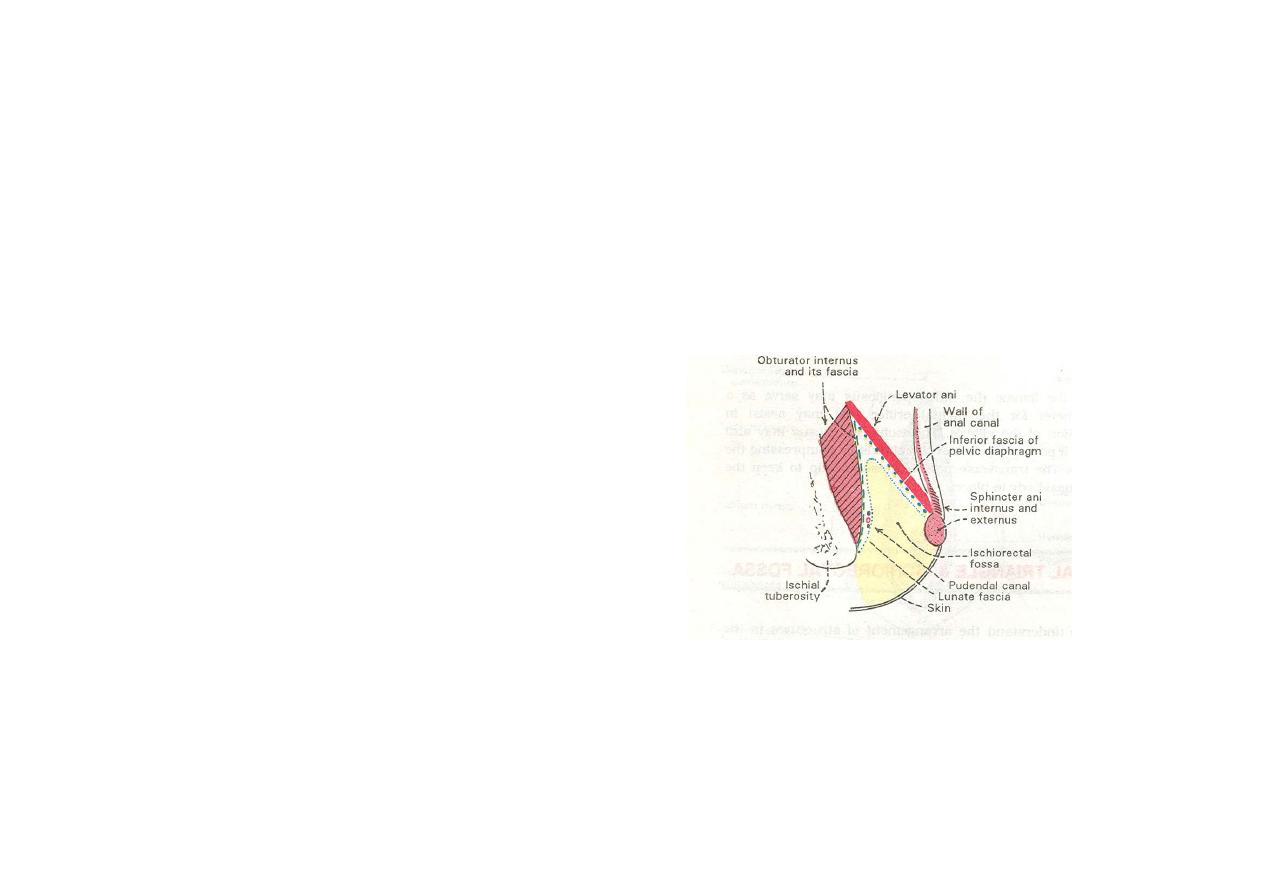
Ischiorectal fossa
•
Wedge shaped space
between obturator internus
& levator ani
•
Base is formed by skin
•
Widest & deepest
Posteriorly, continues with
the lesser sciatic foramen
•
Anteriorly continues with a
narrow space filled with
loose areolar tissue
•
Lateral wall by fascia
covering obturator internus
•
superomedial wall formed
by fascia covering levator
ani & external anal sphincter
Contents of Ischiorectal fossa
• vessels & nerves
that enter thru
lesser sciatic
foramen and run in
pudendal canal
• Perineal branch of
the fourth sacral
nerve
• Perforating
cutaneous
branches of S2 &3
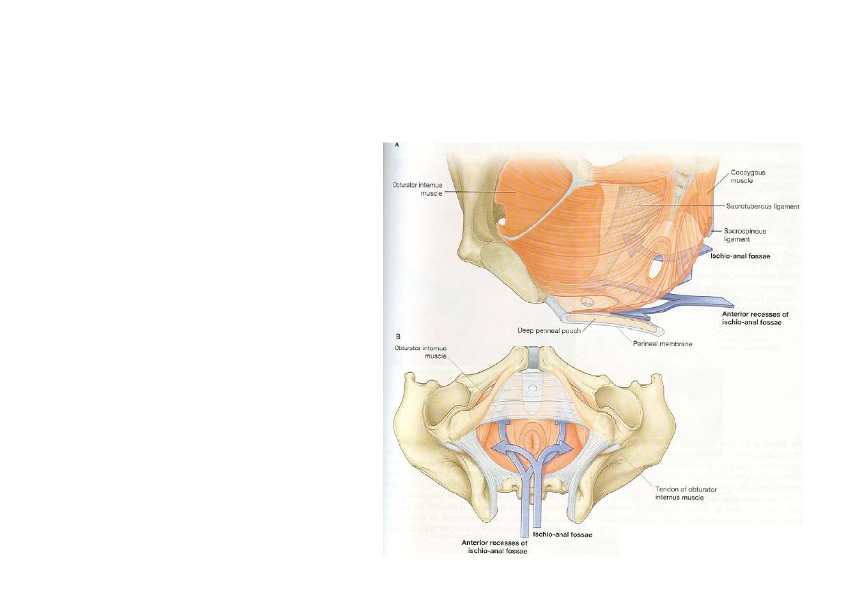
Recesses of Ischiorectal fossa
• Anterior recess
• Posterior recess
• Horseshoe recess

Applied anatomy
• Ischiorectal abscess
• Anorectal fistula and external sinus
• Prolapse of rectum
• Ischiorectal hernia
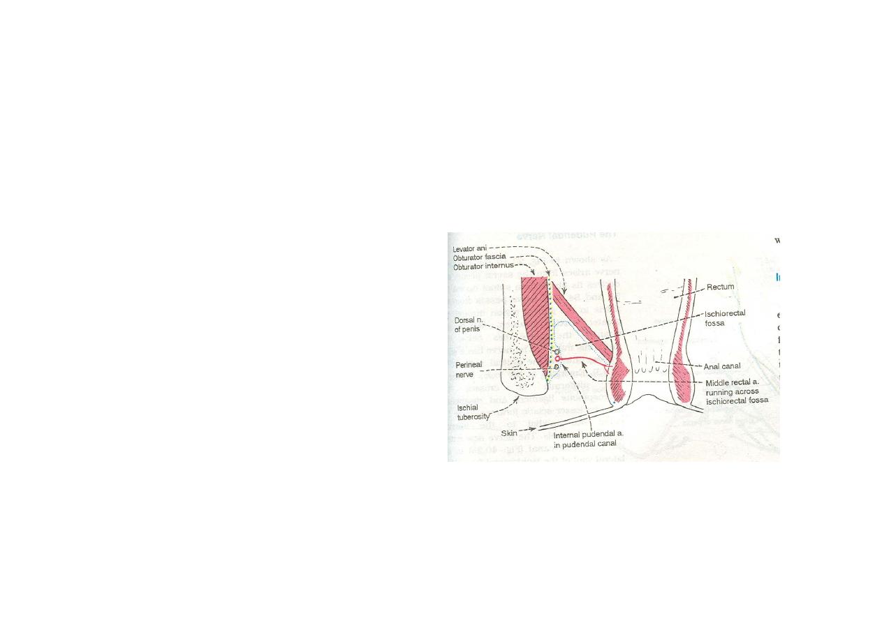
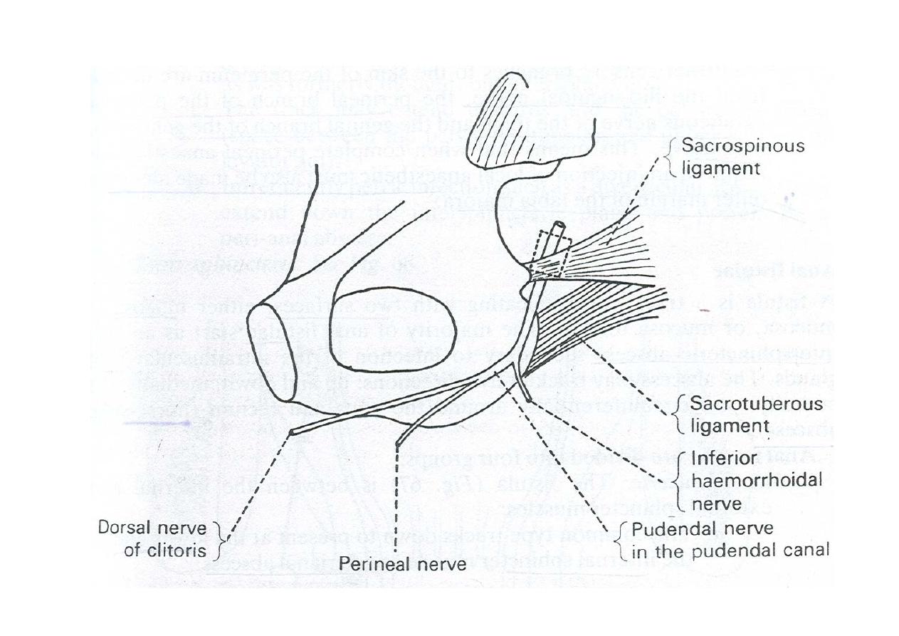
PUDENDAL CANAL
•
seen in the lateral wall of
Ischiorectal fossa
• bounded laterally by
obturator fascia
• Medially by lunate fascia
• Begins Posteriorly near
lesser sciatic foramen
• Inferiorly related to
sacrotuberous ligament
• Extends to the posterior
border of perineal
membrane
Contents
• Pudendal nerve S234
• Internal pudendal
vessels
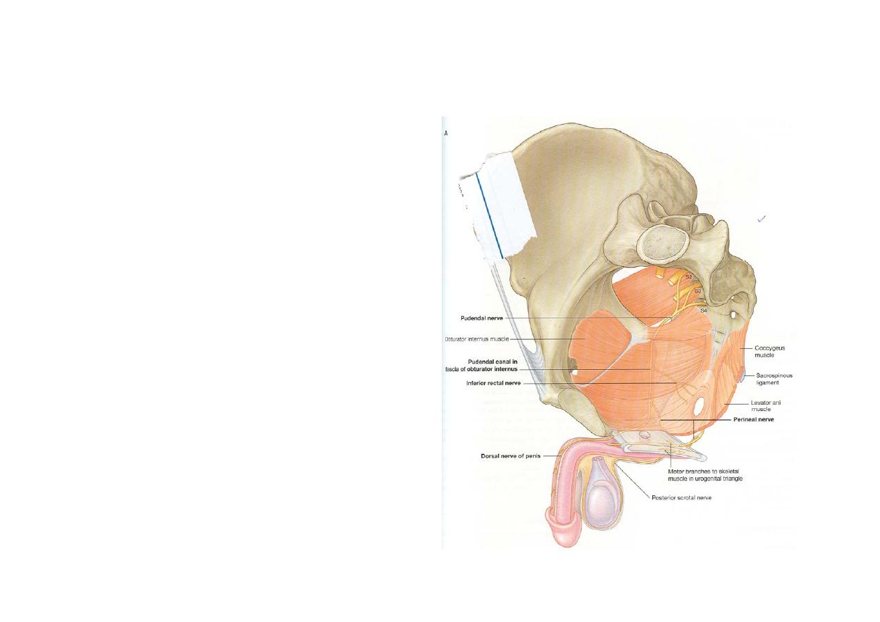
Pudendal nerve
• Chief nerve of
perineum & external
genitalia
• Derived from sacral
plexus
• Branches
Inferior rectal nerve
Perineal nerve
Dorsal nerve of
penis
Applied – pudendal
nerve block
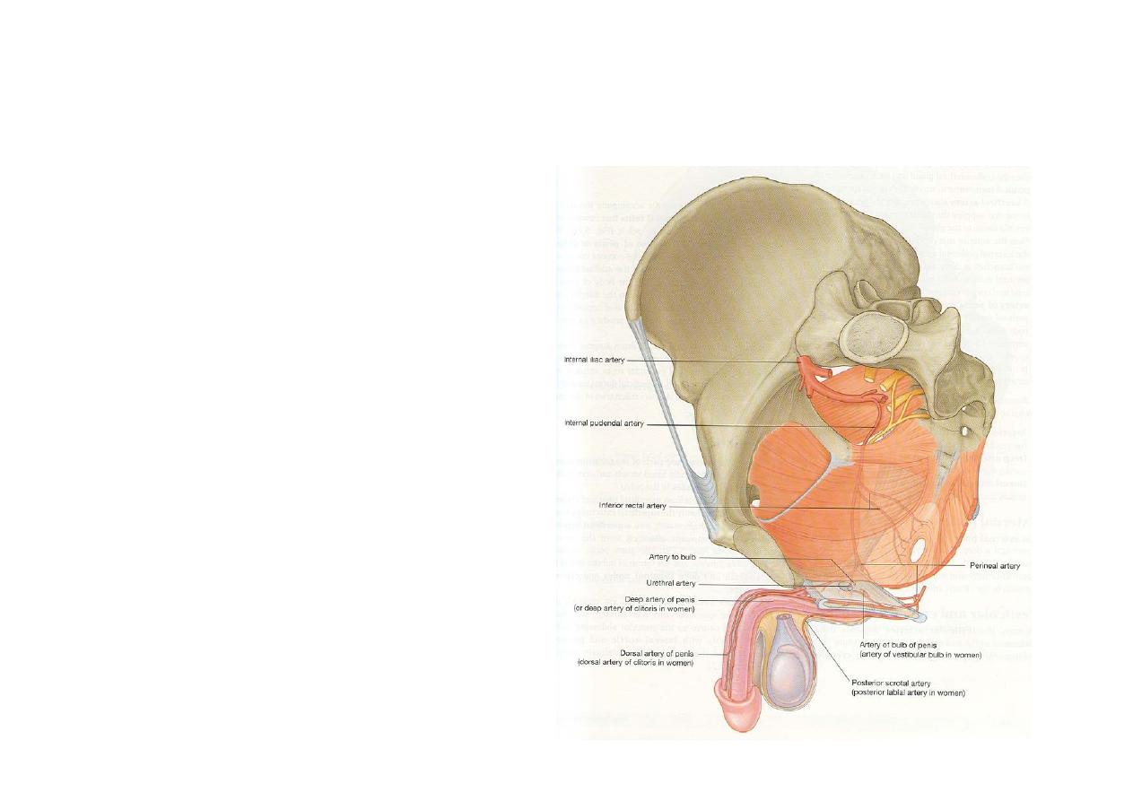
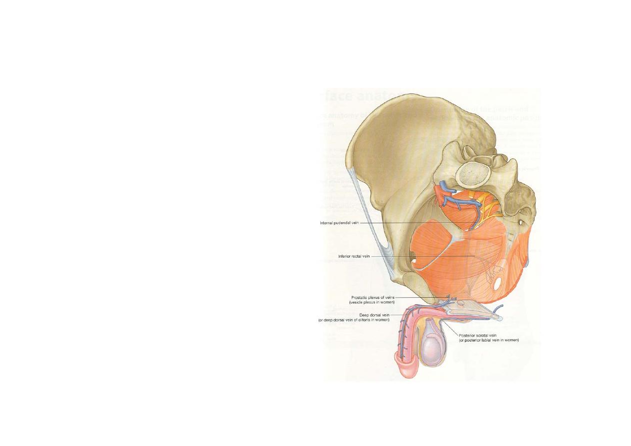
Internal pudendal artery
• Chief artery of
perineum
• Terminal branch of
anerior division of
internal iliac artery
• Branches
• Inferior rectal artery
• Perineal atrery
• Artery of penis/clitoris