Salivary Glands
[د.زيد م/4]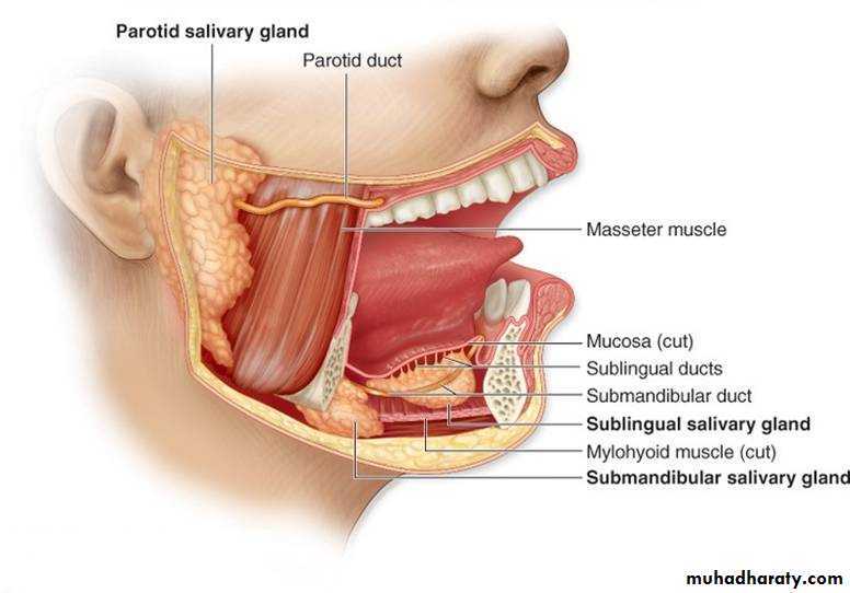
Anatomy of salivary glands:
Two submandibular glands
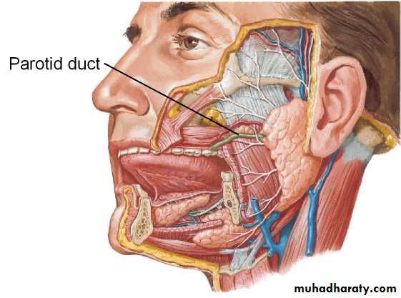
Two parotid glands
Two sublingual glands
Approximately 450 minor salivary glands
Common disorders of minor salivary glands
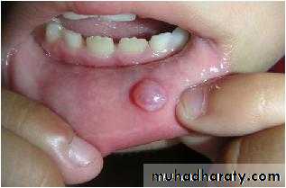
1- Extravasation CystsCommon
trauma to the overlying mucosa.
affect lower lip producing painless swelling and usually translucent .
resolve spontaneously
most require formal surgical excision +overlying mucosa +underlying minor salivary gland.
Recurrence is rare.
Tumours of minor salivary glands
90 % malignant.
anywhere in the upper aerodigestive tract
common sites : upper lip, palate and retromolar regions.
Less common sites nasal and pharyngeal cavities.
Benign minor salivary gland tumors present as painless, firm, slow- growing swellings.
Overlying ulceration is extremely rare.
Treatment: excision of the tumor +overlying mucosa+ primary closure
Malignant minor salivary gland tumours
rare.Firm
discoloration overlying mucosa (pink to blue or black) .
late necrotic with ulceration.
Treatmentwide excision +/- partial or total maxillectomy + reconstruction.
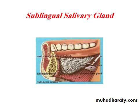
Common disorders of the sublingual glands:
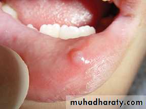
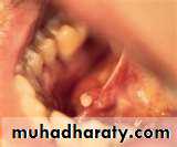
1- Cysts(ranula)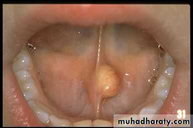
mucous extravasation cyst that arises from a sublingualgland. translucent swelling ‘frog’s belly’ .
resolve spontaneously.
many require formal surgical excision of the
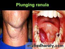
2- Plunging ranula
rare mucous retention cystarise from sublingual and submandibular s g.
Mucus collects within the cyst, which perforates through the mylohyoid muscle diaphragm to enter the neck.
dumb-bell-shaped swelling
fluctuant
Painless
Soft
in the submandibular or submental region of the neck .
Diagnosis : ultrasound or magnetic resonance imaging (MRI).
Treatment:
- Excision transcervical ( removing the cyst+ submandibular+ sublingual glands).
- Smaller plunging ranulas transoral sublingual gland excision+/- marsupialisation.
Tumors of sublingual:
- extremely rare85 per malignant.
hard or firm
painless swelling
in the floor of the mouth.
Treatment:
wide excision + overlying mucosa+ neck dissection. +reconstruction
THE SUBMANDIBULAR GLANDS
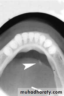
Ectopic/aberrant salivary gland tissue\Stafne bone cyst- most common ectopic salivary tissue .
Asymptomatic
clearly demarcated radiolucency of the angle of the mandible.
below the inferior dental neurovascular bundle.
No treatment is required.
Inflammatory disorders of the submandibular gland(sialadenitis)
acute, chronic or acute on chronic.
Common causes are:Acute submandibular sialadenitis:
1- Bacterial sialadenitis
more common than viral sialadenitis
secondary to obstruction.
antibiotics, if chronically inflamed formal excision.
2- Viral. The paramyxovirus (mumps)
Usually parotitis.occasionally submandibular glands
painful tender swollen glands.
Other viral infections rare.
Chronic submandibular sialadenitis.
Obstruction and trauma to submandibular gland
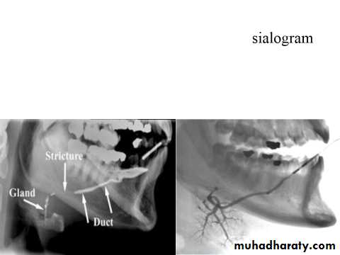
Stone formation (sialothiasis): most common cause of obstruction within the submandibular gland is within the gland and duct system.
80%of all salivary stones occur in the submandibular glands because highly viscous secretions .
80% submandibular stones are radio-opaque
Clinical symptoms
acute painful swelling in the region of the submandibular gland
precipitated by eating
completely obstruct(less common) opening of the submandibular ductswelling develop rapidly 1–2 hours after the meal resolves spontaneously
partial obstruction(more common) (hilum of the gland or within duct in the floor of the mouth)
- infrequent symptoms
- minimal discomfort and swelling
- not confined to mealtimes.
- examination enlarged firm submandibular gland, tender on bimanual examination.
Pus from the sublingual papilla .
Management
stone within the submandibular duct in the floor of the mouth anterior to the point at which the duct crosses the lingual nerve (second molar region) incising over the duct+stone delivered+ leave the wall of the ductstone is proximal to the lingual nervesubmandibular gland excision a removal of the stone + ligation of the submandibular duct
endoscopic retrieval of stone, lithotripsy(sialadenoscope)
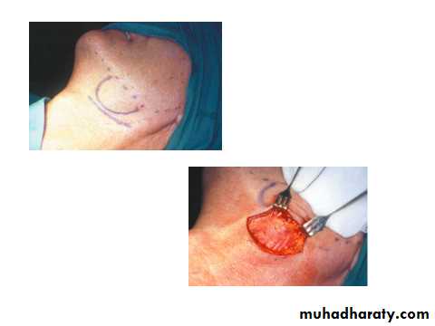
Submandibular gland excision
Indication:sialadenitis
Salivary tumours.
Complications:
• haematoma
• wound infection
• marginal mandibular nerve injury
• lingual nerve injury
• hypoglossal nerve injury
• transection of the nerve to the mylohyoid muscle producing submental skin anaesthesia
Tumours of the submandibular gland
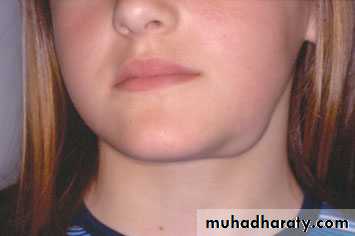
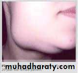
uncommon(benign and malignant)present as a slow-growing, painless swelling within the submandibular triangle.
50 % of submandibular gland tumours are benign.
pain is not a reliable indication of malignancy as benign tumours often present with pain in the affected gland, presumably due to capsular distension or outflow obstruction.
Clinical features of malignant salivary tumours
• facial nerve weakness
• rapid enlargement of the swelling• induration and/or ulceration of the overlying skin
• cervical node enlargement.
Investigation
1-Computed tomography (CT) and MRI scanningthe extension, circumscribed (benign, or diffuse, invasive and probably malignant).
2- Biopsy
Open surgical biopsy is contraindicated as this may seed the tumour into surrounding tissues, making it impossible to eradicate microscopic deposits of tumour cells.
Fine-needle aspiration biopsy : no risk of seeding viable
Management of submandibular gland tumours
surgical excision with a cuff of normal tissue is the goal.- Small tumor+ localised ( entirely within the submandibular gland parenchyma)
intracapsular submandibular gland excision is
- Benign tumors (large and beyond the submandibular gland) suprahyoid neck dissection
(preserving the marginal mandibular branch of the facial nerve, lingual nerve and hypoglossal nerves).
- Overt malignancymodified neck dissection /radical neck dissection .
(may sacrifice of the lingual and hypoglossal nerves )