Histology
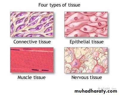
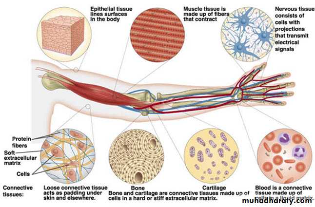
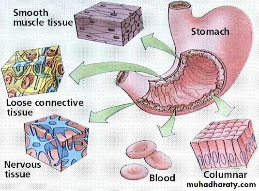
All human structures are composed of just four basic types of tissue:Epithelial tissue Connective tissue Muscular tissue Nervous tissue
These tissues, which are formed by cells and molecules of the extracellular matrix
The main characteristics of these basic types of tissue are shown in table - ATissue
CellsExtracellular
Main
matrix
function
Nervous
Intertwining
None
Transmission
Elongated
of nervousProcesses
impulsesEpithelial
Aggregated
Very small
Lining of
polyhedral cells
amountsurface or
body cavities
glandular
secretion
Muscle
Elongated
Moderate
Movement
contractile cell
amount
Connective
Several types ofAbundant
Support and
fixed and wondering cells
amountprotection
Epithelial tissues
Are composed of closely aggregated polyhedral cells with very little extracellular substance. These cells have strong adhesion due to adhesion molecules, membrane interdigitation, and intercellular junctions.These features allow the cells to form cellular sheets that cover the surface the body and line it cavities or are arranged as three-dimensional secretory units.
The principal functions of epithelial tissues are
Covering and lining of surfaces e.g. skin
Absorption e.g. intestines
Secretion e.g. glands
Sensation e.g. neuroepithelial cells
Contraction e.g. myoepithelial cells
Protection skin
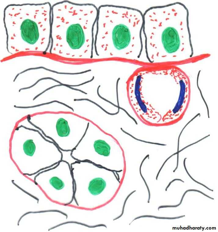
Basal lamina and basement membrane:
most epi cells are separated from the c.t. by sheet of extracellular material called basal lamina, this structure is visible only with E.M. where it appears as a dense layer, consisting of a delicate network of very thin fibrils lamina densa, in addition, basal lamina may have an electron-lucent layer on one or both sides of the lamina densa, called lamina Lucida.
Between cell layers without intervening C.T. such as in lung alveoli, renal glomerulus. The basal lamina is thicker as a result of fusion of the basal lamina of each epithelial cell layer.
The main components of basal lamina are type IV collagen, the glycoprotein [lamina and entactine] and proteoglycans.
Basal laminae are attached to the underlying C.T. by anchoring fibrils formed by type VII collagen.
These components are secreted by epithelial, muscle, adipose and Schwann cells.
Podocyte
Laminin
Lamina densa
Lamina lucid
Basal lamina
Reticular lamina
Anchoring fibril
Basal lamina
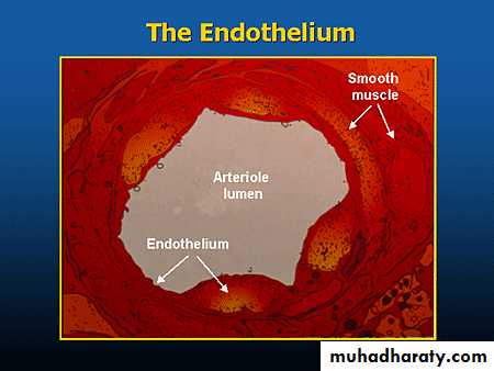
Endothelium
Basement membrane
Basement membrane
Is usually formed by the association of either two basal laminae or a basal lamina and a reticular lamina and is therefore thicker.This layer visible with L.M. when used to specify a periodic acid-Schiff (PAS)-positive layer.
Basal lamina
Types of epithelia
Epithelia are divided into two main groups according to their structure and functioncovering epithelia
Glandular epithelia
In covering epithelia the cells are organized in layers that cover the external surface or line the cavities of the body.
They can be classified according in the number of cell layers and the morphological features of the cells in the surface layer.
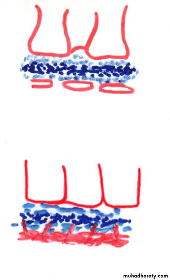
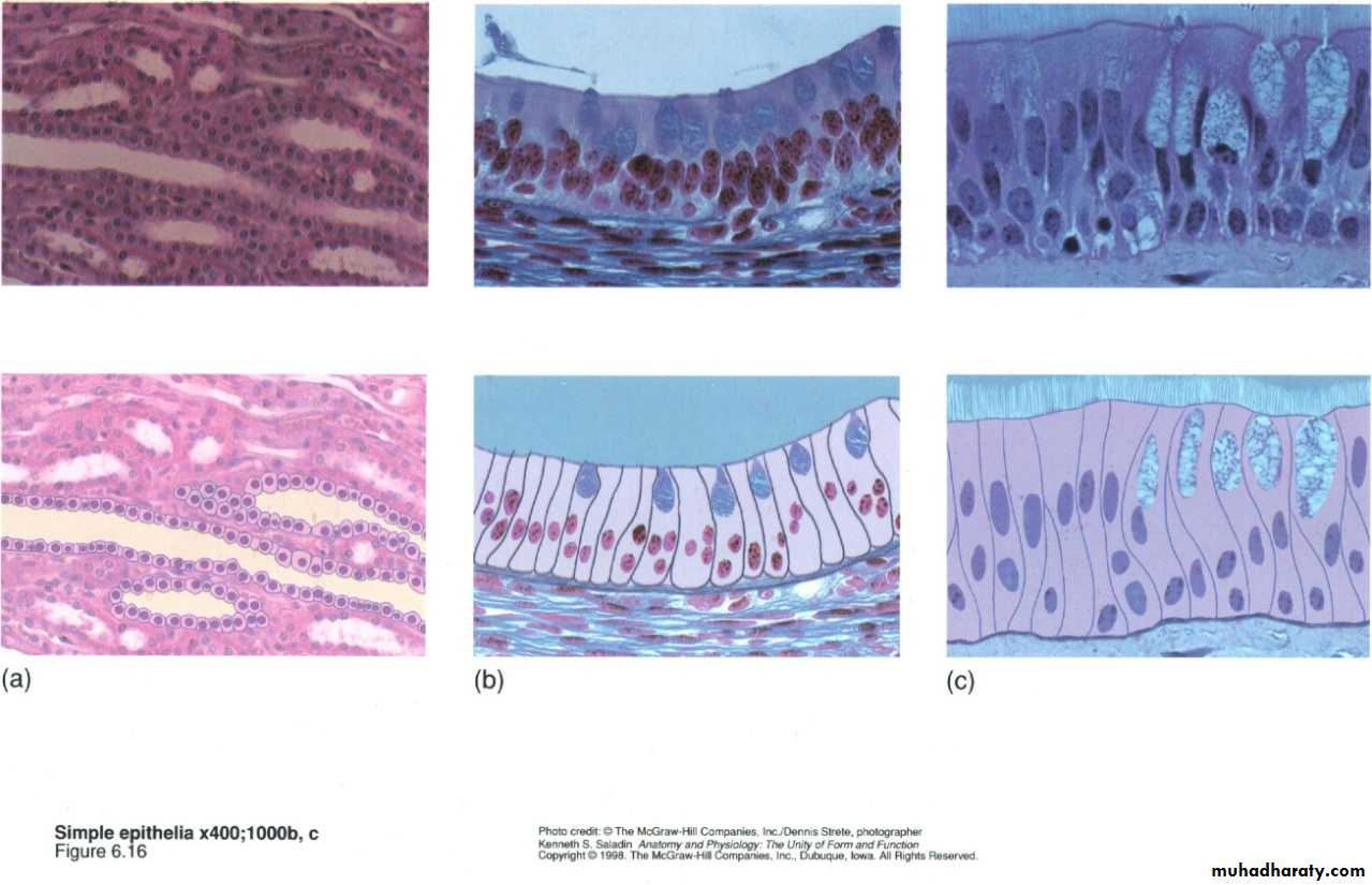
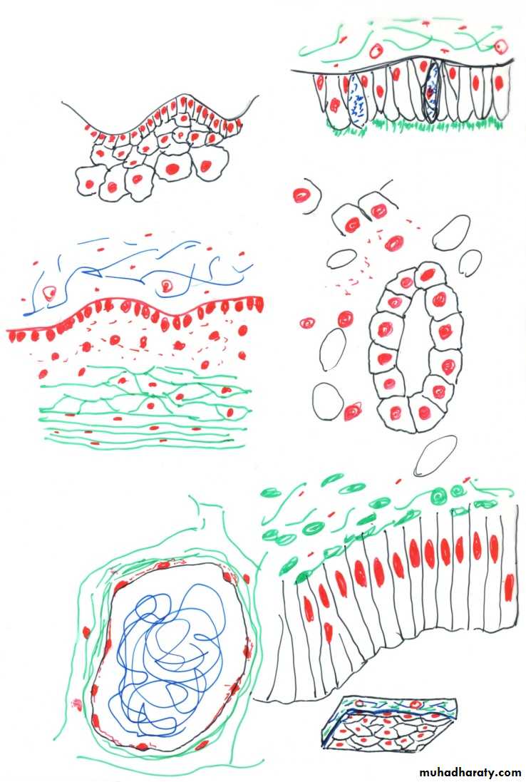
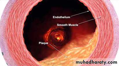
Simple epithelium contains only one layer of cells stratified epithelium contains than one layer based on cell shape, simple epi. can be squamous, cuboidal, columnar, pseudostratified endothelium: squamous that lines blood and lymph v. mesothelium: that lines certain body cavities such as the pleural and peritoneal cavities and covers the viscera.
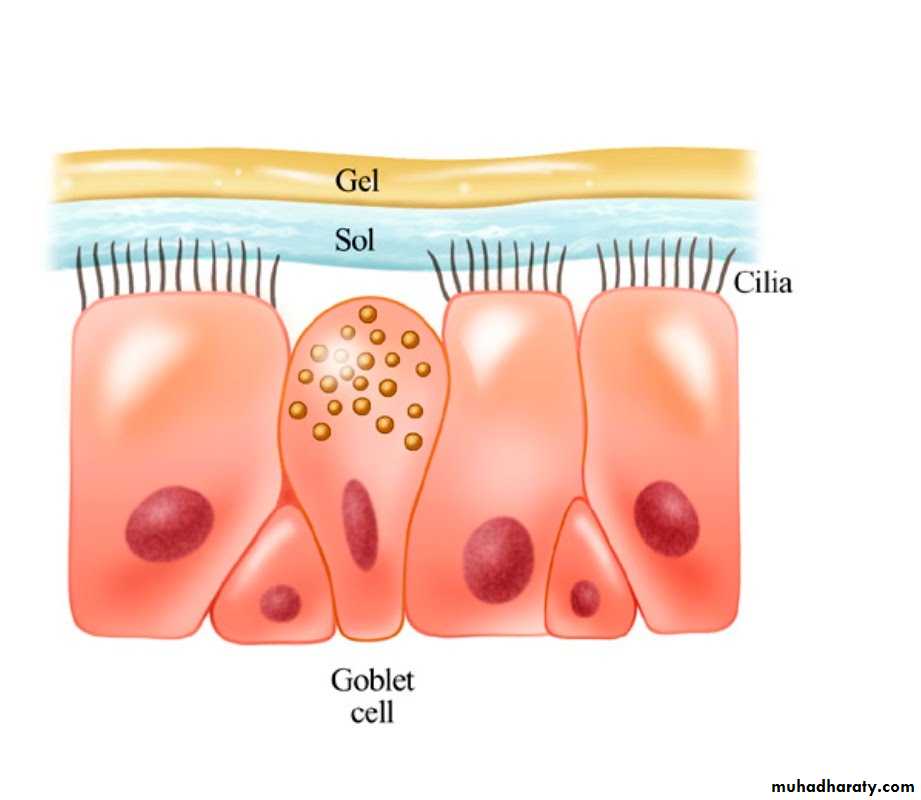
Psudostratified: the nuclei appear to lie in various layers all the cells attached to the basal lamina.
Common types of covering epithelia in the human body
TypeCell form
Example of distribution
Main function
Simple
Squamous
Lining of vessels lining of body cavities. lining bowman’s capsule
Facilitates the movement of the viscera, diffusion
cuboidal
Thyroid, covering the ovary, tubules of kidneyCovering, secretion
Columnar
Lining of intestine, gall bladder
Protection, lubrication, absorption, secretion
Pseudostratified
Some columnar some cuboidal
Lining. Trachea, bronchi, nasal cavity
Protection, secretion cilia-mediated transport of particles trapped in mucus
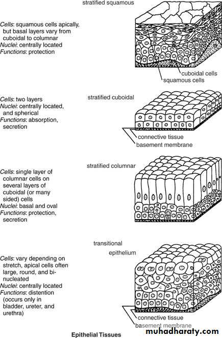
Stratified
Squamous keratinized
Epidermis
Protection, prevent water loss
Squamous nonkeratinized
Mouth, esophagus vagina, larynxProtection, secretion prevent water loss
Cuboidal
Duct of sweat gland ovarian folliclesProtection, secretion
Transitional
Bladder, ureter, part of urethraProtection, distensibility
Columnar
Conjunctiva large duct of salivary gland
Protection
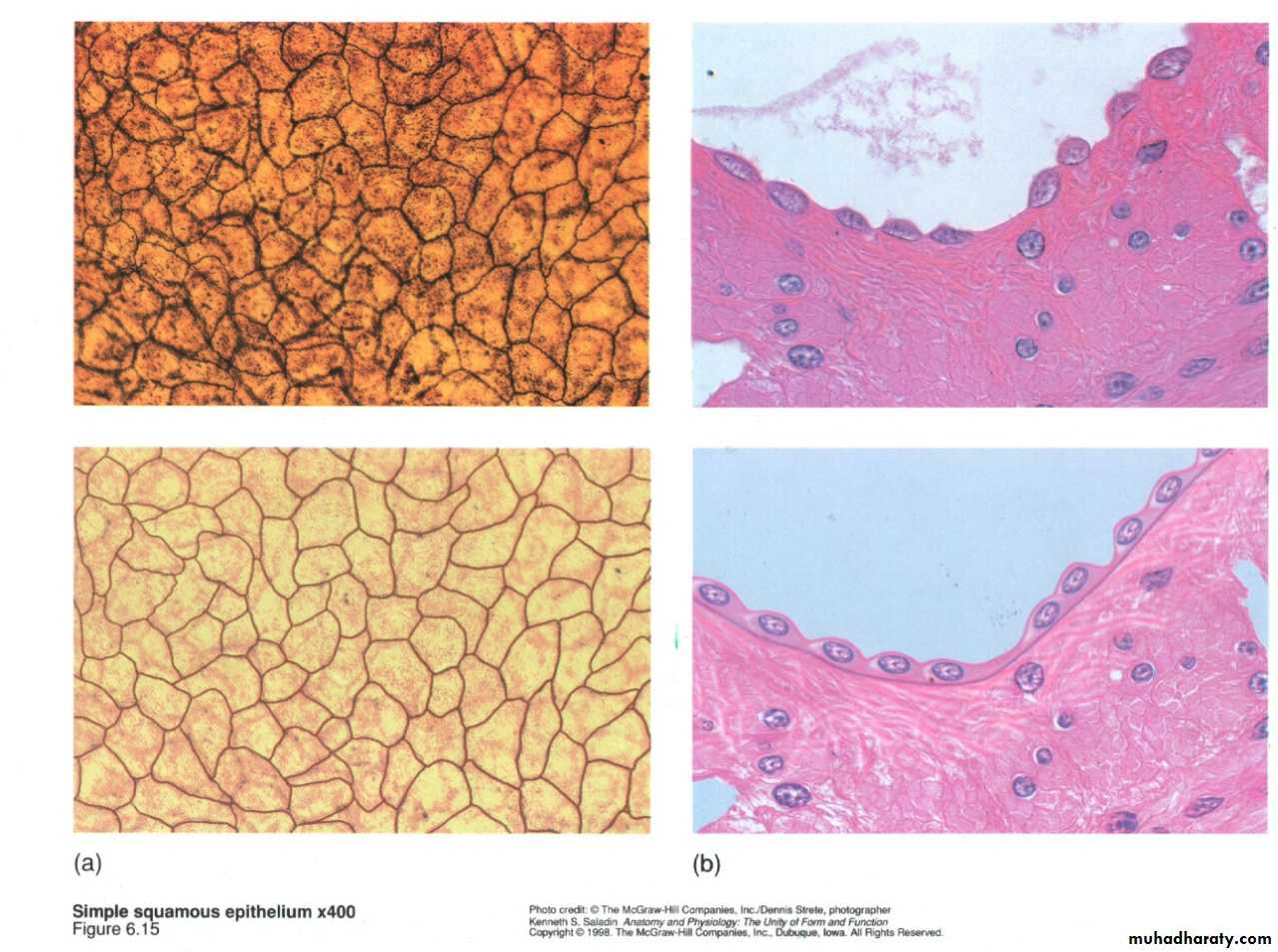
Mesothelium : simple squamous epithelium lining the cavities of the body.
Endothelium: simple squamous epithelium lining the blood vessels.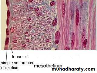
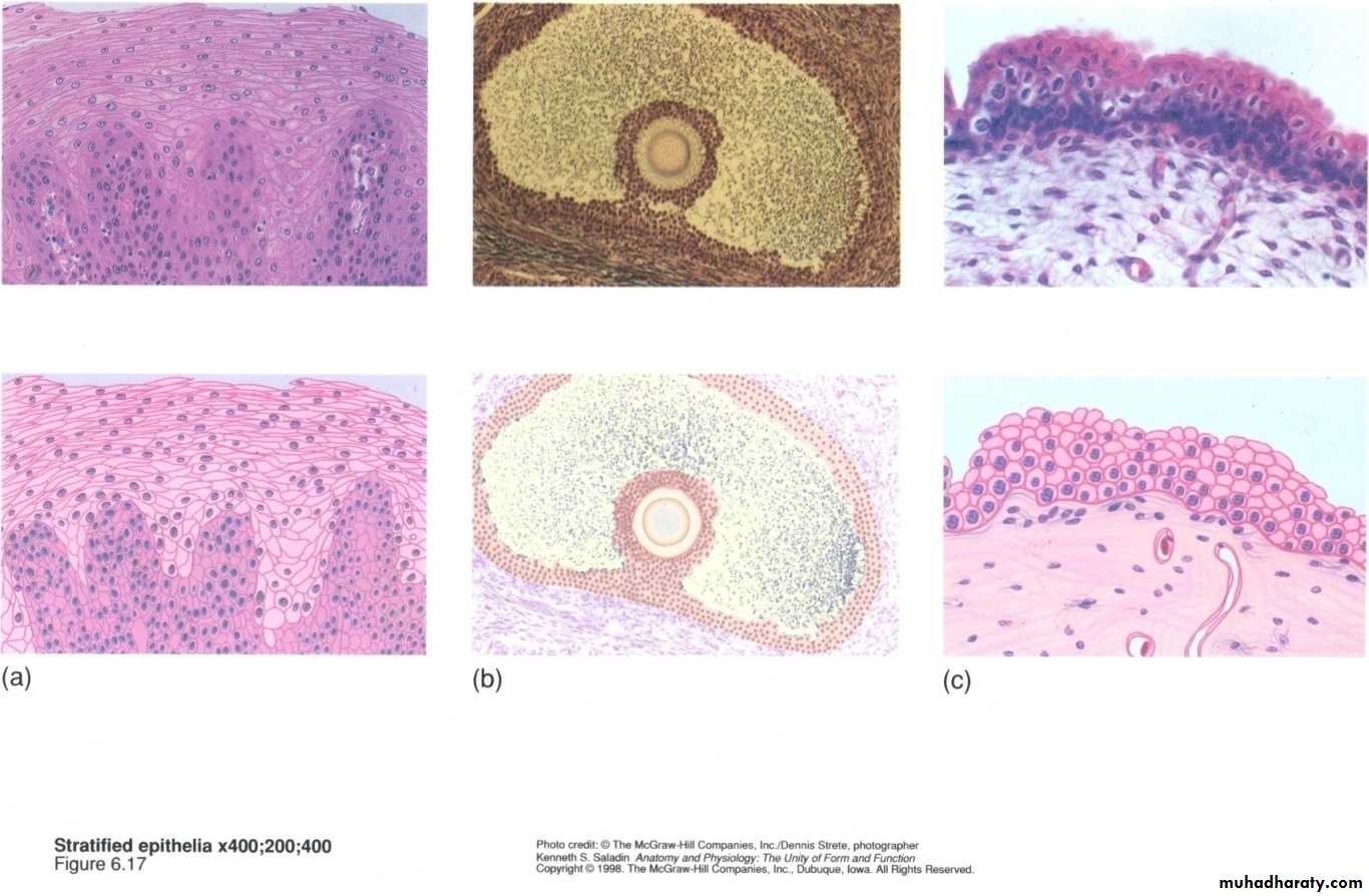
Stratified epithelial
Stratified squamous epi.:Keratinize
Non keratinize
Keratinize: The surface cells have died after having secreted a large amount of the tough protein keratin.
This type of st. epi. Resists abrasion there for well suited to the skin surface and the passages subject to abrasion by the swallowing of food and passage of feces.
Exfoliation: The separation of or desquamation surface cells from the surface.
Exfoliate cytology:The study of exfoliated cells .St. cuboidal epi.: has cuboidal or rounded surface cells. It lines follicles in the ovaries and sperm-producing ducts called seminiferous tubules in the testes , sweat gland duct.
St. Columnar. Epi.: Is a rare type in which col. Surface cells rest on cuboidal basal cell it is found in short transitional zones where a st. Epi. Grades into a columnar or pseudst. Type as in limited region of the pharynx, larynx, anal canal, and male urethra.
Transitional: This type of epi. Is adapted to stretching when the bladders empty the epi. Is up to six cells thick, However as the bladder becomes distended with urine the epi. cells slide over each other the epi. Becomes thinner (only 2 or 3 cells thick) and the surface cells flatten.
Two other of epi
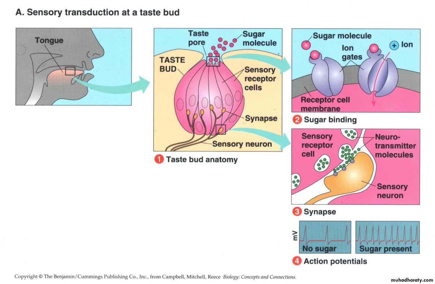
Neuroepithelial cells: Are cell of epi. Origin with specialized sensory function (cell of taste buds)
Myoepithelial cells
Metaplasia
Under certain abnormal conditions are type of epi. T. may undergo transformation in to another type. This eversible process is called mataplassia in heavy cigarette smokers. The ciliated pseudostratified epi. Lining the bronchi can be transformed into stratified sq. epi. Metaplasia is not restricted to epi t. it also occurs in c.t.Benign and malignant tumors can arise from most type of epi cells. Carcinoma is a malignant. Malignant tumors derived from glandular epi. Called adenocarcinoma.
Neuroepithelial cell: Are cells of epithelial origin with specialized sensory function cells of taste buds and of the olfactory mucosa.
Myoepithelial cells: Are branched cells that contain myosin and a large number of action filaments. They are specialized for contraction mainly of the secretary
units of the mammary, sweat and salivary glands.