Muscle tissue
The three types of muscle:Three types of muscle tissue can be identified histologically: skeletal muscle, cardiac muscle and smooth muscle. The fibres of skeletal muscle and cardiac muscle exhibit cross striations at the light microscope level and they are both referred to as striated muscle.
Skeletal muscle
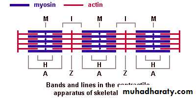
Skeletal muscle constitutes the muscle that is attached to and upper part of the esophagus. (Some people use that moves the term visceral striated muscle in the foregoing examples, but since it is identical in structure to the the skeleton and controls motor movements and posture. There are a few instances where this type of muscle is restricted to soft tissues: the tongue, pharynx, diaphragm muscle the skeleton,.)Skeletal muscle fibres (cells) are actually a multinucleated formed by the fusion of individual small muscle cells or myoblasts, during development. They are filled with longitudinally arrayed subunits called myofibrils. The myofibrils are made up of the myofilaments myosin (thick filaments) and actin (thin filaments). The striations reflect the arrangement of actin and myosin filaments and support structures. The individual contractile units are called sarcomeres. A myofibril consists of many sarcomeres arranged end to end. The entire muscle exhibits cross-striations because sarcomeres in adjacent myofibrils and muscle fibers are in register. The most obvious feature in longitudinal sections of skeletal muscle is the alternating pattern of dark and light bands, called respectively the A (anisotropic) and I (isotropic) band. The I band is bisected by a dense zone called the Z line, to which the thin filaments of the I band are attached.
The nuclei are located peripherally, immediately under the plasma membrane (sarcolemma). The thickness of individual muscle fibres varies (depending for example on location in the body and exercise) but each fibre is of uniform thickness throughout its length. Skeletal muscle fibres do not branch.
Connective tissue elements surround muscle fibres. Individual muscle fibres are surrounded by a delicate layer of reticular fibres called the endomysium. Groups of fibres are bundled into fascicles by a thicker CT layer called the perimysium. The collection of fascicles that constitutes one muscle is surrounded by a sheath of dense CT called the epimysium, which continues into the tendon
. Blood vessels and nerves are found in the CT associated with muscle. The endomysium contains only capillaries and the finest neuronal branches.
Summary: Skeletal muscle fibres bear obvious striations, have many peripherally located nuclei, are of the same thickness throughout their length and do not branch.
.

. .
Cardiac muscle
Cardiac muscle is the type of muscle found in the heart, and at the base of the venae cavae as they enter into the heart. Cardiac muscle is intrinsically contractile but is regulated by autonomic and hormonal stimuli.Cardiac muscle exhibits striations because it also has actin and myosin filaments arranged into sarcomeres. Generally these striations do not appear as well-defined as in skeletal muscle. Cardiac muscle also has a much greater number of mitochondria in its cytoplasm.
At the light microscope level, a number of features distinguish cardiac from skeletal muscle. Cardiac muscle cells have only one or two nuclei, which are centrally located. The myofibrils separate to pass around the nucleus, leaving a perinuclear clear area . This clear area is occupied by organelles, especially mitochondria (which are of course not visible in LM). As in skeletal muscle, individual muscle fibres are surrounded by delicate connective tissue. Numerous capillaries are found in the connective tissue around cardiac muscle fibres.
Cardiac muscle cells are joined to one another in a linear array. The boundary between two cells abutting one another is called an intercalated disc. Intercalated discs consist of several types of cells junctions whose purpose is to facilitate the passage of an electrical impulse from cell to cell and to keep the cells bound together during constant contractile activity. Unlike skeletal muscle fibres, cardiac muscle fibres branch and anastomose with one another. Although made up of individual fibres, heart muscle acts as a functional syncytium during contraction for the efficient pumping of blood.
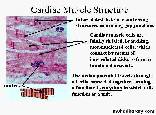
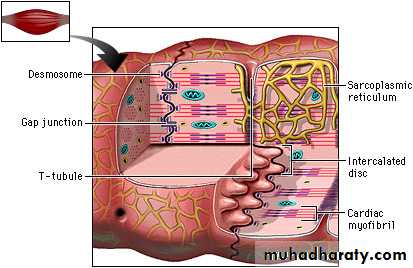
Summary: Cardiac muscle fibres are striated, have one or two centrally located nuclei, branch and anastomose with other fibres, and are joined to one another by intercalated discs.
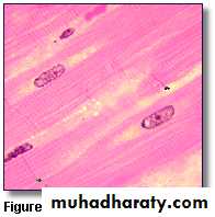
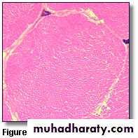
Figure 3 HYPERLINK "http://www.courseweb.uottawa.ca/medicine-histology/english/ss_basictissues/muscle_tissue.htm" \l "Muscle Tissue"
shows a longitudinal section of cardiac muscle. Two nuclei belonging to cardiac muscle fibres can be clearly seen (lower right and middle left). Both have a prominent nucleolus and a delicate pattern in the remainder of the nucleus. The perinuclear area is also evident around both nuclei. The other two nuclei are not very clear, but appear to lie in connective tissue and probably belong to fibroblasts. Fibroblast nuclei tend to be more flattened and darker staining. Two intercalated discs are indicated , others, not quite so prominent, can also be seen. To the left of the upper intercalated disc indicated, a muscle fibre appears to be branching.
.
Smooth muscle
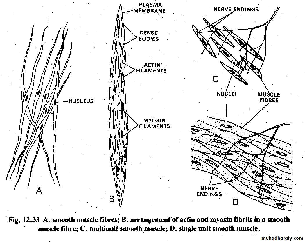
Smooth muscle is the intrinsic muscle of the internal organs and blood vessels. It is also found in the iris and ciliary body of the eye and associated with hair follicles (arrector pili). No striations are present in smooth muscle due to the different arrangement of actin and myosin filaments. Like cardiac muscle, smooth muscle fibres are contractile but responsive to autonomic and hormonal stimuli. They are specialized for slow, prolonged contraction.
Smooth muscle fibres are generally arranged in bundles or sheets. Each fibre is fusiform in shape with a thicker central portion and tapered at both ends. The single nucleus is located in the central part of the fibre. Fibres do not branch. , . Smooth muscle fibres lie over one another in a staggered fashion (tapered part of one fibre over thicker part of another). In longitudinal sections, it is often not possible to distinguish the fibre boundaries, and smooth muscle may closely resemble connective tissue (bundles of collagen). Where smooth muscle bundles are interlaced with bundles of connective tissue (eg. in the uterus), one can distinguish the smooth muscle by the orientation of the nuclei (all oriented in the same direction), smooth muscle cell has a nucleus, fibroblast nuclei are more scattered in bundles of CT). .
One distinguishing physiological feature of smooth muscle is its ability to secrete connective tissue matrix. In the walls of blood vessels and the uterus in particular, smooth muscle fibres secrete large amounts of collagen and elastin.
Summary: Smooth muscle fibres are fusiform with tapered ends, have a single centrally located nucleus, and do not exhibit striations.
.
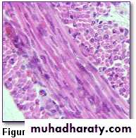
Figure 6
shows a layer of smooth muscle in longitudinal section lying between two layers in cross section. Notice how the shape of the fibres in longitudinal section is harder to distinguish than when individual fibres are seen,
Smooth muscle is an involuntary non-striated muscle. It is divided into two sub-groups; the single-unit (unitary) and multiunit smooth muscle. Within single-unit smooth muscle tissues, the autonomic nervous system innervates a single cell within a sheet or bundle and the action potential is propagated by gap junctions to neighboring cells such that the whole bundle or sheet contracts as a syncytium (i.e., a multinucleate mass of cytoplasm that is not separated into cells). Multiunit smooth muscle tissues innervate individual cells; as such, they allow for fine control and gradual responses, much like motor unit in skeletal muscle.
ر

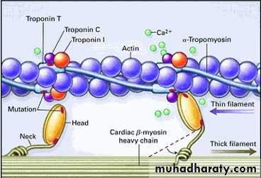
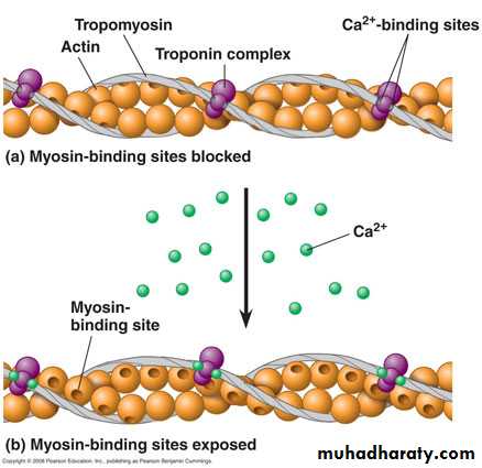
Troponin is a complex of three regulatory proteins that is integral to muscle contraction[1] in skeletal and cardiac muscle but not smooth muscle
)
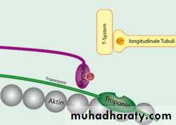
Tropomyosin and troponin bound to actin blocking myosin binding
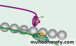
Troponin binds to calcium, releasing Tropomyosin from actin allowing myosin binding
Tropomyosin is an actin-binding protein that regulates actin mechanics. It is important, among other things, for muscle contraction. Tropomyosin, along with the troponin complex, associate with actin in muscle fibers and regulate muscle contraction by regulating the binding of myosin. In resting muscle, tropomyosin overlays the myosin binding sites on actin, with a single tropomyosin molecule spanning 7 actin subunits, and is "locked" down in this position by troponin T (tropomyosin binding troponin) and troponin I (inhibitory troponin). Upon release of calcium from the sarcoplasmic reticulum calcium binds to troponin C (calcium binding troponin). This "unlocks" tropomyosin from actin, allowing it to move away from the binding groove. Myosin heads can now access the binding sites on actin. Once one myosin head binds, this fully displaces tropomyosin and allows additional myosin heads to bind, initiating muscle shortening and contraction. Once calcium is pumped out of the cytoplasm and calcium levels return to normal, tropomyosin again binds to actin, preventing myosin from binding.