Light-Related Disorders
Sunlight has profound effects on the skin and is associated with a variety of diseases.Ultraviolet (UV) light causes most photobiologic skin reactions and diseases.UV light is divided into: UVA (320 to 400 nm), UVB (290 to 320 nm), and UVC (100 to 290 nm).UVA: is further subdivided into UVA I (long wave) and UVA II. The ratio of UVA to UVB is 20:1, and two thirds of this UVA is UVA I.
More than 90% of UV radiation may penetrate clouds!!!
UV radiation generates reactive oxygen species that damage skin.
UVA : UVA causes immediate and delayed tanning and contributes little to erythema and burning. Constant throughout the day and throughout the year. The longer wavelengths of UVA can penetrate more deeply, reaching the dermis and subcutaneous fat. Chronic exposure causes connective tissue degeneration (photoaging), photocarcinogenesis, and immunosuppression.
UVA augments the carcinogenic effects of UVB
UVA penetrates window glass and interacts with topical and systemic chemicals and medications.
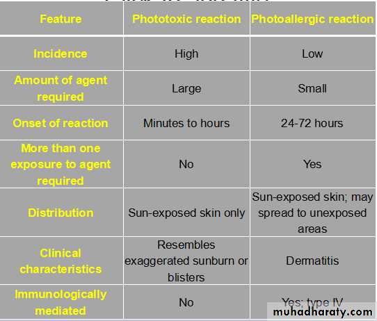
It produces photoallergic and phototoxic reactions.
UVB: UVB produces the most harmful effects and is greatest during the summer. Snow and ice reflect UVB radiation. UVB delivers a high amount of energy to the stratum corneum and superficial layers of the epidermis. It is primarily responsible for sunburn, suntan, inflammation, delayed erythema, and pigmentation changes. Chronic effects include photoaging, immunosuppression, and photocarcinogenesis.
It is most intense when the sun is directly overhead between 10 am and 2 pm.
UVB is absorbed by window glass.
Prior exposure to UVA enhances the sunburn reaction from UVB.
UVC: UVC is almost completely absorbed by the ozone layer and is transmitted only by artificial sources such as germicidal lamps.
So what happens?
DNA is mutated by UVB.
Absorption of UVA leads to the release of reactive oxygen species.These reactive oxygen species cause oxidation of lipids and proteins that affect DNA repair, produce dyspigmentation, and cause photoaging and carcinogenesis.
NORMAL AGING :The skin begins to show signs of aging by ages 30 to 35. Aged skin is thin, fragile, and inelastic. The epidermis becomes thin.There is a gradual loss of blood vessels, dermal collagen, subcutaneous fat, and the number of elastic fibers .There is a reduction in the density of hair follicles, sweat ducts, and sebaceous glands, resulting in a reduction in perspiration and sebum production. The skin becomes atrophic and fragile when subcutaneous tissue is lost. Loss of elastic fibers results in fine wrinkles that disappear by stretching. The skin is easily distorted, but it recoils slowly. Potent steroids should not be used on aged skin.
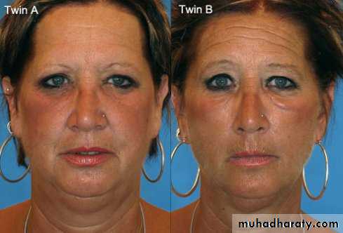
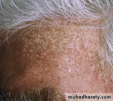
PHOTOAGING:Photoaging refers to those skin changes superimposed on intrinsic aging by chronic sun exposure.Unprotected, chronically exposed children can acquire significant actinic damage by the time they reach 15. Sun-damaged skin is characterized by elastosis (a coarsening and yellow discoloration of the skin), irregular pigmentation, roughness or dryness, telangiectasia, deep wrinkling, follicular plugging, and a variety of benign and malignant neoplasms. The epidermis thickens.
Solar Elastosis: Is a sign highly characteristic of severe sun damage.There is massive deposition in the upper dermis of an abnormal, yellow, amorphous elastotic material that does not form functional elastic fibers. This altered connective tissue does not have the resilient properties of elastic tissue.
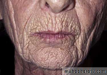
Wrinkling becomes coarse and deep rather than fine, and the skin is thickened. These wrinkles do not disappear by stretching.
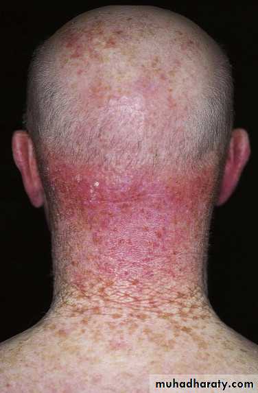
Sun-induced wrinkling on the back of the neck shows a series of crisscrossed lines that form a rhomboidal pattern (cutis rhomboidalis nuchae).
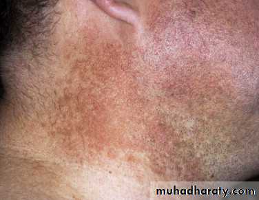
Reddish-brown, reticulate pigmentation with atrophy and telangiectasia is seen on the sides of the neck (poikiloderma of Civatte). مامطلوب
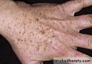
Slightly elevated seborrheic keratoses occur on the back of the hands and may be misdiagnosed as solar lentigines.
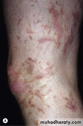
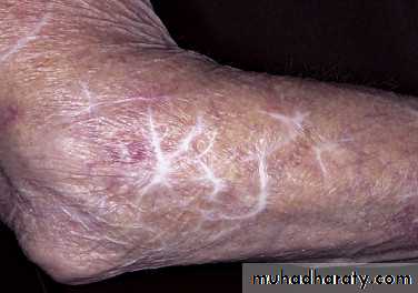
Blood vessels diminish in number, and the walls of the remaining vessels become thin. Bleeding occurs with the slightest trauma to the sun-damaged surfaces of the forearms and hands but not to the unexposed surfaces. Fragile sun-damaged skin is easily torn and heals with haphazard scars called stellate pseudoscars.
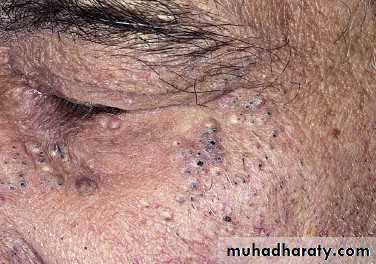
Actinic comedones. Open and closed comedones are present in the periorbital areas. Acne-like inflammation does not occur.
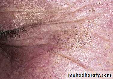
Actinic comedones may become very large but can easily be expressed. with a comedone extractor.
Treatment of photoaging
1.Photoaging is treated with either topical treatments (tretinoin cream) or resurfacing through chemical peels, dermabrasion, or lasers.2.Sun protection: Sunscreens are topical agents that absorb, scatter, or reflect UV radiation and visible light.
SPF???مطلوب(sun protection factor) : amount of erythema caused by exposure to certain amount of UV radiation in the prescense of sun cream to amount of erythema caused by exposure to certain amount of UV radiation in the abscense of sun cream
3.Estrogen replacement.
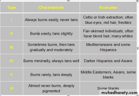
Skin phototypes: مهم الجدول
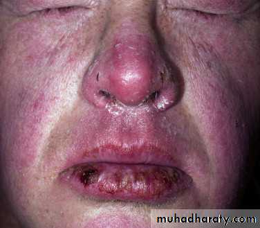
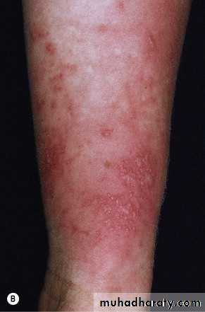
PHOTOTOXIC REACTIONS :
Phototoxicity occurs when a photosensitizer is absorbed into the skin either topically or systemically in appropriate concentrations and is exposed to adequate amounts of specific wavelengths of light, usually UVA.There is a variety of topical and systemic agents like perfumes, plants {phytophotodermatitis} (eg., lime juice), and drugs (eg., antibacterials, NSAIDs, diuretics, retinoids, antifungals).
Doxycycline-induced phototoxicity