Structures passing in & out:
Applied anatomy:
The PPG is the ganglion of seasonal allergy.
Allergic rhinitis & conjunctivitis involve mainly m.m. supplied by this ganglion
whose sensory root is derived from Vb.
The nose:
The External Nose:
Skeleton:
1-The bony part:
It is formed by the two nasal bones, it forms the upper part of the external nose.
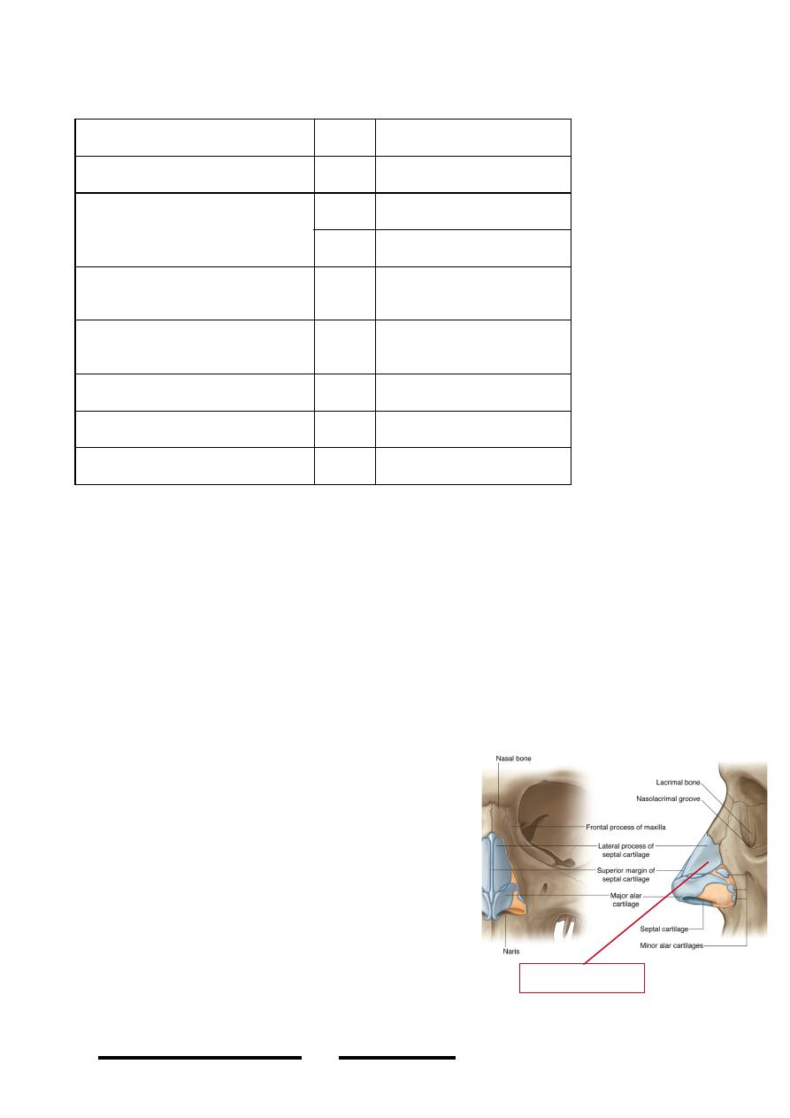
2-The cartilagenous part:
- Formed by two lateral nasal & two alar
cartilages
- Forms the lower part of the nose.
- Lateral nasal cartilage; is a triangle whose
borders articulate with the nasal bone
above, septal cartilage medially & alar
cartilage inferiorly.
- Alar cartilage; thin flexible cartilage,
hooks around the nostrils from lateral
rounded part to the medial hook-shaped
part , the medial ends of the two cartilages
are connected by loose tissue in the midline
below the septum
THROUGH
STRUCTURE
Foramen Rotundum
in
Vb
Pterygomaxillary fissure
In
Maxillary artery
out
Post. Sup. Alv. V.&N.
Inferior orbital fissure
In-out
Connection between
IOV & pt. plexus
Sphenopalatine foramen
out
PSLN V.&N.
Nasopalatine V.&N.
Pterygoid canal
in
Vidian nerve
Palatine foramina
out
Palatine V.&N.
Pharyngeal canal
out
Pharyngeal V.&N.
!
82
Head & Neck Dr. Nawfal K. Al-Hadithi
Lateral nasal cartilage
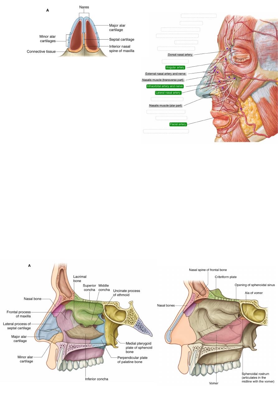
Arterial supply:
1-Lateral nasal branch of facial artery.
2-Nasal branches of infraorbital artery.
3-Dorsal nasal branch of inferior
ophthalmic artery.
Veins:
Accompany arteries.
Nerves: p80
1-Bridge; infratrochlear (Va)
2-Midline shin below the bridge;
external nasal (Va)
3-Lateral side; infraorbital (Vb)
The Nasal Cavity:
Skeleton:
1- Lateral wall:
*Upper part: from before backward the bones are:
Nasal bone, frontal process of maxilla, lacrimal bone, ethmoid, body of sphenoid
*Lower part: from before backward the bones are:
Maxilla, inferior concha, palatine bone, pterygoid plate
2-Roof: cribriform plate of ethmoid.
3-Floor: hard palate (palatal process of maxilla & horizontal plate of palatine).
4-Medial wall (septum):
Posterosuperior; perpendicular plate of ethmoid
Posteroinferior; vomer
Anterior; septal cartilage
!
83
Head & Neck Dr. Nawfal K. Al-Hadithi
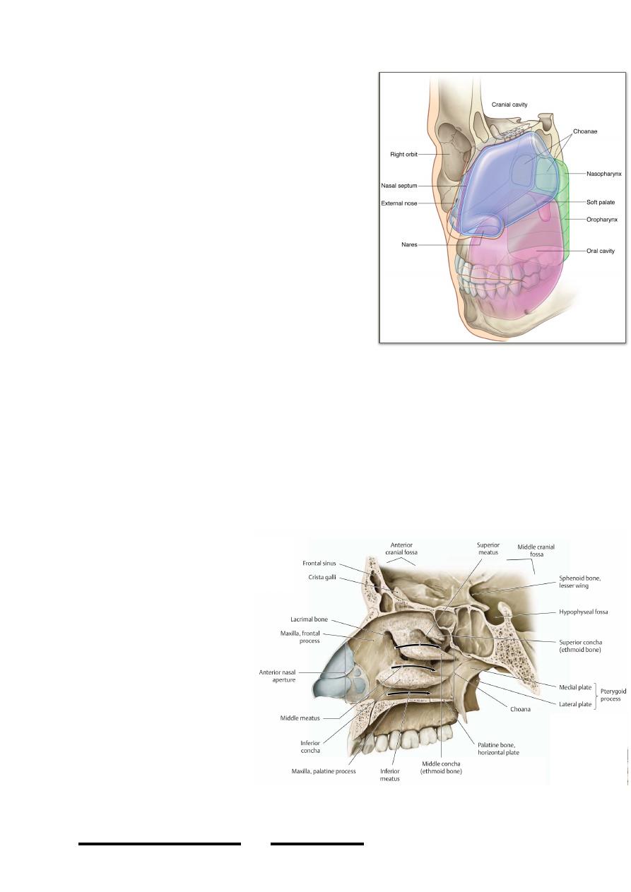
Parts of the nose:
• Vestibule; is the skinny part of the nasal
cavity at the nostrils, it carries coarse hairs
with other skin derivatives
• Choanae; are the posterior nasal apertures
which open to the nasopharynx, they are 2 X
1.5 cm in dimensions & separated from each
other by the posterior part of the septum
• Conchae:
- Are the three scroll-like projections in the
lateral wall of the nose
- Their size increase as we descend downward
- The upper two are parts of the ethmoid while
the lower is a separate bone
- Their medial ends almost reach the septum
- Their action is to increase surface area of
m.m. to humidify & warm air
- They also act as shelves for the underlying
meatuses
- Their covering m.m. is highly vascular & contain erectile tissue so their size
may increase or decrease according to the situation
• Meatuses:
- Are the groove-like passages underneath the corresponding conchae
- The superior meatus receives the opening of the posterior ethmoidal air sinus
- The middle meatus show a bulging (bulla ethmoidalis) formed by the middle
ethmoidal sinus on which opens the sinus itself.
- B e l o w t h e b u l l a &
parallel to it lies the
uncinate process of the
ethmoid converting the
area between it & the
bulla into a smilunar
h i a t u s
( h i a t u s
semilunaris) into its
anterior end opens the
f r o n t o n a s a l d u c t
(frontal sinus), just
behind it opens the
anterior ethmoidal sinus
& in the posterior part
of the hiatus opens the
maxillary sinus.
- The inferior meatus
receives the opening of
the nasolacrimal duct.
!
84
Head & Neck Dr. Nawfal K. Al-Hadithi
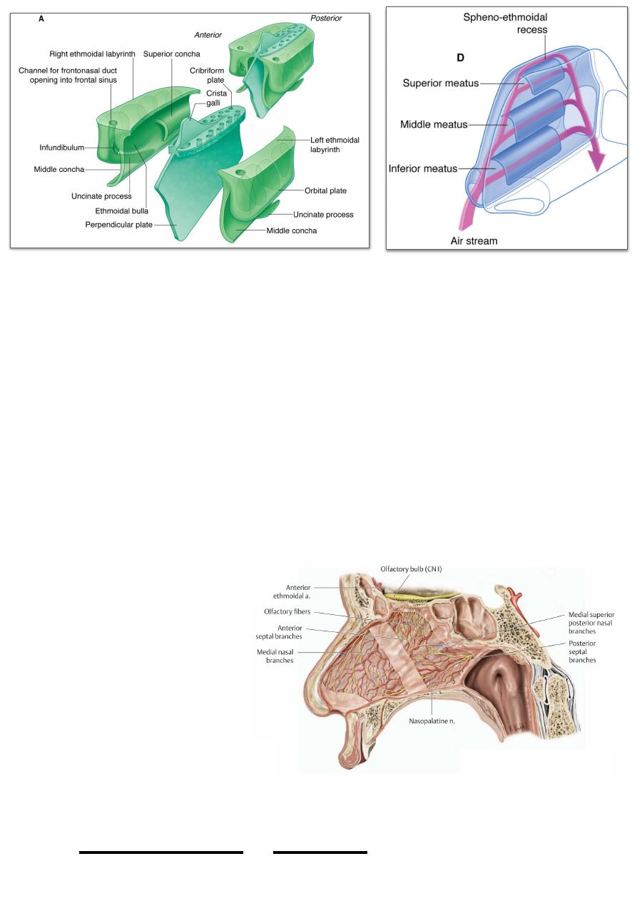
• Spheno-ethmoidal recess:
Is the part of the lateral nasal wall between the superior concha & the body of
sphenoid, it receives the sphenoidal ostia
Mucous membranes:
- The nasal cavity is lined with m.m. except for the vestibule
- m.m. of the nose is firmly bounded to the periosteum & perichondrium of the
underlying structures (mucoperiosteum & mucoperichondrium)
- M.m. is continuous with other m.m. of the chambers with which the nasal
cavity is continuous like the nasopharynx, paranasal sinuses, conjunctiva, ..
Nerve supply: page 80
1- Lateral wall:
According to nerve supply the lateral wall of the nose could be divided into four
quadrants:
• Anterosuperior: anterior ethmoidal n.
• Posterosuperior: posterior superior lateral nasal n.
• Anteroinferior: anterior superior alveolar n.
• Posteroinferior: greater & lesser palatine n.
2- The septum:
- The nasopalatine
branch of the PPG
(Vb) descends on
each side of the nasal
septum in an antero-
i n f e r i o r l y i n t h e
d i r e c t i o n o f t h e
incisive foramen of
the palate.
- The part below the
course of this nerve is
supplied by the same
nerve
- The part above it is supplied by the medial nasal branches
of the anterior ethmoidal n. (Va)
!
85
Head & Neck Dr. Nawfal K. Al-Hadithi
3- The roof:
The roof is lined by olfactore neroepithelium & is supplied by the olfactory nerve.
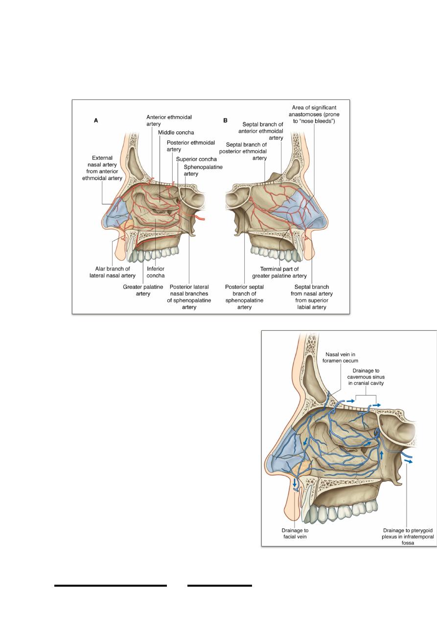
Arterial supply:
Follows the same way of nerve supply for both lateral wall & the septum.
Venous drainage:
- Starts in the cavernous plexus on the middle
& inferior conchae & lower part of the
septum
- From the lateral wall:
*Anterior ½; anterior facial vein
*Posterior ½; pterygoid venous plexus
- From the septum like lateral wall.
- connections:
*veins of the roof; superior sagittal sinus
*anterior ethmoidal; superior ophthalmic v.
Applied anatomy:
• Deviation of nasal septum is a common
problem involving usually the septal
cartilage commonly associated with part of
the perpendicular plate of ethmoid & / or
vomer.
• Epistaxis, bleeding from the nose most often
takes place in Little’s area on the lower part
of the septum where the cavernous venous
plexus & capillaries often injured by the fingers & foreign bodies.
!
86
Head & Neck Dr. Nawfal K. Al-Hadithi
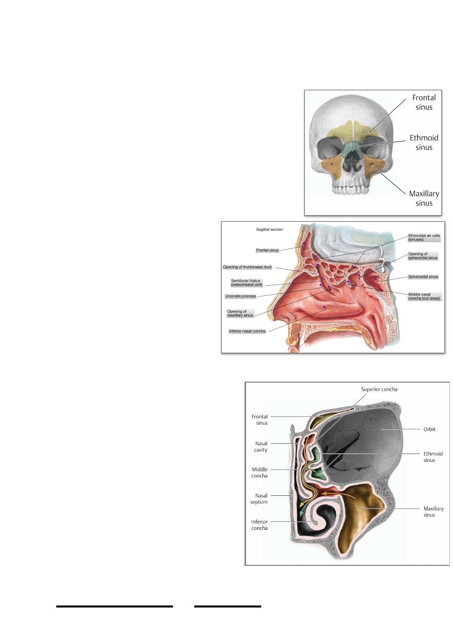
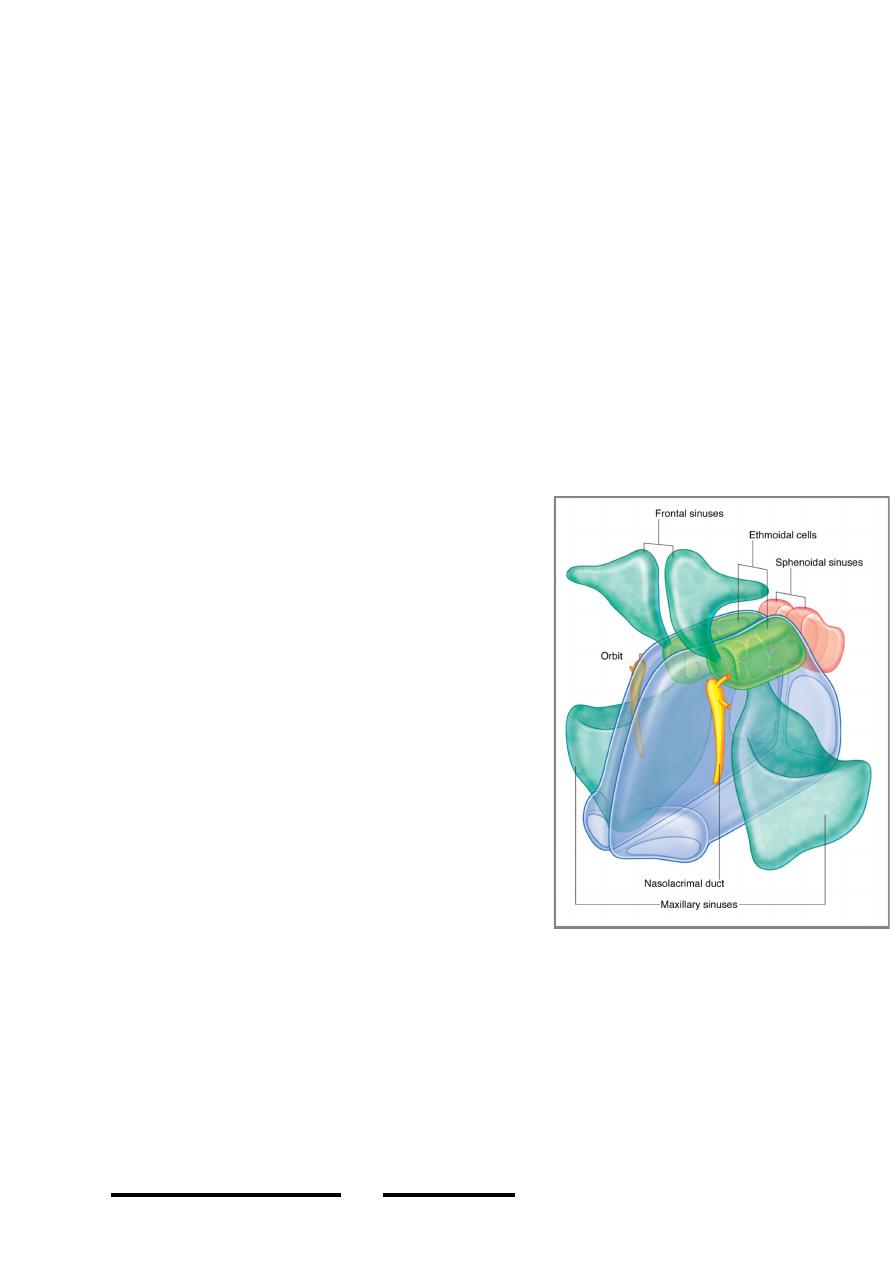
The paranasal sinuses:
• PNS are pneumatic areas in the frontal, ethmoidal, sphenoidal & maxillary
bones.
• They are lined with m.m. which is continuous with
that of the nasal cavity through the sinus apertures
in the lateral wall of the nose.
• The maxillary sinus is well developed at birth, the
frontal & sphenoidal exhibit a definite cavity at the
7th year of life while the ethmoidal develop late at
puberty.
• The definite function of the PNS is not well known,
theories about lightening the skull & resonating the
voice exist but still there is more
to be studied.
The frontal sinuses:
• These two sinuses are located on
each side of the midline in the
f r o n t a l b o n e b e h i n d t h e
superciliary ridges.
• They are rarely symmetrical.
• They are roughly triangular in
sagittal section with a maximum
vertical length of 2.5 cm &
maximum AP depth of 2 cm in
the orbital plate of the frontal
bone.
• It drains through the frontonasal duct
to the lateral wall of the nose where
its ostium opens in the most anterior
part of the hiatus semilunaris.
• Supraorbita, supratrochlear & anterior
ethmoidal nerves & vessels supply the
sinus.
The ethmoidal sinuses:
• These three groups of sinuses are
located in the lateral mass of the
ethmoid in its labyrinth. They are
named; anterior, middle & posterior
sinuses (air cells).
• The walls of these spaces are very thin
& completed by other bones like the
lacrimal, sphenoid, palatine, frontal &
maxillae.
!
87
Head & Neck Dr. Nawfal K. Al-Hadithi
• Medial to them lies the nasal cavity, laterally lies the orbit, superiorly the ACF
& inferiorly the nasal cavity & maxillary antrum.
• The anterior opens in the anterior part of the hiatus, the middle in the summit
of the bulla & the posterior in the superior meatus.
• The anterior & middle sinuses are supplied by anterior ethmoidal vessels &
nerves while the posterior is supplied by the posterior ethmoidal vessels &
nerves.
The sphenoidal sinuses:
• These two sinuses are located on each side of the midline in the body of
sphenoid separated from each other by thin plate of bone.
• They lie behind the posterior ethmoidal cells, in front of the dorsum sellae,
inferior to the hypophyseal fossa superior to the nasopharynx & bounded on
each side by the MCF.
• Their ostia open in the spheno-ethmoidal recess.
• They are supplied by the posterior ethmoidal vessels & nerves & by the
pharyngeal branch of the PPG.
The maxillary sinuses:
• These two sinuses occupy most of the
maxillary body extending from the lateral
nasal wall medially to the zygomatic process
of the maxilla laterally & from the floor of the
orbit superiorly to the alveolar process of
maxilla inferiorly.
• They are pyramidal cavities with their bases
open in the lateral nasal wall & apices in the
direction of the zygomatic process.
• The big opening in the lateral nasal wall
produced by the maxillary sinus is blocked for
most of its size by the overlapping inferior
concha.
• The sinus opens in the posterior part of hiatus
semilunaris.
• It is supplied by vessels & nerves of the
region, i.e; superior alveolar, infraorbital,
zygomaticofacial …
Applied anatomy:
• Infection of nasal mucosa as in flu leads to blockage of the draining system of
one or more of the PNS with the consequent accumulation of secretion in the
sinus & superadded infection resulting in sinusitis. So this condition could not
be treated unless the draining system is restored & normal physiology of its
cilia is resumed either medically or surgically.
• Pain from sinusitis is usually referred to areas supplied by the same nerves &
adjacent areas like the orbit, upper teeth, forehead & nose.
• The root of the upper second premolar tooth sometimes perforates the floor of
the maxillary sinus & its extraction may lead to oro-antral fistula.
!
88
Head & Neck Dr. Nawfal K. Al-Hadithi