The oral cavity:
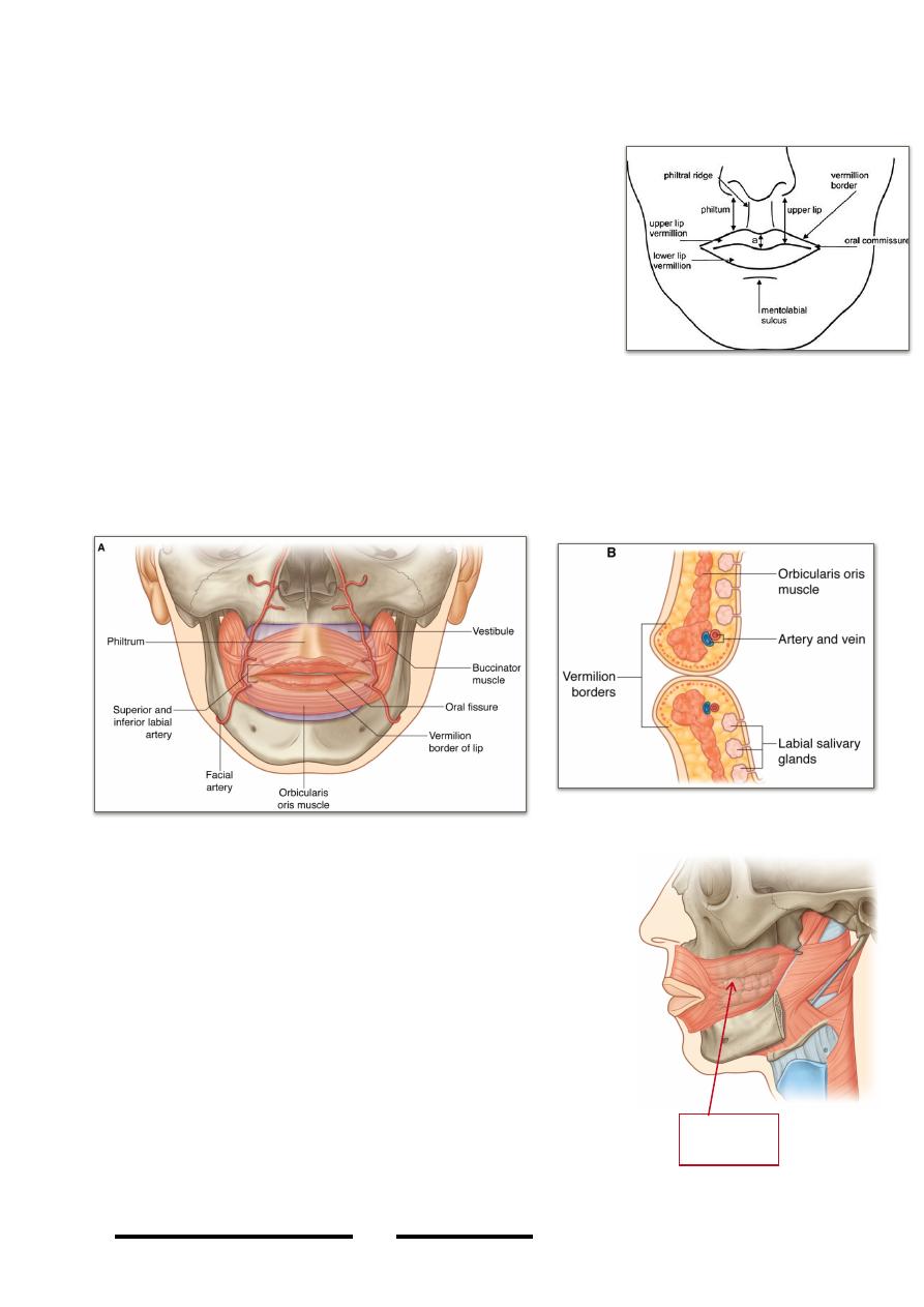
The lips:
- Are two muscular folds covered by skin, lined by
m.m & formed mainly by muscles including the
constrictors & dilators of the oral fissure
- The upper extends laterally to the nasolabial fold &
the lower extend inferiorly to the mentolabial fold
- The philtrum are two skin ridges end below at the
labial tubercle & above at the nasal septum
- The red margin (vemilion border) is covered by dry
transparent m.m which give the underling red color
of the highly vascular organ
- The upper lip is supplied by the infraorbital n. & the lower by the mental n.,
both are supplied by facial artery
- Labial glands are mucous gland in the submucosa & open by small individual
ducts to the surface of m.m
The cheeks;
- Resemble the lips in structure but their main muscle
is buccinator
- The fatty subcutaneous tissue (buccal pad of fat) is
very loose & transmits the parotid duct
- The buccal glands simulate labial glands
The gingivae;
- Consist of dense fibrous connective tissue firmly
attached to the underlying alveolar process
- Covered by smooth highly vascular m.m
- The gingivae also surround the necks of teeth
- The main blood supply to the upper gum are the
palatine arteries & to the lower is the lingual artery
!
89
Head & Neck Dr. Nawfal K. Al-Hadithi
Buccinator
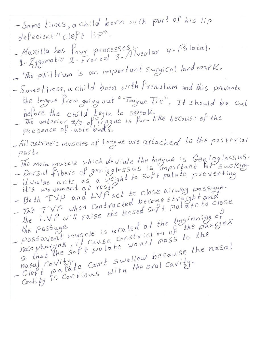
The oral cavity:
• The mouth consists of two parts:
1- The vestibule; is the narrow cavity in the
interval between the gums & teeth on one side
and the lips & cheeks on the other side.
2- The mouth proper; is the part of the cavity
within the alveolar arches roofed by the palate,
floored by the mylohyoid muscle & contains the
tongue.
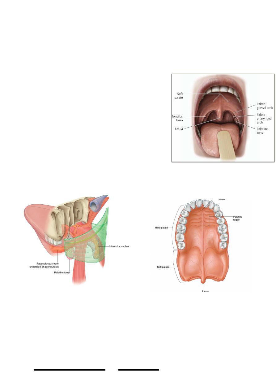
The roof of the mouth:
1- The hard palate;
- Forms the anterior 2/3 of the roof, it is
formed in its anterior 2/3 by the palatal
process of maxilla & in its posterior 1/3
by the horizontal plate of the palatine
bone.
- The m.m is firmly bounded to the
periosteum (mucoperiosteum) especially
in the anterior part therefore any
injection in this area is severely painful.
- It is supplied by the greater palatine &
terminal parts of nasopalatine vessels &
nerves.
!
90
Head & Neck Dr. Nawfal K. Al-Hadithi
Soft palate
2- The soft palate; will be discussed later.
The tongue:
- The tongue is a highly mobile muscular organ important for mastication,
swallowing, taste & speech.
- At its root it is fixed by its connection to the palate (palatoglossus), pharynx
(superior constrictor) & epiglottis (glosso-epiglottic folds), while its anterior part is
free for movement
- It is partially separated into two symmetrical halves by a median septum of areolar
tissue
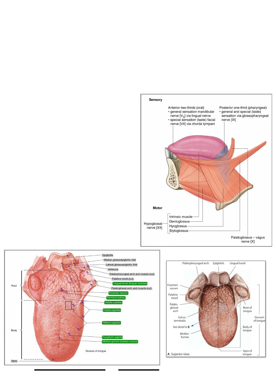
- Tongue is composed embryologically of
two different parts which possess
different structure, appearance & in nerve
supply & lymphatic drainage, they are
the oral part (anterior 2/3) & pharyngeal
part (posterior 1/3).
- The anterior 2/3 is separated from the
posterior 1/3 by a v-shape sulcus
terminalis whose apex is directed
posteriorly at foramen caecum of the
tongue
- The m.m of the anterior2/3 is fur-like &
adheres to the underlying muscles, it is
also characterized by the presence of the
filiform, fungiform & vallate papillae,
while m.m of the posterior 1/3 is smooth
& movable over the muscle & shows
multiple round elevations produced by
the underlying lymphoid follicles (lingual
tonsil).
!
91
Head & Neck Dr. Nawfal K. Al-Hadithi
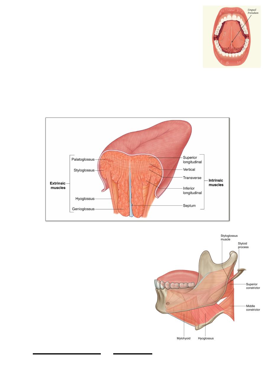
- Frenulum linguae is a m.m fold in the midline connects the
undersurface of the tongue to the floor of the mouth
Muscles of the tongue:
I. Intrinsic muscles;
- These are muscle fibers arranged within the tongue in three
directions, antero-posterior (superior & inferior longitudinal),
horizontal & vertical
- They are attached by their ends to the m.m. & to the midline lingual septum
- Their contraction changes the shape of the tongue, the size of the tongue mass
is static, any change in one dimension affects other dimensions, e.g; flattening
of the tongue (contraction of vertical group) is always associated with increase
side to side length of it an d so on.
II) Extrinsic muscles;
There are three muscles which are related to the
tongue, they connect the tongue to three
different bones & their contraction alters the
p o s i t i o n & d i r e c t i o n o f t h e t o n g u e .
Palatoglossus, thogh it is a palatal muscle it will
be discussed here for its important action.
Hyoglossus:
Origin; upper border of greater horn & body of
hyoid
Insertion; fibers ascend anteosuperiorly to be
inserted in the posterior part of the lateral border
of the tongue intermingling with other muscles.
Action; retracts the tongue & depresses its sides.
It also elevates the hyoid bone.
Genioglossus:
!
92
Head & Neck Dr. Nawfal K. Al-Hadithi
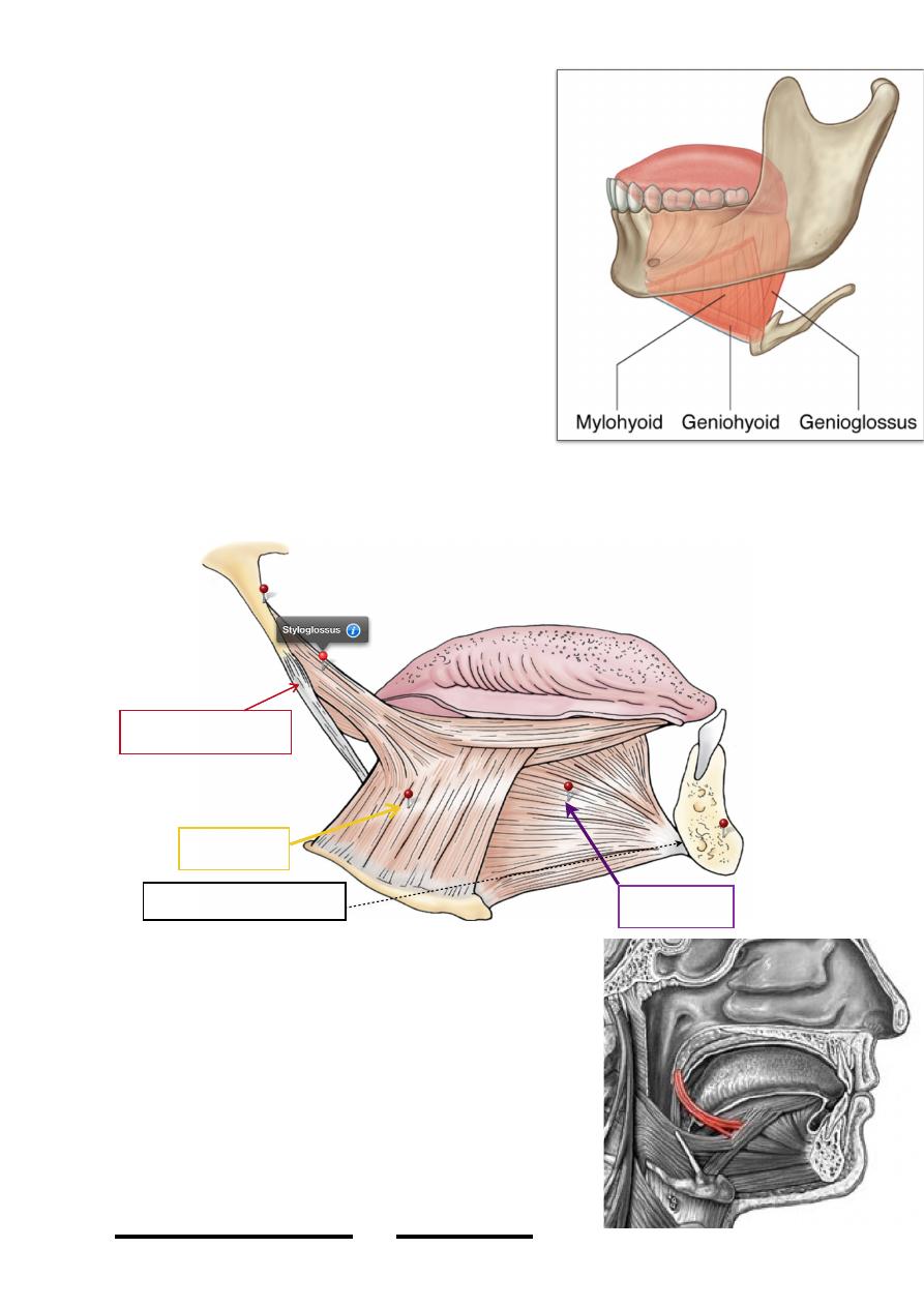
Origin; inferior genial tubercle
Insertion; fibers go back to enter the substance of
the tongue contributing to its mass, the superior
fibers are inserted into the tip of the tongue, middle
fibers into the dorsum & the lowest fibers are
inserted inferiorly.
Action; -protracts the tongue
-the superior fibers brings the tip of the
tongue in contact with the floor of the mouth
-the dorsal fibers cause cupping of the
tongue
Styloglossus;
Origin; lower part of styloid process anteriorly
Insertion; fibers descend anteroinferiorly &
medialward to be inserted into the posterior part of
hyoglossus, the fibers entr the tongue & pass forward along its side
Action; retracts the tongue & deviates it laterally
Palatoglossus:
Origin;-oral surface of the palatine aponeurosis
Insertion; fibers arch down under the m.m of the mouth
raising the anterior pillar of the fauces (palatoglossal
arch) to be inserted into the side of the tongue.
Action; It is the opponent of LVP:
-sphincter of the fauces
-depresses the soft palate
-raises the tongue
!
93
Head & Neck Dr. Nawfal K. Al-Hadithi
Hyoglossus
Genioglossus
Inferior genial tubercle
Stylohyoid ligament
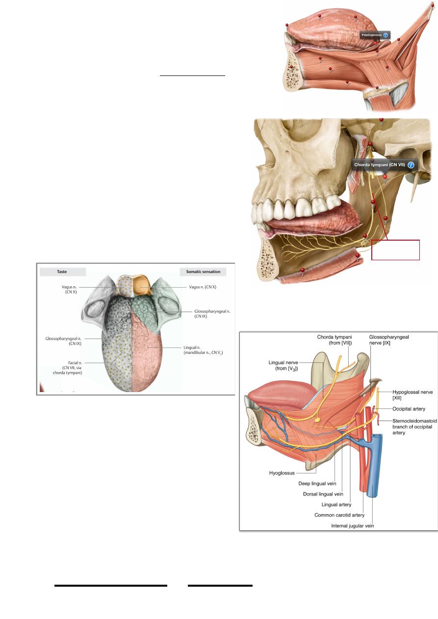
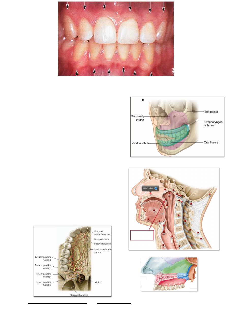
Nerve supply of lingual muscles:
All muscles of the tongue are supplied by the
hypoglossal nerve except palatoglossus which is
supplied (as a palatal muscle) by the pharyngeal plexus.
Sensory nerve supply of the tongue:
• Anterior 2/3;
-lingual n.; common sensation
-chorda tympani; taste sensation
Chorda tympani is an autonomic nerve derived
from nervus intermedius (VII?), it joins the Vc
just below the base of the skull in the ITF &
runs in its lingual branch to supply
parasympathetic power to the submandibular
ganglion & give taste fibers to the anterior 2/3
of the tongue
• Posterior 1/3;
glossopharyngeal nerve supplies it with both
types of senses
Arterial supply of the tongue:
Lingual artery:
This branch of ECA provides the tongue with
all its blood, at its origin (level with the tip of
grater horn of hyoid) it is crossed externally
by the XII nerve & posterior belly of
digastric & stylohyoid muscles. It passes
forward to lie deep to hyoglossus between it
& the septum of the tongue 5 mm deep to the
inferior surface of the tongue, it gives:
a. Dorsal lingual branches to the tongue
mass.
b. Sublingual branches to the sublingual
gland & floor of the mouth
Veins of the tongue:
!
94
Head & Neck Dr. Nawfal K. Al-Hadithi
Lingual n.
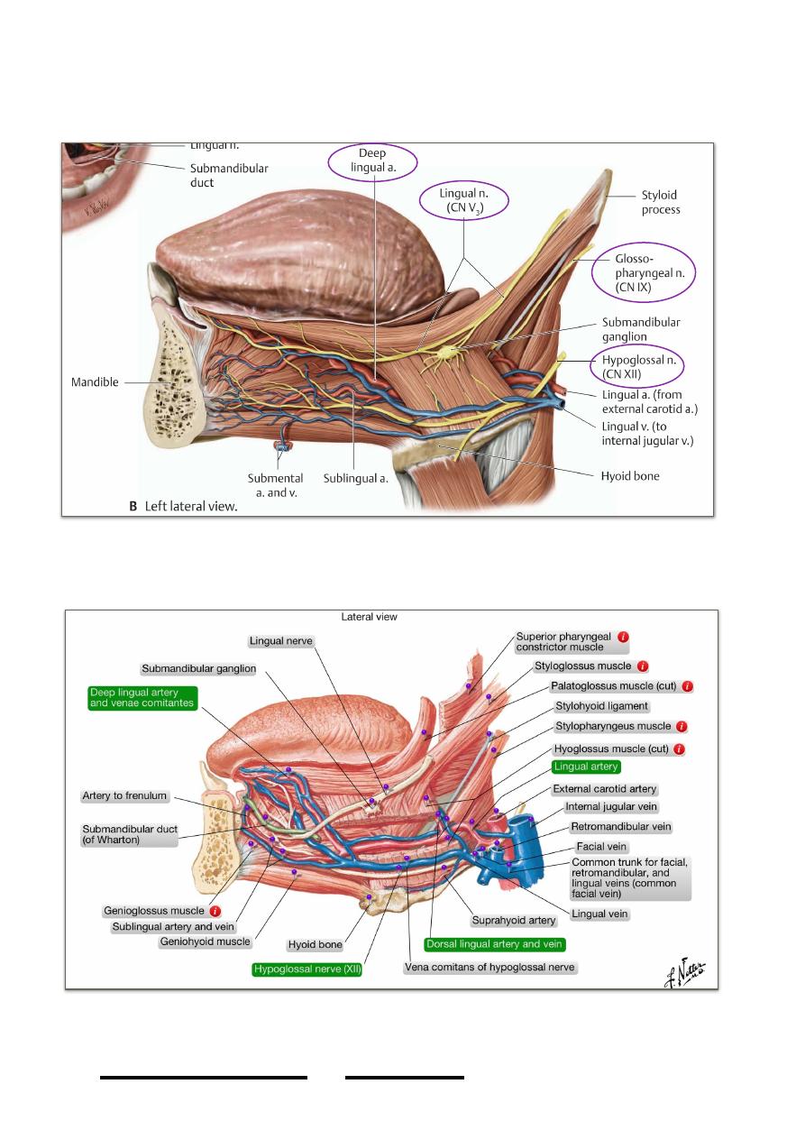
1- Deep lingual veins; two in number, accompany the lingual artery & receive similar
tributaries, they empty in the IJV
2- Veni comitans nervi hypoglossi; accompany the XII, bring blood from the tip, they
are larger than the deep veins & empty in the facial vein.
The floor of the mouth:
- The floor of the mouth is mylohyoid
!
95
Head & Neck Dr. Nawfal K. Al-Hadithi
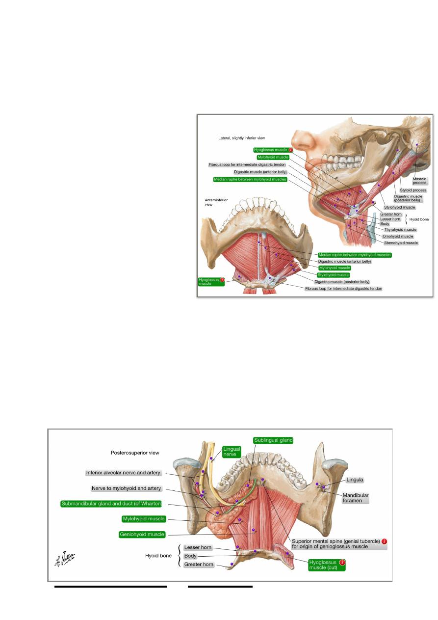
Origin; mylohyoid line of the mandible
Insertion;
- The fibers descend downward & backward, those of anterior 2/3 interdigitate
with the opposite one in a midline raphe which extends from the symphysis
menti to the hyoid bone
- The posterior fibers reach
the hyoid bone leaving a
posterior free border for the
muscle which connects the
o r a l m o u t h t o t h e
submandibular triangle
Nerve supply; mylohyoid nerve,
branch of the inferior alveolar
nerve
Action;
A. An essential swallowing
muscle, by wavy elevation
of the tongue from anterior
to posterior direction against
the palate it compresses the
bolus backward
B. Moves the tongue changing
its position & direction
C. Elevates the hyoid & eventually the larynx
Structures in the floor of the mouth:
- A coronal section through the mid-mouth reveals a slit like cavity in the floor
of the mouth between mylohyoid laterally & the side of the tongue
(hyoglossus) medially.
- This cavity is covered with m.m of the floor of the mouth & contain important
structures in relation to the lateral surface of hyoglossus, these are:
*Lingual nerve above (hooks around the submandibular duct)
*Submandibular duct in the middle
!
96
Head & Neck Dr. Nawfal K. Al-Hadithi
*XII nerve is the lowest
- Structures deep to hyoglossus are:
*Deep lingual artery page 95
*Stylohyoid ligament page 93
*IX nerve
The soft palate:
- Is a triangular fold of m.m containing
aponeurosis of tensor palati, muscle fiber,
mucous glands, vessels & nerves
- It hangs from the posterior border of the hard
palate where its anterior surface will face
forward to the oral cavity & its posterior
surface faces backward
- The tip of the triangle contains a rounded mass
of muscle fibers (musculus uvulae) with
mucous glands it is called the uvula
- The main function of the soft palate is to act as
policeman between the airway & foodway
regulating swallowing in relation to berathing.
- Muscles of the soft palate are; tensor veli palatini, levator veli palatini,
palatoglossus, palatopharyngeus & musculus uvulae
Tensor veli palatini:
- One should think of this muscle as two triangles, a muscular one lies in the ITF
& an aponeurotic one in the oral cavity
- These two triangles are united to each other by a tendon which get access to
the mouth through the fibro-osseous canal produced by attachment of the
pterygomaxillary ligament between the pterygoid hamulus & maxillary
tuberosity
!
97
Head & Neck Dr. Nawfal K. Al-Hadithi
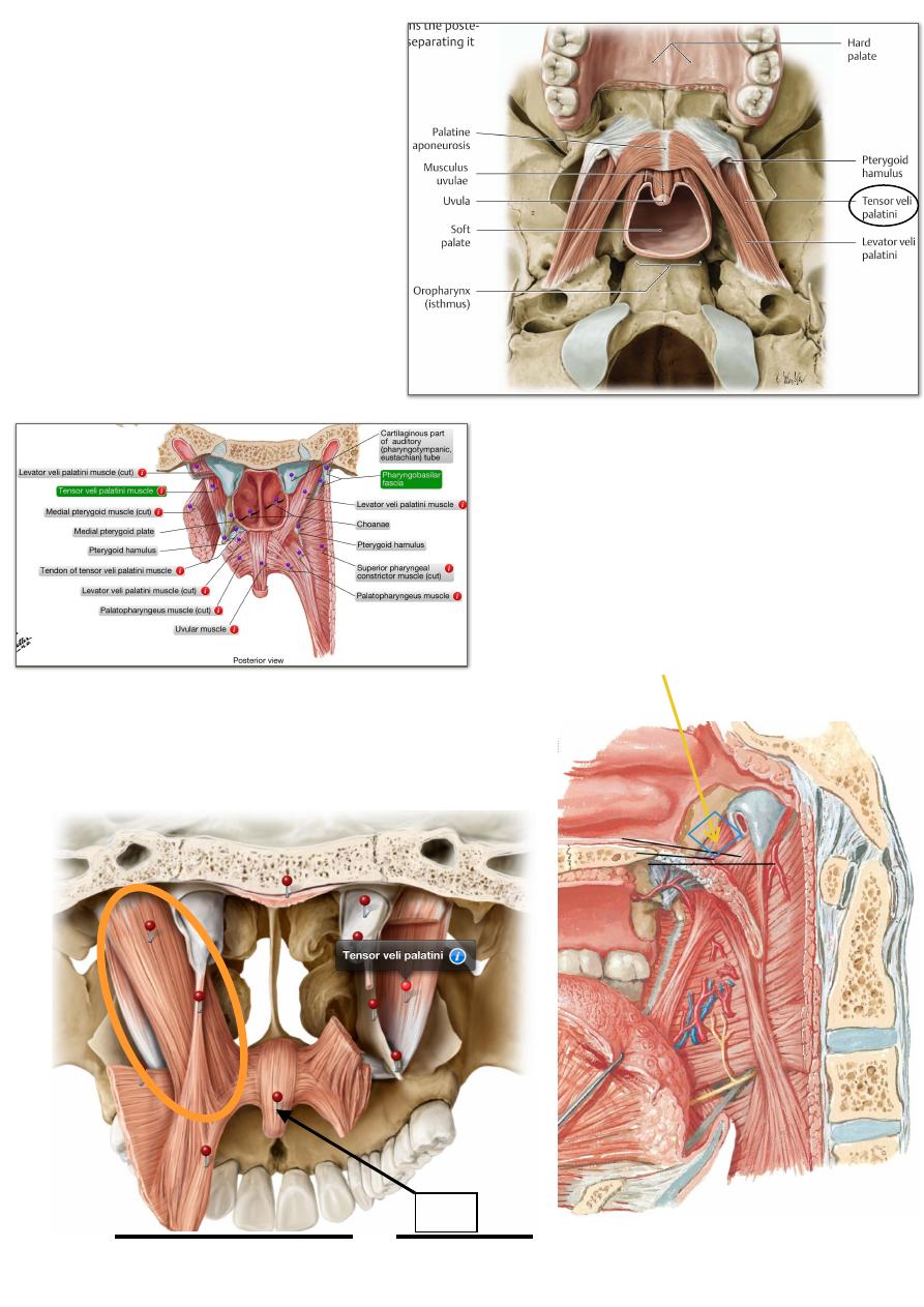
Origin:
The muscular triangle in the ITF
arises by two of its limbs;
- from the roof of ITF (from the
spine of sphenoid to the scaphoid
fossa)
-from the posterior border of
medial pterygoid plate passing
over the cartilagenous part of the
auditory tube
The third border of the muscle is
free
The muscle will lie in the fossa
between the medial pterygoid plate &
muscle to taper as it approaches the
maxillary tuberosity into a slender
tendon
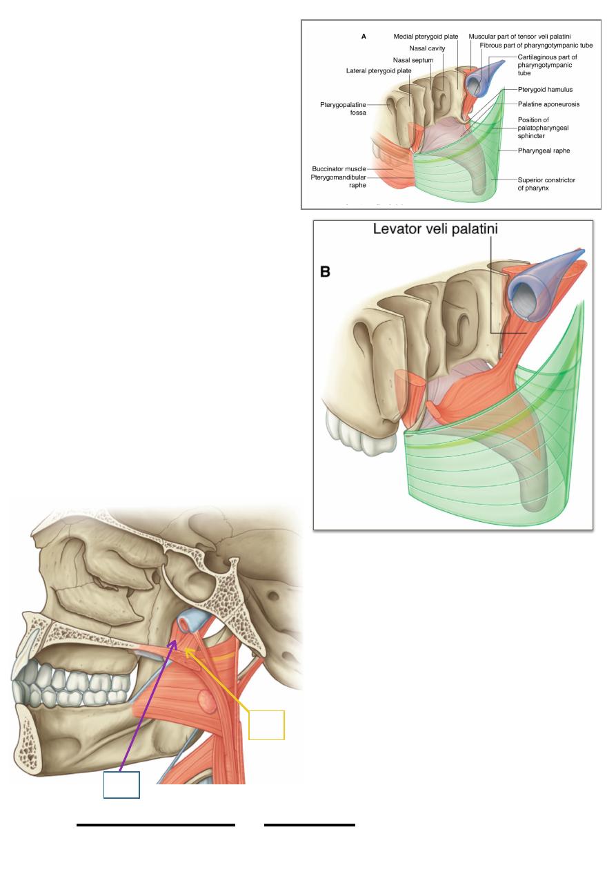
Insertion:
The slender tendon after entering the
fibro-osseous canal expands into
aponeurotic triangle with three borders:
-One is attached to the posterior
border of the hard
palate
- The other will fuse with the opposite fellow
- The third will hang freely inside the
oropharynx suspending the uvula
Action:
!
98
Head & Neck Dr. Nawfal K. Al-Hadithi
Uvula
Contraction of TVP causes tension of
the soft palate (aponeurosis of the
muscle) which becomes straight &
lower down to be elevated by the
levator muscle to fit the Passavant
ridge in the junction between the oro-
& nasopharynx separating the two.
Levator veli palatini:
Origin: From the petrous apex &
cartilagenous part of the auditory tube
Insertion: the pencil-like muscle
descends deep to the m.m of the
nasopharynx elevating a ridge near the
tube orifice to be inserted into the dorsal
surface of the palatine aponeurosis
Action: elevate the tense palatine
aponeurosis closing the naso from
oropharynx
Both TVP & LVP contraction opens the
auditory tube since part of their origin is
taken from it & since their contraction
occurs mainly during swallowing this
process will open the auditory tube
equalizing pressure on both sides of the
eardrum
Palatoglossus:”discussed”
Palatopharyngeus:
Origin:
- Nasal side of palatine aponeurosis
- Posterior part of the hard palate
Insertion: fibers arch down behind
palatoglossal fibers to raise the posterior
pillar of the fauces (palatopharyngeal
fold) & inserted in the posterior border
of the thyroid cartilage
Action: - depresses the soft palate
- elevates the larynx in the early
stage of deglutition
Nerve supply of palatal muscles:
All palatal muscles are supplied by the
!
99
Head & Neck Dr. Nawfal K. Al-Hadithi
LVP
TVP
pharyngeal branch of X nerve in the pharyngeal
plexus except TVP which is supplied by nerve to
medial pterygoid from the main trunk of Vc.
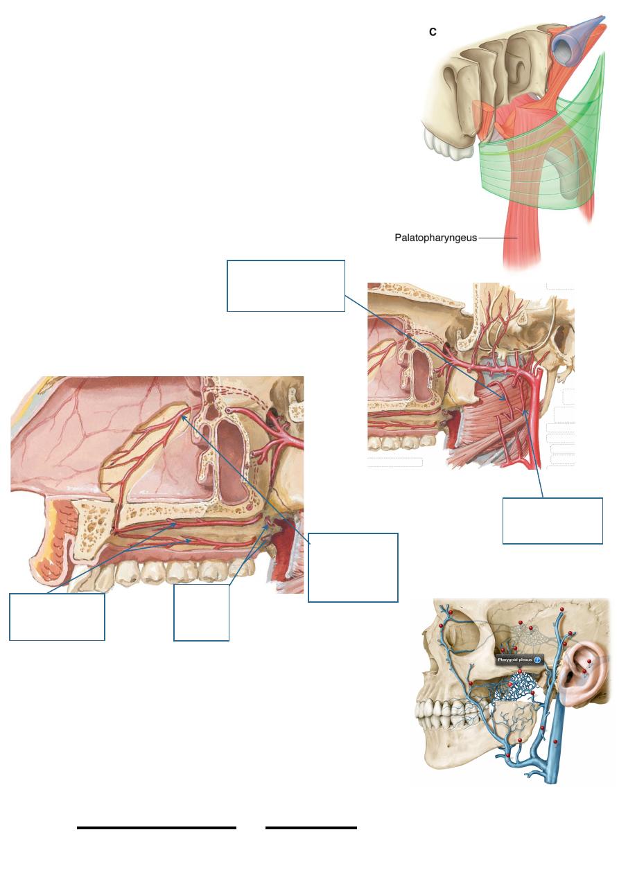
Arteries of the palate:
1- Greater palatine; hard palate
2- Long sphenopalatine; hard palate
3- Lesser palatine; soft palate
4- Ascending palatine br. of facial a.; soft palate
5- Palatine br. of ascending pharyngeal; soft palate
Veins of the palate:
Similar to arteries; pterygoid venous plexus
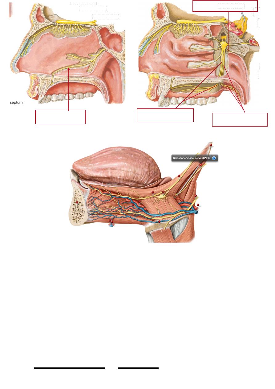
Nerves of the palate:
1- Greater palatine n.
2- Nasopalatine n.
3- Lesser palatine n.
4- Pharyngeal branch of
PPG --> pterygo palatine ganglion
5- IX nerve.
!
100
Head & Neck Dr. Nawfal K. Al-Hadithi
Rt. & lt. Greater
palatine arteries
Lesser
palatine
arteries
Long
Sphenopalatine
aretry
Ascending
pharyngeal artery
Ascending palatine
artery
ﻭﻻ ﻛﻞ ﻛﻞ ﺍﻟﺨﺎﻟﻌﻴﻦ ﺍﺭﺍﺫﻝٌﻖﻳﺪﺻ ﺎﻬﺑﻮﺛ ﺔﻗﺍﺪﺼﻟﺍ ﺲﺒﻟ ﻦﻣ ﻞﻛ ﺎﻣﻭ
!
101
Head & Neck Dr. Nawfal K. Al-Hadithi
Nasopalatine nerve
Greater palatine nerves
Lesser palatine nerves
Pharyngeal branch of PPG