
Head & Neck
Ear
Larynx
Oral Cavity
Nose
Done by:
Abbas Fadel
Group C
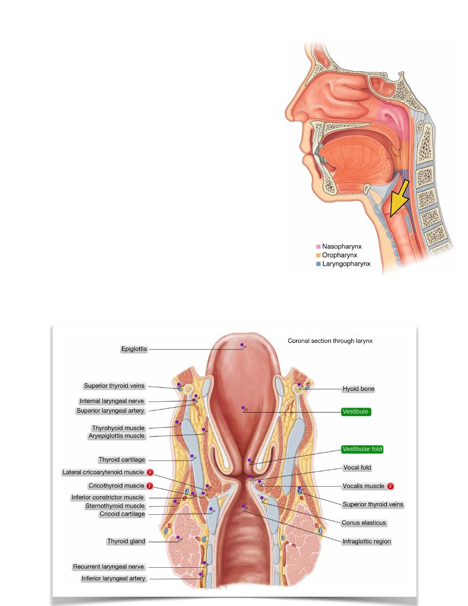
The Larynx:
The larynx lies in the anterior part of the neck in the
midline opposite to C4-C6 vertebrae forming the
laryngeal prominence (Adam’s apple)
At this level the L. is triangular in cross section
while lower down at the level of the cricoid
cartilage it is circular in cross section
Relations:
- Anterolaterally; Thyroid gland & strap muscles
- Laterally; Carotid sheath
- Posteriorly; Pharynx
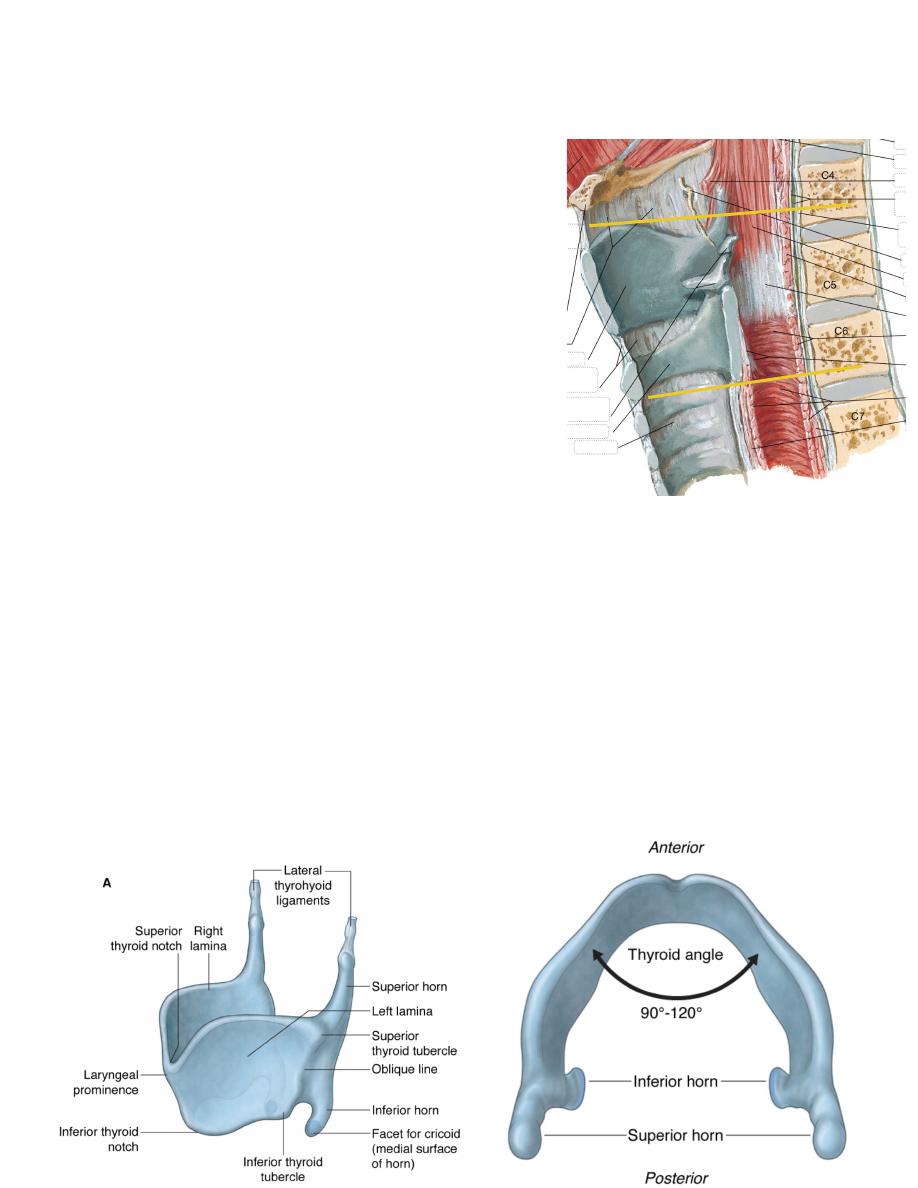
Skeleton:
The thyroid cartilage:
• This hyaline cartilage is composed of two
quadrilateral laminae meet in the midline
• The meeting angle is 90O in male & 120O in
female (therefore the L is more prominent in
male)
• The superior thyroid notch is a V-shape notch just above the prominence
• The posterior border of the cartilage is thick & rounded & extends above &
below the laminae as the superior & inferior horns
• The superior & inferior borders are characterized by superior & inferior
tubercles between them the oblique line of the cartilage extends which gives
attachment to thyrohyoid, sternothyroid & thyropharyngeus
• The upper border is attached to the thyrohyoid membrane which is thickened
in the midline as the median thyrohyoid ligament & thickened laterally
between the superior horn & tip of greater horn of the hyoid as the lateral
thyrohyoid ligament
• The inferior thyroid notch lies opposite to the superior one, from its deep
surface the root of the epiglottis arises
1
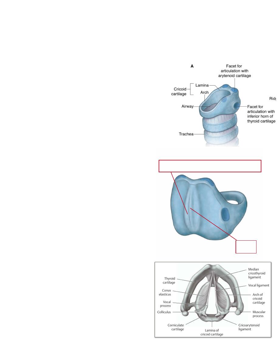
The cricoid cartilage: the only complete ring in the respiratory pathway
• This signet ring-like cartilage is characterized by an anterior arch &
posterior broad lamina
• It lies at the level of C6 vertebra & forms the
foundation on which the rest of the larynx is
built
• The posterior lamina is marked in the
midline by a ridge on either side of which
lies a shallow depression for the posterior
crico-arytenoid muscle
• The arch which is 5 mm in vertical height
gives attachment anterolaterally to crico-
thyroid muscle & posteriorly to the
cricopharyngeus just above which the lateral
crico-arytenoid arises
• The upper border of the arch gives
attachment to the conus elasticus
• The sloping shoulders of the lamina
provides a synovial joint for the
arytenoid cartilages
• At the junction of the arch & lamina is
a synovial joint for articulation of the
inferior horns of the thyroid cartilages
The arytenoid cartilage:
• This is a three sided pyramidal hyaline
cartilage whose apex projects
posteromedially & carries the
corniculate cartilages
• The base of the pyramid carries three
processes
• The anterior sharp process is the vocal
process & to which the upper free end
of the conus elasticus is attached as the
vocal fold
• The lateral process is the muscular
process to which the lateral &
posterior crico-arytenoids are attached
• The medial surface of the pyramid is
flat & faces the opposite one
2
Ridge
Depression for the post. Crico-arytenoid
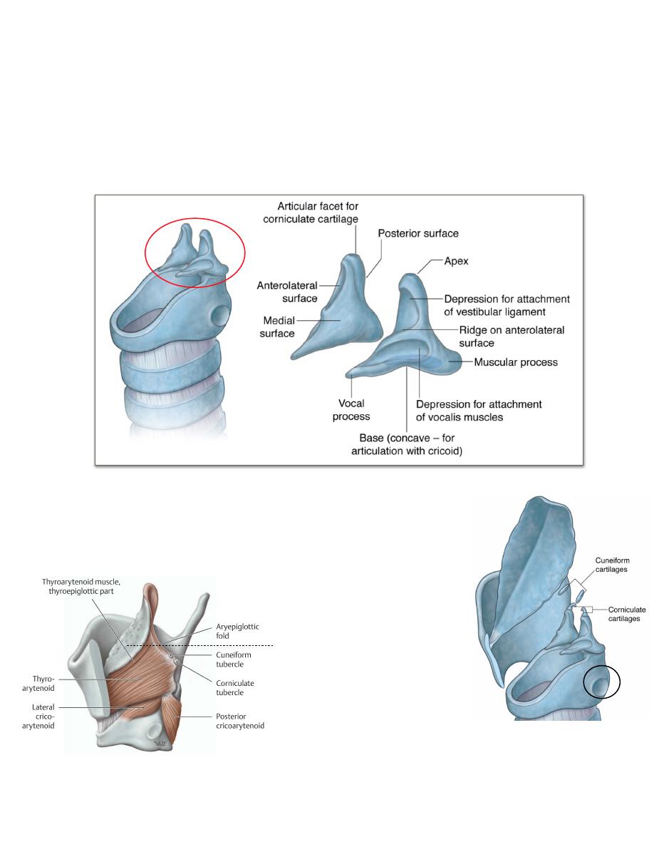
• The anterolateral surface is curved & gives attachment for the thyro-
arytenoid muscle
• The posterior surface is smooth & gives attachment for the transverse
arytenoid muscle
• The arytenoid cartilage sits on the elongated facet on the sloping shoulder of
the cricoid lamina forming the crico-arytenoid synovial joint
The corniculate cartilage:
• This small nodular elastic cartilage lies on the apex of
the arytenoid to prolong it backward & medialward
• They are enclosed by the ary-epiglottic folds
The cuneiform cartilage:
•A r e r o d l i k e e l a s t i c
cartilages lies on the previous
ones in the ary-epiglottic
folds
The epiglottis:
•This elastic, leaf-like
cartilage is attached by its
lower 1/2 to the back of the
thyroid cartilage by the
thyro-epiglottic ligaments in
the midline, its upper 1/2 stands erect behind the
posterior 1/3 of the tongue & the hyoid bone
3
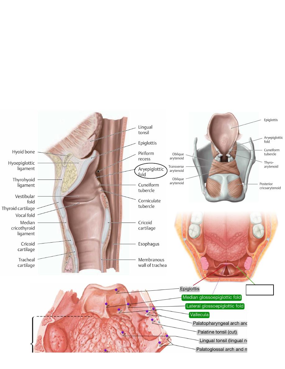
• The anterior surface of the epiglottis is attached to the hyoid bone by the
hyo-epiglottic ligament
• The m.m of the E. is reflected on the posterior 1/3 of the tongue as three
folds, the median & two lateral glosso-epiglottic folds which mark the
division between the oro- & laryngo-pharynx, on each side of the median
one lies a vallecula
• The cartilage is well pitted to receive the multiple mucous glands which
cover it
• To the margins of the free upper ½ is attached the quadrate membrane
4
Vallecula
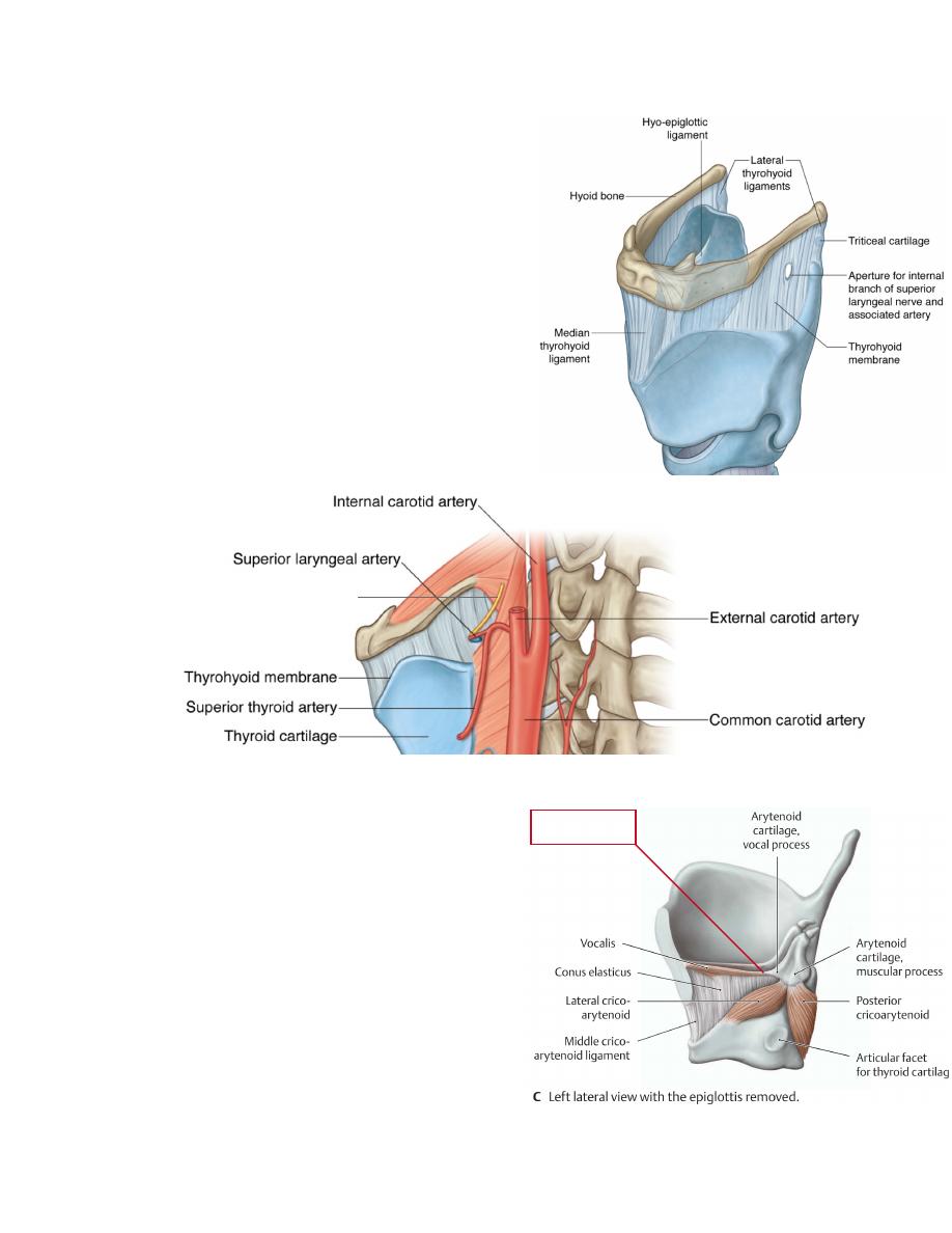
Laryngeal membranes:
1-Thyro-hyoid membrane:
- Suspends the thyroid cartilage to the
hyoid bone
- It passes from the upper border of the
thyroid cartilage to the upper border of
the hyoid bone passing behind the bone
separated from it by a bursa
- It shows one median & two lateral
ligaments of the same name
- It is pierced by the superior laryngeal
artery & internal laryngeal nerve.
- It forms the lateral boundary of the
piriform recess
2- Conus elasticus:
- Is a half-circle ligament whose lower
attachment is to the whole length of
the upper border of the cricoid arch
- Its free upper border is attached on
either side to the vocal process of the
arytenoid cartilage forming the vocal
fold (true vocal cord) which contains
in its free border muscle fibers
(vocalis)
- Anteriorly the membrane is attached
to the back of the thyroid cartilage in
the angle between the two laminae in
5
Internal laryngeal nerve
Vocal fold
the midline midway between the superior & inferior notches converting the
curved membrane to V-shape membrane
- Its thickening in the midline anteriorly produces the median crico-thyroid
ligament
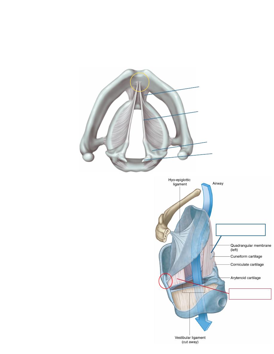
3- Quadrate membrane:
- Is a weak membrane whose posterior
border is attached to the anterior
surface of the arytenoid cartilage & its
anterior border is attached to the sides
of the lower half of the epiglottis
- Its upper free border will extend
between the epiglottis & the arytenoid
cartilage forming the ary-epiglottic fold
which involves in its substance the
corniculate & cuneiform cartilages
- Its lower free border will be parallel to
the upper free border of the conus (true
vocal cords) forming the vestibular fold
(false cords)
- It forms the medial boundary of the
piriform recess
The interior of larynx:
The laryngeal inlet:
- Opens in the anterior wall of the
6
Crico-thyroid ligament
Vocal ligament
Arytenoid cartilage
Corniculate cartilage
Ary-epiglottic fold
False vocal fold
pharynx in a vertical plane
- Is an inverted triangle whose base is formed
by the epiglottis antero-superiorly & its apex
is the narrow interval between the two
arytenoids postero-inferiorly
- The sides of the triangle is the ary-epiglottic
olds
The laryngeal vestibule:
- Is the triangular cavity beyond the laryngeal
inlet until the rima glottidis
- It is bounded on each side by the quadrate
membrane
- It shows three features:
1- The vestibular folds; are the lower free border
of the quadrate membranes
2- The laryngeal ventricle; is a sac like mucosal
herniation between the true & false vocal folds
whose upper border extends up & may reach the
upper border of the thyroid cartilage, it is filled
with mucosal glands for lubrication
7
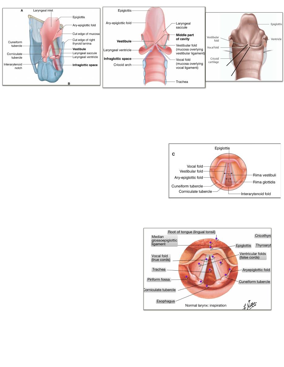
3- The rima vestibuli; is the opening between the vestibular folds, it is wider than
the rima glottidis.
The vocal folds:
- Are formed by the free upper border of the conus stretched between the
thyroid cartilage anteriorly & the arytenoids posteriorly
- The anterior 3/5 are true components of the conus while the posterior 2/5 are
formed by the vocal process of the
arytenoids
- They contain in their free edge the vocalis
muscle which increases the apposed
surface area of the cords during phonation
- The opening between them is called the
rima glottidis
- Glottis, is a term applied to the two vocal
cords & the rima glottidis as they are the
main structure involved in phonation
The rima glottidis:
- Is the interval between the two
vocal cords
- Is 23 mm long in male & 17 mm
in female
- It could be opened either in V-
shape or diamond shape manner
a c c o r d i n g t o t h e t y p e o f
movement of the arytenoid
- Downward movement of the
a r y t e n o i d o n t h e s l o p i n g
shoulders of the cricoid lamina
pulls the two ends of the conus
downward separating them &
opens the rima in a V-shape manner, here the vocal processes of the
arytenoids are parallel to each other, this occurs in quite respiration
8
- Rotation of the arytenoids around their vertical axes
pulls the free ends of the conus away & opens the
rima in a diamond shape manner, the back of the
diamond is formed by the vocal processes of the
arytenoids which become perpendicular on each
other, this occurs in forced respiration
- During phonation the folds come in contact with
each other & the rima becomes slit like
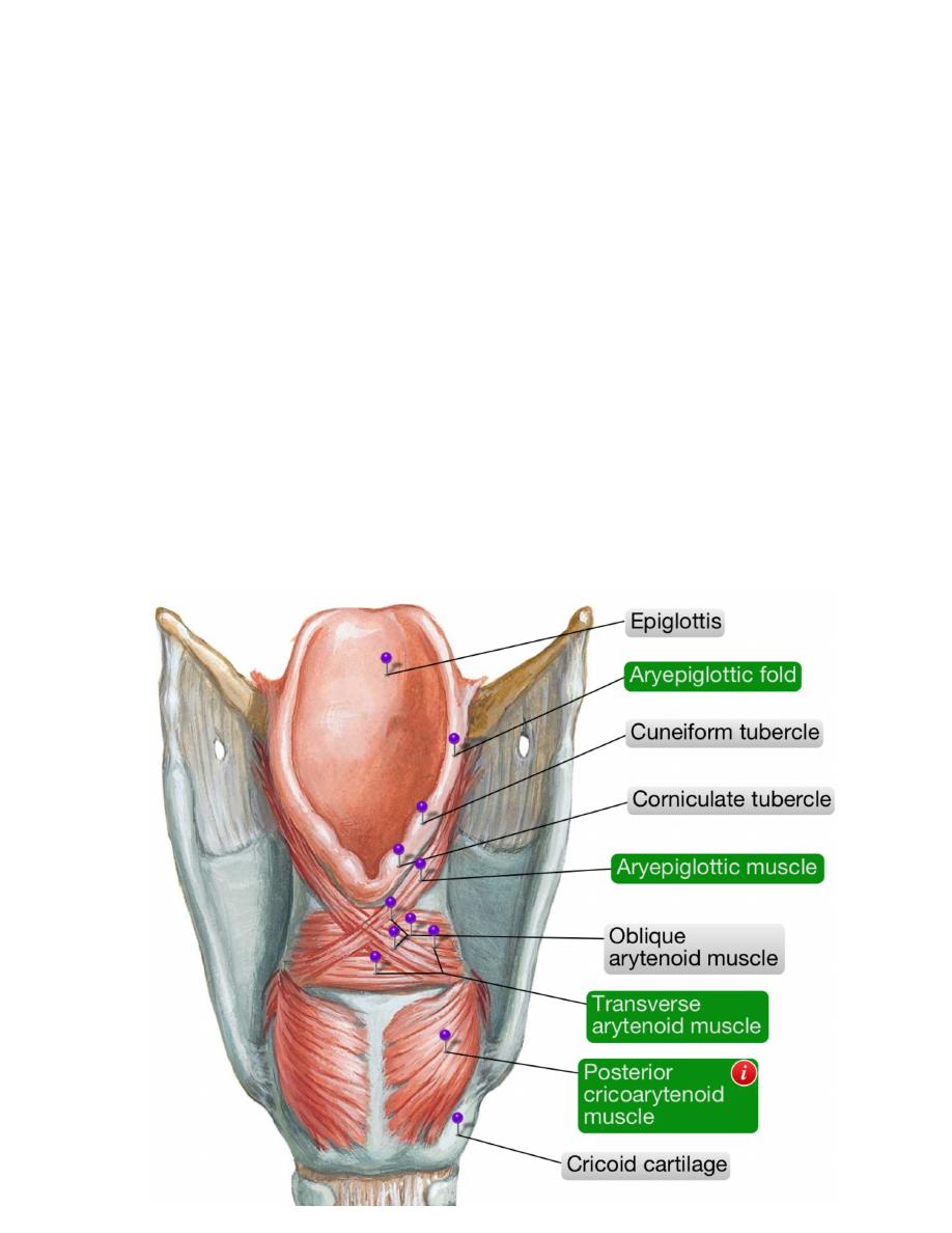
Intrinsic muscles:
The ary-epiglottic muscle:
- Extends in the ary-epiglottic fold from the
arytenoids to the lateral border of the epiglottis
- The oblique inter-arytenoids are regarded as the
continuation of the muscle to the vocal process of
the opposite arytenoid
- Contraction of both brings the arytenoids near each
other & oppose the ary-epiglottic folds & pull the
epiglottis as a shelf over the constricted laryngeal
inlet producing an effective sphincteric action for
the inlet
The posterior crico-arytenoid: the only muscles open
vocal folds
Origin; from the back of cricoid lamina from the fossa on
each side of the midline ridge
Insertion; upper fibers go horizontally to the vocal process of the arytenoid while
the lower fibers go vertically to the to the same process
Action; upper fibers rotate the arytenoids so they open the rima glottidis in a
diamond shape while the lower fibers pull the
arytenoids away from each other so they open the
rima in a V-shape
The transverse arytenoid:
- This is a muscle formed of fine & short
fibers stretched between the two arytenoids
deep to the oblique one
- Its contraction opposes the vertical fibers
of the posterior c-a muscle ﻣﻬﻢ
The lateral crico-arytenoid:
- Arises from the posterior part of the
cricoid arch
- Inserted into the vocal process of the
9
Thyroarytenoid
Aryepiglottic
Late
ral cri
co a
ryten
oid
arytenoid
- Its contraction opposes the horizontal fibers of the posterior c-a muscle
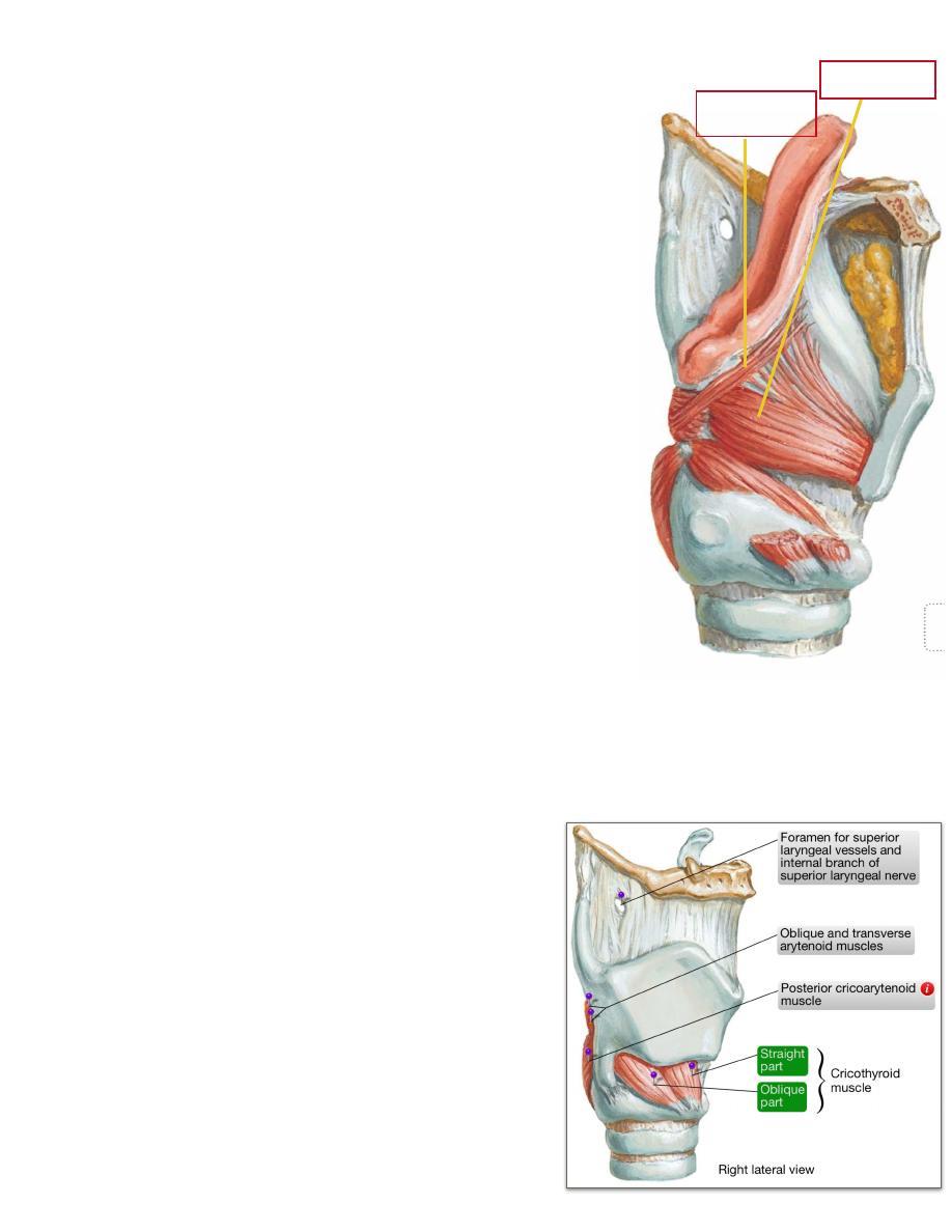
The crico-thyroid:
- This muscle arises from the anterolateral surface of the cricoid arch
- Its fibers radiate upward & backward to be inserted into the lower border &
medial aspect of the thyroid cartilage
- Its contraction approximates the thyroid & cricoid cartilages diminishing the
area between them, this will bring the thyroid cartilage away from the
arytenoid increasing the length & hence tension of the vocal cords affecting
consequently the type of the voice
The thyro-arytenoid: page 9
- This muscle arises from the inner surface of the thyroid lamina
- Its fibers pass backward to be inserted into the muscular process of the
arytenoid
- Its contraction approximates the thyroid & arytenoid cartilages diminishing
the area between them, this will decrease the length & hence tension of the
vocal cords affecting consequently the type of the voice, this movement also
acts as a laryngeal sphincter
10
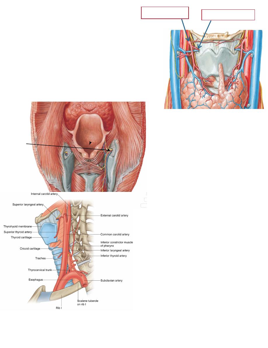
Arteries of the larynx:
1- Superior laryngeal artery;
- A branch of the superior thyroid a.
- pierces the thyrohyoid membrane together
with the internal laryngeal nerve to lie
underneath the m.m of the floor of the
piriform recess
- supplies the larynx to supply mucosa down
to the level of the vocal cords.
2 -
Inferior laryngeal artery;
-A branch of the inferior thyroid
artery
-enters the lower part of the
larynx deep to the inferior
pharyngeal constrictor
-supplies it up to the vocal cords
(vocal cords are supplied by the
inferior one).
-
-Nerves of the larynx:
-Motor:
-All muscles of the larynx are
supplied by the recurrent laryngeal
n. except cricothyroid which is
supplied by the external laryngeal
branch of the superior laryngeal nerve
(X nerve).
-Sensory:
•Above the vocal folds : internal
laryngeal branch of superior laryngeal
nerve (X nerve) accompanies the sup.
laryngeal artery.
•Below the vocal folds: recurrent
laryngeal nerve accompanies the
inferior laryngeal artery
11
Sup. thyroid artery
Sup. laryngeal artery
Internal
branches
of sup.
laryngeal
nerve ,
artery and
vein

12