The Reproductive system
in males, 22 pairs of autosomes plus an X chromosome and a Y chromosome;in females, 22 pairs of autosomes plus two X chromosomes.
As a consequence of meiosis during gametogenesis, each normal ovum contains a single X chromosome, but half the normal sperms contain an X chromosome and half contain a Y chromosome .
When a sperm containing a Y chromosome fertilizes an ovum, an XY pattern results and the zygote develops into a genetic male. When fertilization occurs with an X-containing sperm, an XX pattern and a genetic female result (so the sex of the resulting zygot is determined by the fertilizing sperm).
The differentiation of the primitive gonads in utero is genetically determined in humans,
but the formation of male genitalia depends upon the presence of a functional testis. There is evidence that male sexual behavior and, in some species, the male pattern of gonadotropin secretion are due to the action of male hormones on the brain in early development.Development of the Gonads
The gonad develops a cortex and a medulla. Until the sixth week of development, called bipotential gonads which contains primitive germ cells and has both male and female primordial genital ducts.After the 6th week the differentiation started.
In genetic males, the medulla develops into a testis, and the cortex regresses. Leydig and Sertoli cells appear, and testosterone and mullerian inhibiting substance are secreted.
In genetic females, the cortex develops into an ovary and the medulla egresses. It will not secret estrogen inutero, so the secondary sex organs will develop without any hormonal influence.
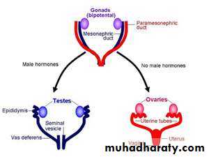
In a normal female fetus, the mullerian duct system then develops into uterine tubes (oviducts) and a uterus. In the normal male fetus, the wolffian duct system on each side develops into the epididymis and vas deferens. The external genitalia are similarly bipotential until the eighth week.
When there are functional testes in the embryo, male internal and external genitalia develop.
The Leydig cells of the fetal testis secrete testosterone (androgens), and the Sertoli cells secrete mullerian inhibiting substance (MIS) .
MIS causes regression of the mullerian ducts, and testosterone fosters the development of the vas deferens and related structures from the wolffian ducts. The testosterone metabolite induces the formation of male external genitalia and male secondary sex characteristics.
Note that the determination of the internal sex organs is not under the influence of androgens, that’s why if we introduce androgen into a genetically female fetus, this does not influence the female pattern of the internal sex organ, but it will affect the external sex organ and it will show a male pattern.
ABERRANT SEXUAL DIFFERENTIATION Chromosomal Abnormalities
Abnormalities of sexual development could be caused by genetic or hormonal abnormalities or other teratogenic causes.An established defect in gametogenesis is nondisjunction, a phenomenon in which a pair of chromosomes fails to separate, so that both go to one of the daughter cells during meiosis.
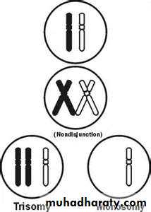
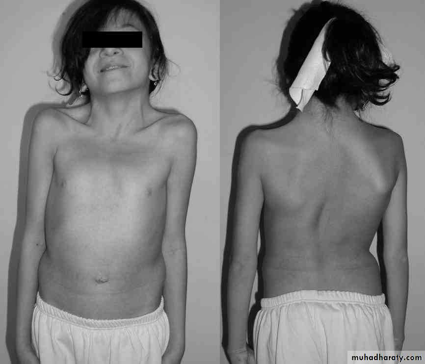
Examples of these abnormalities: 1- a syndrome called gonadal dysgenesis (or agenesis) or, alternatively, ovarian agenesis or Turner's syndrome. In these individuals there is an XO chromosomal pattern, the gonads are rudimentary or absent, there is female external genitalia. Stature is short, webbing of the neck, other congenital abnormalities are often present, and no maturation occurs at puberty.
2- A syndrome known as seminiferous tubule dysgenesis or Klinefelter's syndrome. Individuals with the XXY pattern, the most common sex chromosome disorder, have the genitalia of a normal male. Testosterone secretion at puberty is often great enough for the development of male characteristics. However, the seminiferous tubules are abnormal, and there is a higher than normal incidence of mental retardation, and usually these patients are long.
.
3- The XXX ("superfemale") pattern, it is more common in general population, since it does not seem to be associated with any characteristic abnormalities.
4- The YO combination is probably lethal and incompatible with life.
Nondisjunction or simple loss of a sex chromosome can occur during the early mitotic divisions after fertilization. The result of faulty mitoses in the early zygote is the production of a mosaic, an individual with two or more populations of cells with different chromosome complements.
True hermaphroditism, the condition in which the individual has both ovaries and testes, is probably due to XX/XY mosaicism and related mosaic patterns, although other genetic aberrations are possible.
Chromosomal abnormalities also include transposition of parts of chromosomes to other chromosomes. Sex chromosome abnormalities are, of course, not the only abnormalities associated with disease states; nondisjunction of several different autosomal chromosomes is known to occur. For example, nondisjunction of chromosome 21 produces trisomy 21, the chromosomal abnormality associated with Down's syndrome (mongolism). The additional chromosome 21 is normal, so Down's syndrome is a pure case of gene excess causing abnormalities. In most instances, nondisjunction occurs in the ovary rather than the testis and the incidence of Down's syndrome increases with advancing age of the mother.
Hormonal Abnormalities
Development of the male external genitalia occurs normally in genetic males in response to androgen secreted by the embryonic testes.Genetic females exposed to androgens from some other source during the eighth to the thirteenth weeks of gestation, may develop male genetalia. The syndrome that results is female pseudohermaphroditism. A pseudohermaphrodite is an individual with the genetic constitution and gonads of one sex and the genitalia of the other. After the thirteenth week, the genitalia are fully formed, but exposure to androgens can cause hypertrophy of the clitoris. Its causes either due to congenital virilizing adrenal hyperplasia ,or it may be caused by androgens administered to the mother.
Male pseudohermaphroditism: development of female external genitalia in genetic males. It occurs when the embryonic testes are defective. Because the testes also secrete MIS (mullerian inhibiting substance or mullerian regression factor), genetic males with defective testes have female internal genitalia.
Another cause of male pseudohermaphroditism is androgen resistance, in which, male hormones cannot exert their full effects on the tissues. One form of androgen resistance is a 5α-reductase deficiency, in which the enzyme responsible for the formation of the active form of testosterone is decreased.
This androgen resistance may also due to mutations in the androgen receptor gene and the resulting defects in receptor function ranges from minor to severe. Mild defects cause infertility with or without gynecomastia .
When the loss of receptor function is complete, the testicular feminizing syndrome, now known as complete androgen resistance syndrome, results. In this condition, MIS is present and testosterone is secreted at normal or even elevated rates. The external genitalia are female, but the vagina ends blindly because there are no female internal genitalia. Individuals with this syndrome develop enlarged breasts at puberty and usually are considered to be normal women until they are diagnosed when they seek medical advice because of lack of menstruation .i.e female external genetalia, male internal genetalia
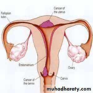
Female reproductive system
Female reproductive functions can be divided into two major phases: (1) preparation of the female body for conception and pregnancy, and (2) the period of pregnancy itself.Ovaries:
From the sixth week of intrauterine life the embryonic ovaries develop which contain the primordial follicles which are about 7000000 follicles, then the number regress during the next stages of life .the female needs only one ovum in each menstrual cycle.
Functions of the ovarian hormones—the ovary secrets 3 hormones:
Estrogens
Progestins
Relaxin
ESTROGENS: It is a steroid synthesized in the ovaries mainly from cholesterol and secreted by the ovarian theca interna cells, corpus luteum, fetoplacental units and small amounts by the adrenal cortices and the testes.
There are three estrogens present in significant quantities in the plasma of the human female: b-estradiol, estrone, and estriol, but The estrogenic potency of b-estradiol is more than the others, it is considered the major estrogen.
They are transported in the blood bound mainly with plasma albumin and with specific estrogen binding globulins.
Functions of the Estrogens:
Growth of the tissues of the sex organs1-Effect of Estrogens on the Uterus and External Female Sex Organs.
Estrogens change the type of cervical mucosa and change the vaginal epithelium from a cuboidal into a stratified type (more resistant to infection), the external genitalia enlarge. The size of the uterus will increase and the excitability of its muscle will increase also. It also cause marked proliferation of the endometrial stroma (especially in the first half of the menstrual cycle).
Estrogen makes the cervical mucosa thinner and more alkaline, these changes promotes survival and transport of sperms.
2-Effect of Estrogens on the Fallopian Tubes: it causes increase in the mobility of the fallopian tubes .
3-Effect of Estrogens on the Breasts.
It causes development of the stroma and the ductile tissue of the breast in addition to that it causes breast growth
4-Effect of Estrogens on the Skeleton.
It stimulates bone growth (and causes a slight increase in total body protein). Also it causes uniting of the epiphyses with the shafts of the long bones. This effect of estrogen in the female is much stronger than the similar effect of testosterone in the male.
After menopause and because of estrogen deficiency there will be decrease of the bone matrix (increase in osteoclastic activity) and osteoporosis usually occurs.
5-Female sexual characteristics: estrogen causes deposition of fat in the breasts and subcutaneous tissues, also in the buttocks and thighs (broad hip), which is characteristic of the feminine figure.
6-Effect of Estrogens on the Skin. Estrogens cause the skin to develop a texture that is soft and usually smooth, also it causes high ratio of scalp hair to the body hair.
7-Effect of Estrogens on Electrolyte Balance. It Causes sodium and water retention by the kidney tubules.
8-Estrogen also causes decrease in the plasma cholesterol level, that’s why the incidence of ischemic heart disease in female is higher after menopause.
PROGESTINS:
The most important of the progestins is progesterone. In the normal nonpregnant female,Secreted mainly during the latter half of each ovarian cycle (secretory phase), by the corpus luteum.
Progesterone is also secreted by the placenta during pregnancy.
Small amounts of progesterone are excreted by the adrenal cortex and testes.
It binds to plasma proteins then almost all the progesterone is degraded by the liver to other steroids that have no progestational effect. The major end product of progesterone degradation is pregnanediol which is excreted in urine after conversion to glucoronide.
Functions of Progesterone
1-Effect of Progesterone on the Uterus.It promotes secretory changes in the uterine endometrium during the latter half of the monthly female sexual cycle, thus preparing the uterus for implantation of the fertilized ovum.
Also it decreases the frequency and intensity of uterine contractions, thereby helping to prevent expulsion of the implanted ovum.
2-Effect of Progesterone on the Fallopian Tubes.
It promotes increased secretion by the mucosal lining of the fallopian tubes.
3-Effect of Progesterone on the Breasts.
Progesterone promotes development of the lobules and alveoli of the breasts, causing the alveolar cells to proliferate, enlarge, and become secretory in nature
4-Progesterone is a thermogenic hormone responsible for rising temperature at time of ovulation.
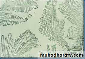
Note: as we said Estrogen makes the cervical mucosa thinner and more alkaline, these changes promotes survival and transport of sperms, while progesterone makes the secretion thick and the mucosa cellular, so the mucosa is thinnest at time of ovulation and rise in an arborizing fern like pattern when a thin layer spread on the slide while after ovulation and during pregnancy, it become thick and fail to form the fern like pattern.
3-Relaxin:
It is the third hormone secreted by from the corpus luteum of pregnancy .It relaxes the symphysis pubis and causes softening and relaxation of the cervix during pregnancy .It may also be secreted by the placenta and the uterus.Menstrual cycle:
Puberty and MenarchePuberty means the onset of adult sexual life.
Menarche means the beginning of the cycle of menstruation.
The period of puberty is caused by a gradual increase in gonadotropic hormone secretion by the pituitary, the onset of puberty and menstruation between ages 11 and 16 years in girls (average, 13 years).
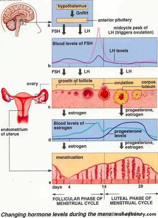
Menstrual cycle is the regular cyclic changes which occur from puberty till menopause .The cycle extends from the first day of the period to the first day of the next.
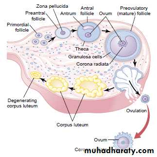
The primordial follicles which were in the ovary from birth begin to ripe under the effect of FSH. There will be accelerated growth of 6 to 12 primary follicles each month.
After a week or more of growth ,one of the follicles begins to outgrow all the others; the remaining 5 to 11 will be atretic .The single follicle reaches a diameter of 1 to 1.5 centimeters at the time of ovulation and called the mature follicle.
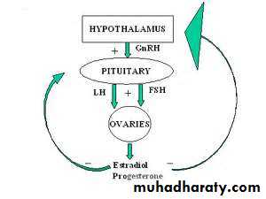
As the follicle grows there will be increase in ovarian secretion of estrogen, it feeds back to the anterior pituitary and stimulate the release of FSH and LH (positive feedback effect) .
By the day 13 of the cycle, the circulating level of LH increases sharply, this LH surge is accompanied to lesser extent by an increase of FSH secretion also, this combination of gonadotropins bring about the final maturation of the of the ovarian follicles and ovulation
Ovulation in a woman who has a normal 28-day female sexual cycle occurs 14 days after the onset of menstruation.
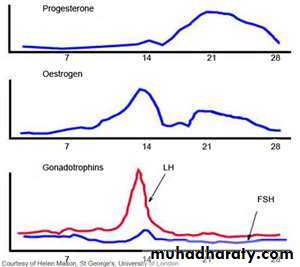
The period between the ovulation and the next cycle is usually 14 days, so any change in the period occurs in the first half of the cycle
If the pregnancy occurs the corpus luteum persists (it persists because of the action of human chorionic gonadotrophin hCG secreted by the embryonic tissues) and continue to secret estrogen and progesterone, but after the 12th week the fetoplacental unit takes the function.
After fertilization the fertilized ovum reaches the uterine cavity in 3-4 days .During this time many or several mitotic divisions occur (it becomes what is called blastocyt about 100 cells), but not reach very large size because the opening of the fallopian tube is small but if it enlarges in the tube for any reason there will be ectopic pregnancy.
But in normal conditions the fertilized ovum implanted in the uterine cavity in the 7th or 8th day after fertilization. Implantation results from the action of trophoblast cells that secrete proteolytic enzymes that digest the adjacent cells of the uterine endometrium.
After implantation the ovulation and menstruation is prevented by the secretion of human chorionic gonadotropin by the newly developing embryonic tissues, the secretion of this hormone can first be measured in the blood 8 to 9 days after ovulation or 6 days after fertilization (or in urine 14 days after fertilization), shortly after the blastocyst implants in the endometrium. Then the rate of secretion rises rapidly to reach a maximum at about 10 to 12 weeks of pregnancy and decreases back to a lower value by 16 to 20 weeks. It continues at this level for the remainder of pregnancy.
The function of human Chorionic Gonadotropin (hCG) is to prevent involution of the corpus luteum at the end of the monthly female sexual cycle. Instead, it causes the corpus luteum to secrete even larger quantities of its sex hormones—progesterone and estrogens—for the next few months.
Regarding the lining of the uterus, it passes through the following stages: (1) proliferation of the uterine endometrium; (2) development of secretory changes in the endometrium; and (3) desquamation of the endometrium, which is known as menstruation.
Proliferative Phase (Estrogen Phase) of the Endometrial Cycle, Occurring Before Ovulation
Under the influence of estrogens, secreted in increasing quantities by the ovary during the first part of the monthly ovarian cycle, the stromal cells and the epithelial cells proliferate rapidly the endometrium increases greatly in thickness, owing to increasing numbers of stromal cells and to progressive growth of the endometrial glands and new blood vessels into the endometrium.Secretory Phase (Progestational Phase) of the Endometrial Cycle, Occurring After Ovulation
Progesterone causes marked swelling and secretory development of the endometrium. The glands increase in tortuosity. Also, the cytoplasm of the stromal cells increases; lipid and glycogen deposits increase greatly in the stromal cells; and the blood supply to the endometrium further increases with the blood vessels becoming highly tortuous. The whole purpose of these endometrial changes is to prepare large amounts of stored nutrients to provide appropriate conditions for implantation of a fertilized ovum.If the ovum is not fertilized, about 2 days before the end of the monthly cycle, the corpus luteum in the ovary suddenly involutes, and the ovarian hormones (estrogens and progesterone) decrease to low levels of secretion, Menstruation follows which lasts usually from 4-7 days (necrosis in the endometrium, especially of the blood vessels → bleeding about 40 ml of blood).