بسم الله الرحمن الرحيم

FEMALE REPRODUCTIVE SYSTEM
Prof.Dr. Huda Al-KhateebHead of Quality Assurence & Academic Performance
Head of Histology section/Dept. of Anatomy
College of Medicine/ Univ. of Baghdad
hmdk54@yahoo.com

objectives
This lecture includes• An introduction on female reproductive system
• Study general histology of ovary
• Study full details of folliculogenesis
• Histology of mature follicle
• Follicular atresia

female reproductive system
The female reproductive system consists of(1) two ovaries
(2) Two oviducts (or uterine tubes)
(3) One uterus
(4) One vagina
(5) One external genitalia

Functions
1.produce female gametes (oocytes)2.provide the environment for fertilization
3. hold the embryo during its complete development through the fetal stage until birth.
4.produce steroidal sex hormones that control organs of the reproductive system and influence other organs of the body.
Definitions
Menarche: the first menstrual cycle. It occurs at 11-13 years.then reproductive system undergoes cyclic changes in structure and functional activity
Menopause is a variable period during which the cyclic changes become irregular and eventually disappear. It occurs at 45-55 years.
Reproductive life: period between menarche and menopause
In the postmenopausal period there is a slow involution of the reproductive organs.

Mammary gland
Although the mammary glands do not belong to the genital system, yet they are included here because they undergo changes directly connected to the functional state of the reproductive system.
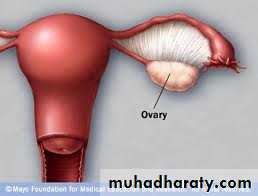
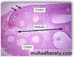
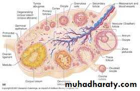
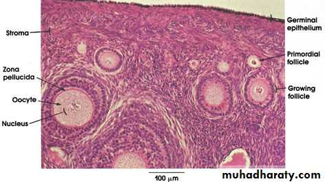
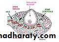
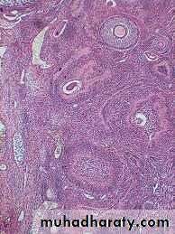
Ovaries
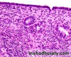
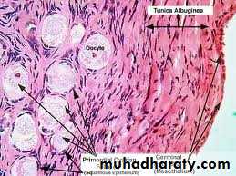
They are almond-shaped bodies (3 x1.5x1 cm).Each ovary is covered by a simple cuboidal epithelium, the germinal epithelium, continuous with the mesothelium and overlying a layer of dense connective tissue capsule the tunica albuginea, responsible for the whitish color of the ovary.
ovaries
It consists of the cortex (stroma) a region filled with a highly cellular connective tissue stroma and many ovarian follicles.The internal part is the medulla, which contains loose connective tissue blood vessels entering the organ through the hilum from mesenteries suspending the ovary.
Early Development of the Ovary
In the first month of embryonic life, a small population of primordial germ cells migrates from the yolk sac to the gonadal primordia.In the gonads these cells divide extensively and differentiate as oogonia.
In the third month, oogonia begin to enter the prophase of the first meiotic division but arrest and called primary oocytes.
Each primary oocyte becomes surrounded by flattened supportive cells called follicular cells forming primordial follicle.
.
No. of primordial follicles
In developing ovaries of atwo-month embryo there are 600,000 oogonia
Five month embryo 7 million oocytes.
At birth 2 million oocytes.
At puberty the ovaries contain 300,000 oocytes.
Because generally only one oocyte resumes meiosis with ovulation during each menstrual cycle (average duration, 28 days) and the reproductive life of a woman lasts about 30–40 years, only about 450 oocytes are liberated from ovaries by ovulation.
All others degenerate through atresia
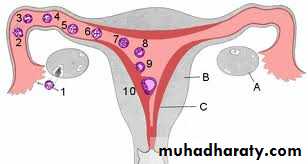
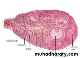
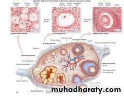
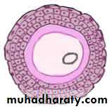
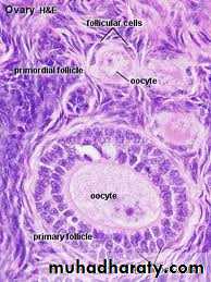
Ovarian Follicles
They consist of an oocyte surrounded by one or more layers of epithelial cells.They are embedded in the cortex of the ovary.
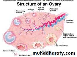
Ovarian follicles are
1.Inactive – Primordial follicles2.Growing follicles – include
Unilaminar primary follicle
Multilaminar primary follicle
Secondary (Antral) follicle
3.Mature (Graafian) follicle
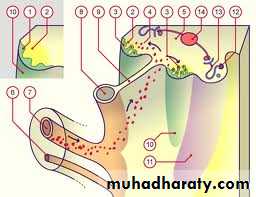
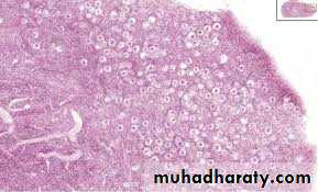
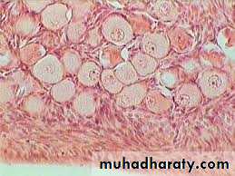
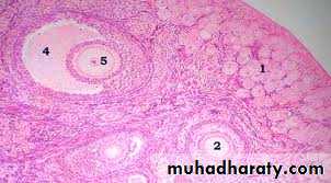
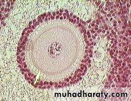
(1)primordial follicles
(1)consist of a primary oocyte enveloped by a single layer of the flattened follicular (granulosa) cells(2)They are inactive follicles.
(3)These follicles are found in the superficial areas of the cortex (under tunica albugenia).
(4)The oocyte in the primordial follicle is a spherical cell about 25 micrometer in diameter, with a large nucleus and in the first meiotic prophase.

Follicular Growth
Beginning in puberty with the release of follicle-stimulating hormone (FSH) from the pituitary, a small group of primordial follicles each month begins a process of follicular growth.
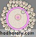
(2)Growing follicles(A) unilaminar primary follicles
(1) Oocyte is larger(2) Follicular (granulosa) cells become cuboidal then columnar

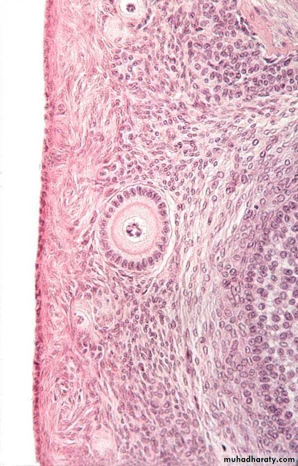
(B) Multilaminar primary follicles
• 1.The follicular cells continue growth, forming a stratified epithelium, termed granulosa cells• 2.the follicle is surrounded by basement membrane.
(B) Multilaminar primary follicles
• 3.Between the oocyte and granulosa cells, a layer of extracellular material called the zona pellucida develops, consist of glycoprotein secreted by the oocyte. Zona pellucida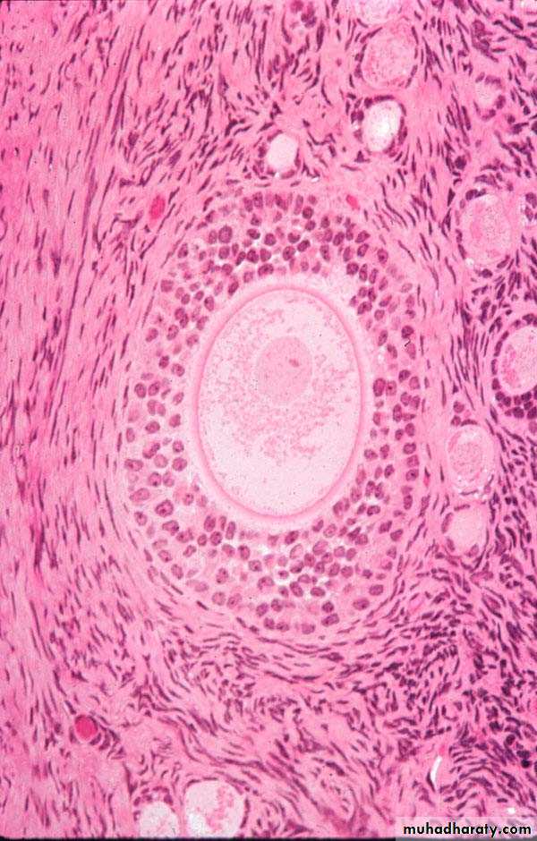
(B) Multilaminar primary follicle
4. the stromal cells immediately around the follicle differentiate to form the follicular theca.This layer subsequently differentiates further as two layers: a well-vascularized endocrine tissue, the theca interna, and a more fibrous outer theca externa containing smooth muscle and fibroblasts
(B) Multilaminar primary follicles

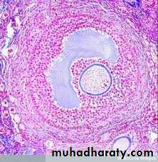
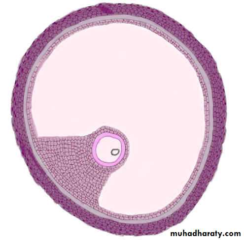
(C)Secondary (antral) follicles
1.They move deeper in the ovarian cortex.2.Small spaces develop within the granulosa layer as the cells secrete follicular fluid (or liquor folliculi). This fluid accumulates, the spaces gradually coalesce, and the granulosa cells reorganize themselves around a larger cavity, the antrum.
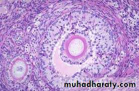
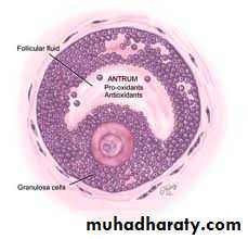
(C)Secondary (antral) follicles
antrum formation pushes oocyte and the surrounding granulosa cells to one side of follicle forming, the cumulus oophorus.The remaining granulosa cells are called membana granulosa
(3) Mature (graafian) follicles
During each menstrual cycle, usually one follicle grows much more than the others and becomes the dominant follicle, while many of the other follicles eventually enter atresia.The dominant follicle usually reaches the most developed stage of follicular growth and may undergo ovulation.
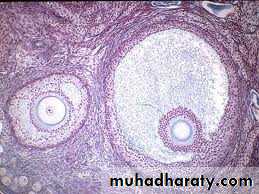
(3) Mature (graafian) follicles
1.reaches a diameter of 20-30 mm or more prior to ovulation,2.large enough to protrude from the surface of the ovary and be detected by ultrasound imaging.
3.The antrum increases greatly in size by accumulating follicular fluid
4.the oocyte adheres to the wall of the follicle through the cumulus oophorus of granulosa cells.
.
(3) Mature (graafian) follicles
5.The membrana granulosa layer becomes thinner.6.The granulosa cells immediately around and linked to the oocyte make up the corona radiata and accompany the oocyte when it leaves the ovary
folliculogenesis
It is a process by which primordial follicle grow on to develop e a mature follicle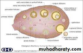

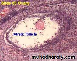
Follicular Atresia
It is adegenerative process of ovarian follicles at any stage of development, including nearly mature folliclesAlthough follicular atresia takes place from before birth until a few years after menopause, it is most prominent just after birth, when levels of maternal hormones decline rapidly, and during both puberty and pregnancy, when qualitative and quantitative hormonal changes occur again.
Follicular Atresia
Atresia of primary follicles (uni- and multi-laminar).First – oocyte degenerate
Followed by follicular (granulosa) cells degeneration
Follicular Atresia
Atresia of secondary (antral) follicles1.starts by apoptosis (programmed cell death) and detachment of the granulosa cells
2.macrophages invade the degenerating follicle and phagocytose the debries3.autolysis of the oocyte and collapse of the zona pellucida.
4.Later fibroblasts occupy the area of the follicle and produce a collagen scar that may persist for a long time.
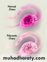
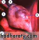
MEDICAL APPLICATION
Polycyctic ovary syndrome
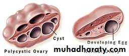
Polycyctic ovary syndrome
Polycyctic ovary syndrome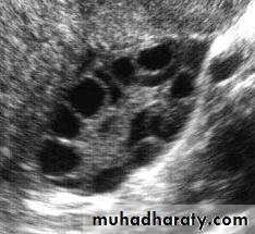
Polycyctic ovary syndrome
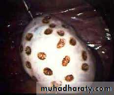
Polycyctic ovary syndrome
Hirsutism

Test tube baby &ivf
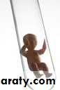

Test tube baby &ivf
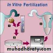
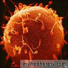
Test tube baby &ivf
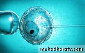

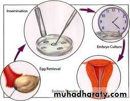
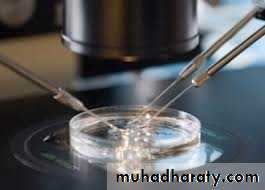
Test tube baby &ivf
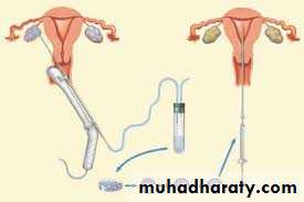
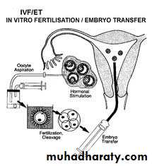
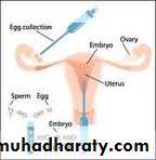

Test tube baby &ivf

Test tube baby &ivf