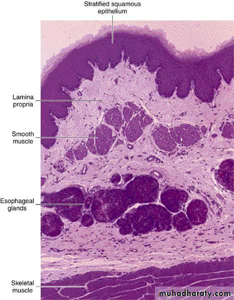
• Photomicrograph of a section of the upper region of the esophagus.
• Mucous oesophageal glands are in the submucosa; striated skeletal muscle• is in the muscularis. PAS and PT stain. Low magnification.
The oesophago-gastric junction is an important site of the pathologic abnormality. Lower oesophagus is an important site of common diseases, particularly ulceration, stricture & cancer.
The epithelium of the oesophagus is protected from exposure to the gastric acid by:-
1. The anatomical arrangement of the oesophago-gastric junction.2. The cardiac sphincter which prevents reflux of the gastric contents into the lower oesophagus.
The oesophago-gastric junction is an important site of the pathologic abnormality. Lower oesophagus is an important site of common diseases, particularly ulceration, stricture & cancer.
Reflux of acid gastric secretions may occur into the lower esophagus causing inflammation & pain. Under the constant irritating effect of reflux of acidic gastric secretions, the epithelium in the lower esophagus changes to a gastric type.
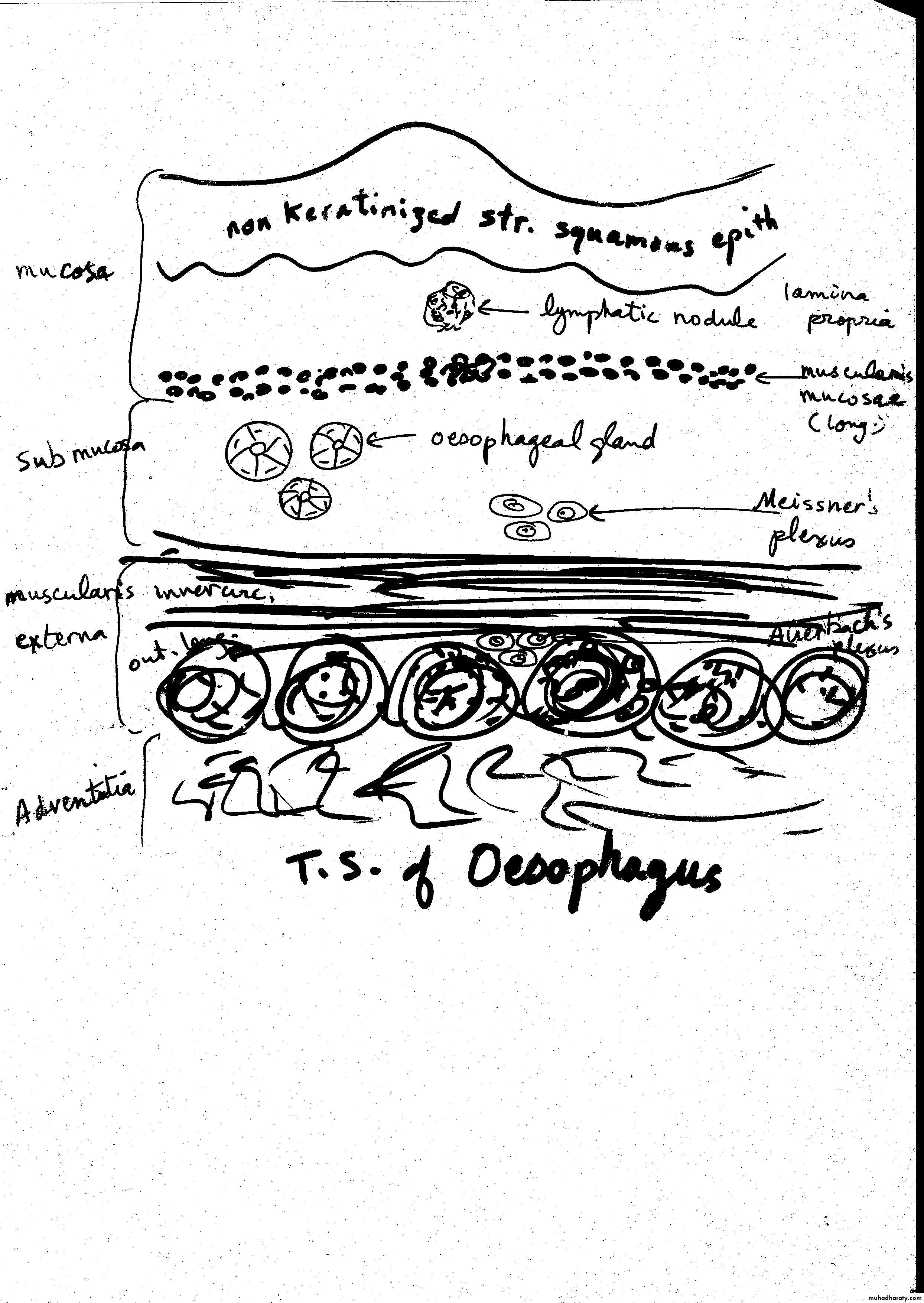
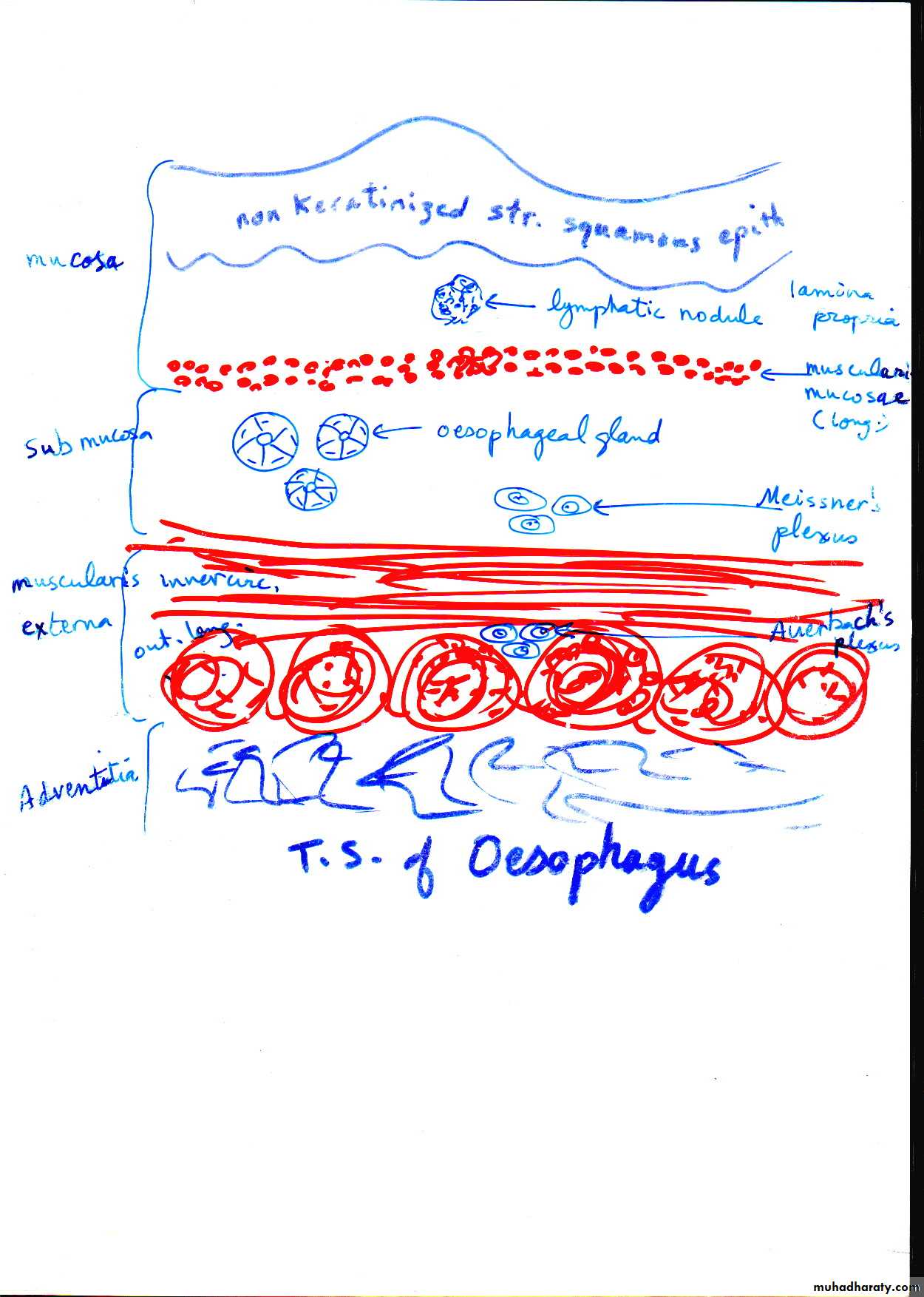
Stratified squamous non-keratinized epithelium→ simple columnar
The columnar epithelium is prone to ulceration & inflammation
and predispose to the development of one type of oesophageal cancer
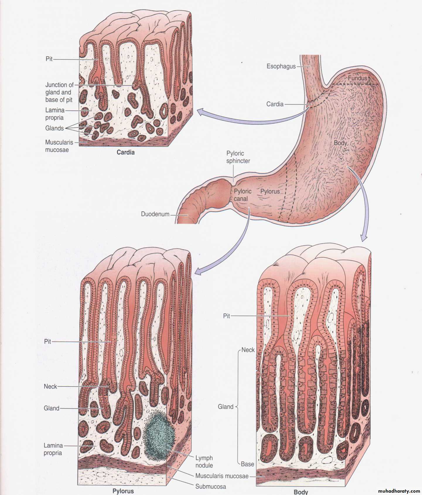
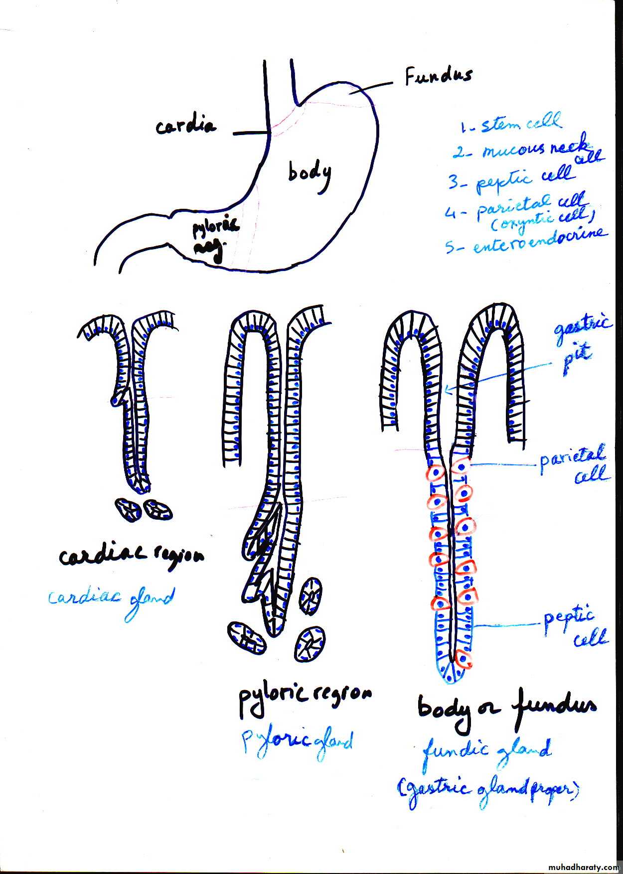
Regions of the stomach
and their histologicalstructure
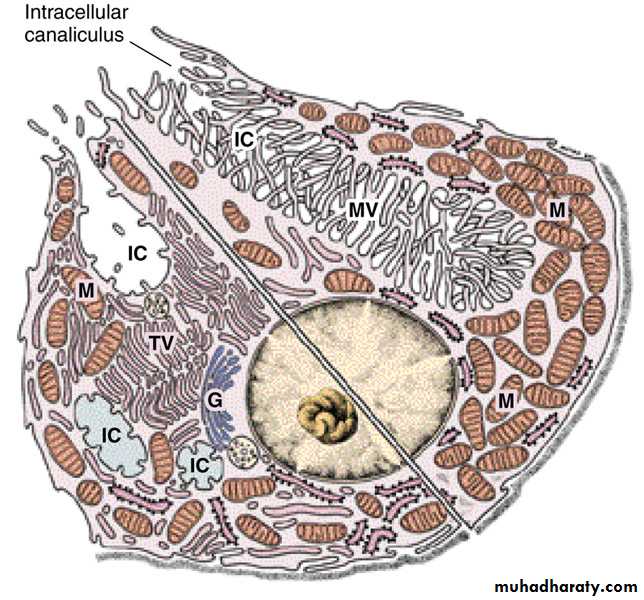
• Composite diagram of a parietal cell, showing the ultrastructural differences between a resting cell (left) and an active cell (right). Note that the tubulovesicles (TV) in the cytoplasm of the resting cell fuse to form microvilli (MV) that fill up the intracellular canaliculi (IC). G, Golgi complex; M, mitochondria. (Based on the work of Ito S, Schofield GC. J Cell Biol 1974; 63:364.)
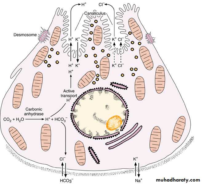
Diagram of a parietal cell, showing the main steps in the synthesis of hydrochloric acid. Active transport by ATPase is indicated by arrows and diffusion is indicated by dotted arrows. Under the action of carbonic anhydrase, blood CO2 produces carbonic acid. Carbonic acid dissociates into a bicarbonate ion and a proton H+, which is pumped into the stomach lumen in exchange for K+. A high concentration of intracellular K+ is maintained by the Na+,K+ ATPase, while HCO3— is exchanged for Cl— by an antiport. The tubulovesicles of the cell apex are seen to be related to hydrochloric acid secretion, because their number decreases after parietal cell stimulation. The bicarbonate ion returns to the blood and is responsible for a measurable increase in blood pH during digestion.
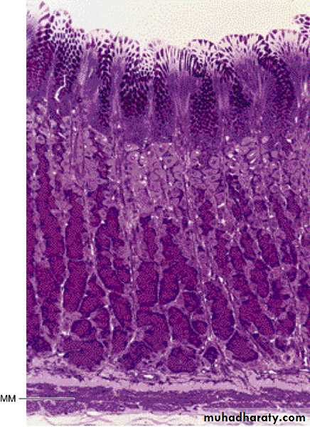
• Photomicrograph of a section of
• the gastric glands in the fundus of• the stomach. Note the superficial
• mucus-secreting epithelium.
• Parietal cells (light-stained)
• predominate in the mid and upper
• regions of the glands; chief (zymogenic)
• cells (dark-stained) predominate in the
• lower region of the gland. MM,
• muscularis mucosae. PT stain.
• Low magnification.
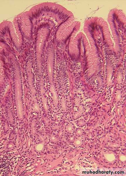
• Photomicrograph of a section of the
• pyloric region of the stomach. Note• the deep gastric pits with short pyloric
• glands in the lamina propria. H&E stain.
• Low magnification. (Courtesy of MF Santos.)