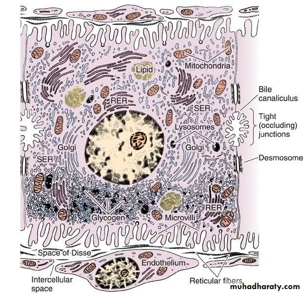
• Ultrastructure of a hepatocyte. RER, rough endoplasmic reticulum; SER, smooth endoplasmic reticulum. x10,000
The contents of space of Disse
1. Microvilli of hepatocyte2. Plasma
3. Reticular fibers
4. Fat storing cell (Ito cell)
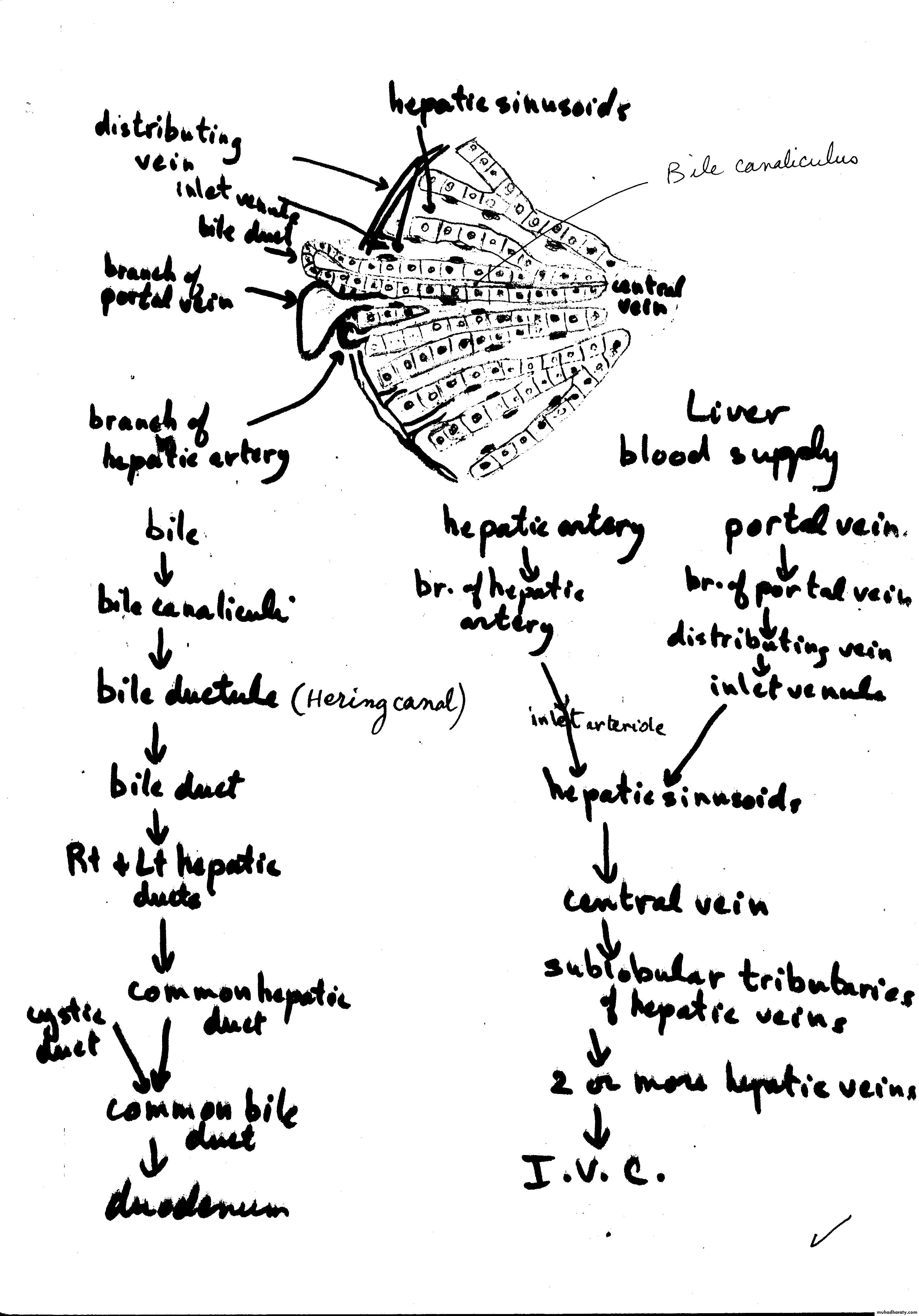

• Schematic drawing of the structure of the liver. The liver lobule in the center is surrounded by the portal space (dilated here for clarity). Arteries, veins, and bile ducts occupy the portal spaces. Nerves, connective tissue, and lymphatic vessels are also present but are (again, for clarity) not shown in this illustration. In the lobule, note the radial disposition of the plates formed by hepatocytes; the sinusoidal capillaries separate the plates. The bile canaliculi can be seen between the hepatocytes. The sublobular (intercalated) veins drain blood from the lobules. (Redrawn and reproduced, with permission, from Bourne G: An Introduction to Functional Histology. Churchill, 1953.
Liver Regeneration
The liver has an extraordinary capacity for regeneration.
The regenerated liver tissue is usually similar to the removed tissue.
If there is continuous or repeated damage to hepatocytes over a long period, the multiplication of liver cells is followed by a pronounced increase in the amount of connective tissue.
Cirrhosis
The excess of c.t. results in disorganization of the liver structure, a condition known as cirrhosis, is a progressive and irreversible process, causes liver failure, and is usually fatal.Cirrhosis is a consequence of any sustained progressive injury to hepatocytes produced by several agents, such as ethanol, drugs or other chemicals, hepatitis virus, and autoimmune liver disease.
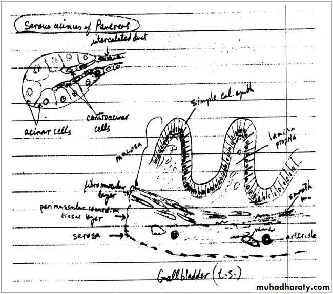
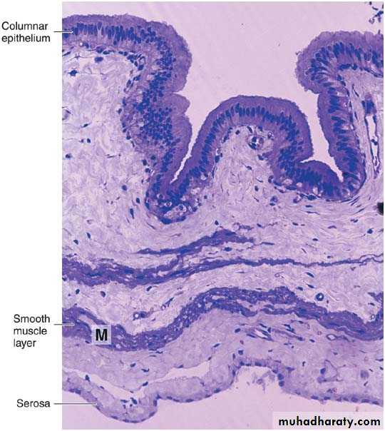
• Photomicrograph of a section of gallbladder. Note the lining of columnar epithelium and the smooth muscle layer (M). PT stain. Low magnification.
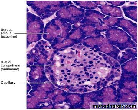
• Photomicrograph of a pancreas showing the exocrine portion (acini) and the endocrine portion (islet of Langerhans). The acini contain secretory cells with basophilic cytoplasm. Different types of endocrine cells are seen in the islet. PT stain. Medium magnification.
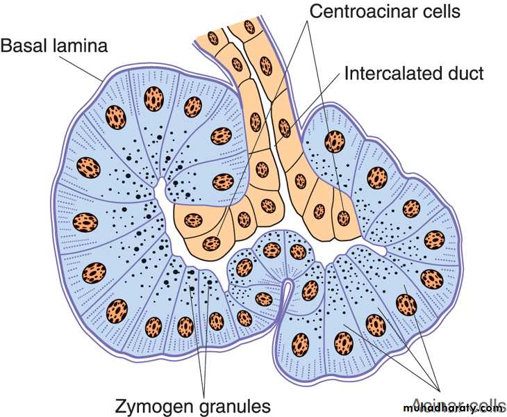
Schematic drawing of the structure of pancreatic acini. Acinar cells are pyramidal, with granules at their apex and rough endoplasmic reticulum at their base. The intercalated duct partly penetrates the acini. These duct cells are known as centroacinar cells. Note the absence of myoepithelial cells
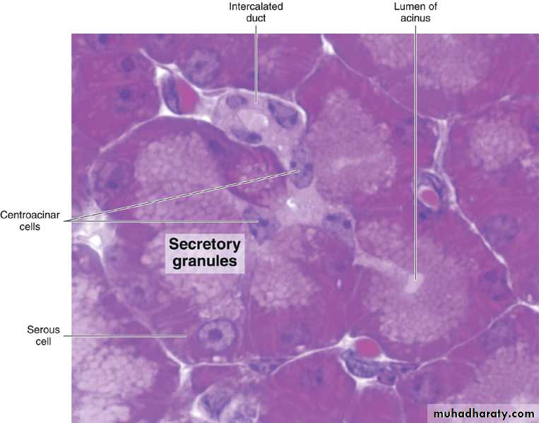
• Section of the exocrine pancreas showing its main components. PT stain. Medium magnification.
• Photomicrograph of a pancreas showing the exocrine portion (acini) and the endocrine portion (islet of Langerhans). The acini contain secretory cells with basophilic cytoplasm. Different types of endocrine cells are seen in the islet. PT stain. Medium magnification.
Cells of islet of Langerhans
1. A cell glucagon2. B cell insulin
3. D cell somatostatin
4. F cell pancreatic polypeptide
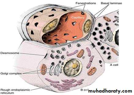
Drawing of the A and B cells; showing their main ultrastructural features. The B cell’s granules are irregular, whereas the A cell’s granules are round