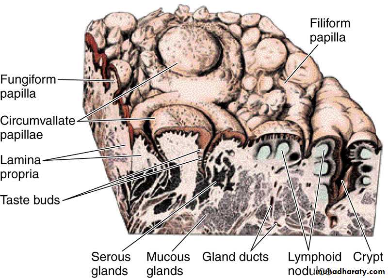
• Surface of the tongue on the region close to its V-shaped boundary,
• between the anterior and posterior portions. Note the lymphoid nodules• (lingual tonsil), glands, and papillae.
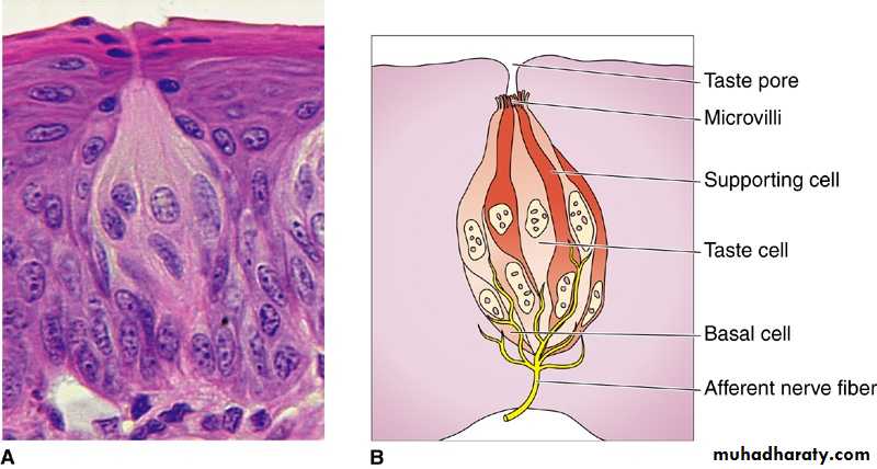
Photomicrograph (A) and drawing (B) of a taste bud, showing the taste cells and the taste pore. The drawing also illustrates several cell types (basal, taste, and supporting) and afferent nerve fibers that, upon stimulation, will transmit the sensory information to the central gustatory neurons. A: Hematoxylin and eosin (H&E) stain.

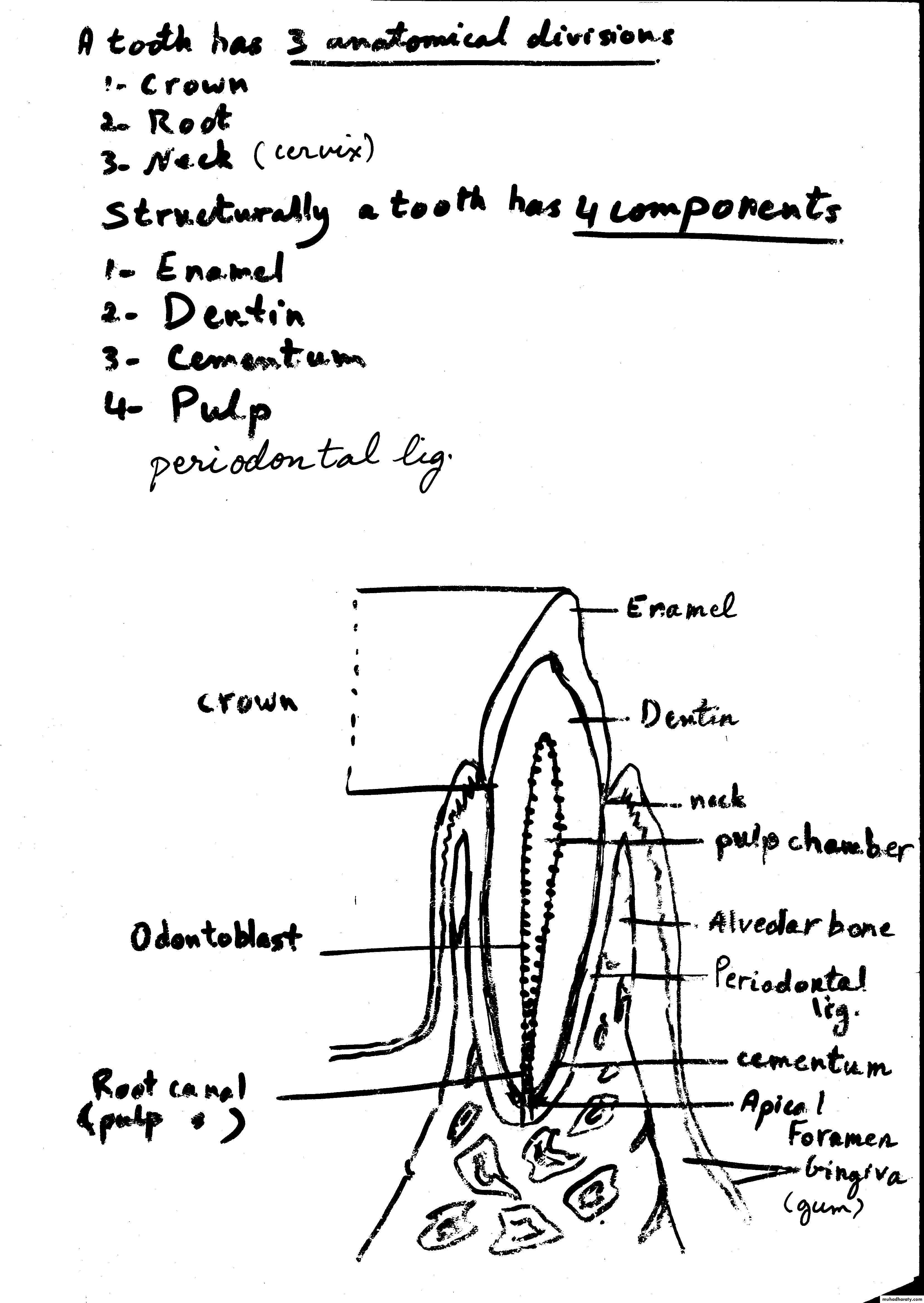
• Diagram of a sagittal section from an incisor tooth in position in the mandibular bone.
• (Redrawn and reproduced, with permission, from Leeson TS, Leeson CR: Histology,
• 2nd ed. Saunders, 1970.)