
1
Lecture: 1
Parasitology
Hemoflagellates
(blood and tissue flagellates)
ﺩ
.
ﻫﻴ
ﻔﺎء ﺩﺍﻭﺩ
*Classification: - Sub-kingdom: Protozoa
-Phylum: Sarcomastigophora
-Sub-phylum: Mastigiphora
-Class: Zoomastigophora
*Flagellates (that infect man) divided into:
1-Intestinal and urogenital flagellates
• Giardia intestinalis
• Chilomastix mesinili
• Trichomonas vaginalis
• Dientamoeba fragilis and other.
2-Hemoflagellates (blood and tissue flagellates)
Two genera within hemoflagellates infect human which are:
• Genus Leishmania
• Genus Trypanosoma
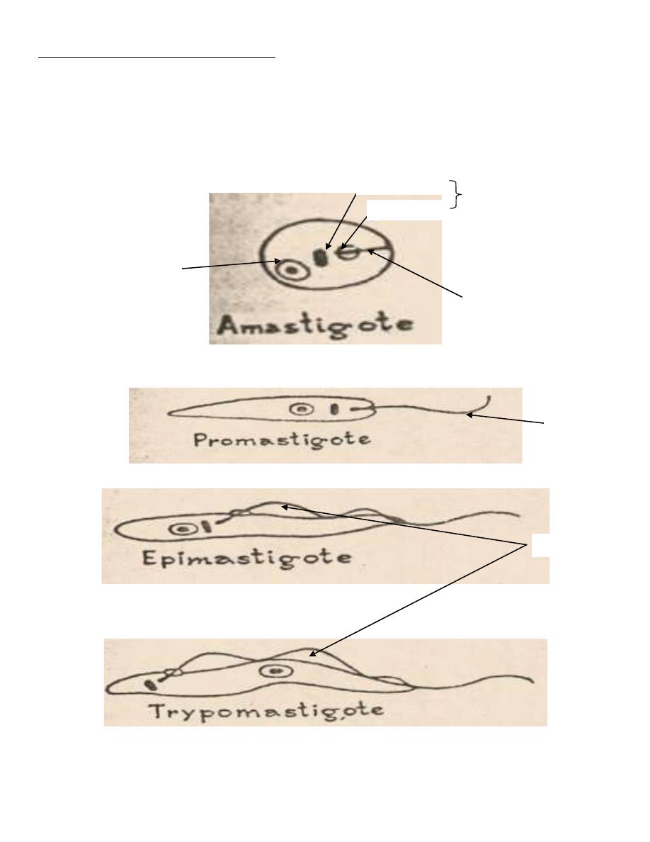
*Morphological forms of hemoflagellates
1-Amastigote (Leishmania) form
Round or oval in shape, 2-5 microns in diameter, surrounded by
delicate cell membrane, have single vesicular nucleus with large
central karyosome, the kinetoplast (which consists from dot-like
blepharoplast and parabasal body beside it) lies at right angle to the
nucleus. Closely located nucleus and kinetoplast known as torpedo
form. The axoneme is a delicate membrane extends from the
kinetoplast to the margin of the body and represents the rest of the

2
flagellum with vacuole lying alongside the axoneme. This form
(amastigote) has no flagellum.
2-Promastigote (leptomonad) form
Elongated (spindle in shape) measuring 15-20 microns X 1-2
microns, have centrally located nucleus and the kinetoplast situated at
the anterior end. The vacuole lying in front of the kinetoplast. From
blepharoplast, single free flagellum projects from the anterior end,
equal or longer than the body length.This form has no undulating
membrane.
3-Epimastigote (crithidia) form
Elongated form, 15-20 microns long and slightly wider than
promastigote, nucleus near middle, kinetoplast is anterior to the
nucleus. From blepharoplast flagellum arise forming the undulating
membrane extending half of the body length, and project from the
anterior end as a free flagellum.
4-Trypomastigote (Trypanosome) form
Elongated form with highly polymorphism from rather short and
stumpy (15micron X 2-4micron) to a long slender from (35micron X
2-4micron). In stained blood film, Trypanosoma cruzi appears as C or
U shape. Nucleus near middle, kinetoplast is at the posterior end, the
flagellum and undulating membrane pass anteriorly along entire body
length and free flagellum extends from anterior end when present.
Genus Leishmania
It includes parasites cause three diseases in human:
1- Cutaneous Leishmaniasisor oriented sore.
2- MucocutaneousLeishmaniasisor Espondia
3- Visceral Leishmaniasis or Kala-azar

3
Disease
Leishmania species
Geographical location
Cutaneous leishmainiasis
-L.tropica complex as
1-L. tropica
2- L. major
3-L. aethiopica
-L. mexicana complexas
L. mexicana and other
species
- Old world (Old world
cutaneous leishmaniasis)
-(New world)New world
cutaneous leishmaniasis
Mucocutaneousleishmaniasis
-L.braziliensis complex as
L.braziliensis and other
species
-(New world)New world
cutaneous leishmaniasis
Visceral leishmaniasis
-L.donovani complex as
1-
L.donovani
2-
L.infantum
3-
L.chagasi
-Old world
-Old world
-New world
Genus Trypanosoma
Cause 2 diseases in human
1- African trypanosomiasis (sleeping sickness)
2- American trypanosomiasis (Chagas’ disease)
Clinical disease
Trypanosome species
Vector
-African
trypanosomiasis
1-
Gamian type
(west African
sleeping skiness)
2-
Rhodesian type
(east African
sleeping sickness)
-T.gambiense
-T. rhodesiense
-Tsetse fly (Glossinia)
-Tsetse fly (Glossinia)
-American
trypanomiasis
T. cruzi
Reduviid or kissing
bug (Triatomid bug)
4
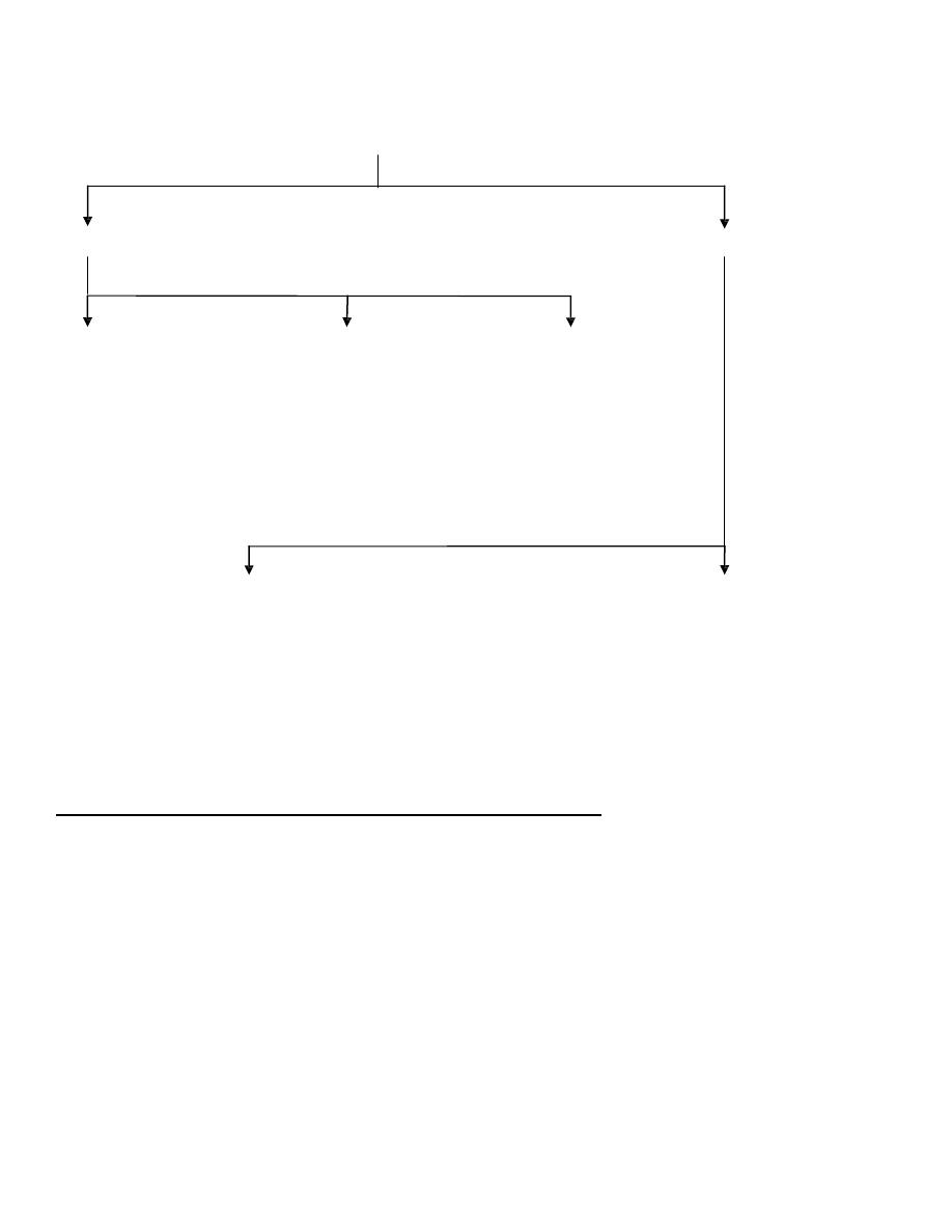
Hemoflagellate
Leishmania
Trypanosoma
Cutaneous leishmaniasis Mucocutaneousleishmaniasis
Visceral leishmaniasis
(Oriental Sore)
(Espondia)
(Kala-azar)
-L. tropica complex
-L. braziliensis complex
-L. donovani complex
-L. mexicana complex
Americantrypanosomiasis
African trypanosomiasis
(Chagas’ disease)
(Sleeping sickness)
-T. Cruzi
-T. Gambiense
-T. rhodesiense
General characters of genus Leishmania
1- Life cycle is indirect and completed in tow hosts, vertebrate (human,
dog, rodent) as a final host and invertebrate; blood sucking insect
(female of sand fly) as an intermediate host (vector).
2- Tow developmental forms are found, amastigote and promastigote
,amastigote in the final host (human) and promastigote in the vector
(sand fly).
3- The vector is sand fly of genus Phlebotomus in Old World and
genus Lutzomyia in New World.

5
4- Promastigote is the infective stage to final host (man) and
amastigote is infective stage to sand fly (vector).
5- The parasite infects the reticuloendothelial cells of skin, mucus
membrane or viscera (as liver, spleen and bone marrow) of the final
host (man).
6- The parasite multiplies by binary fission (asexual).
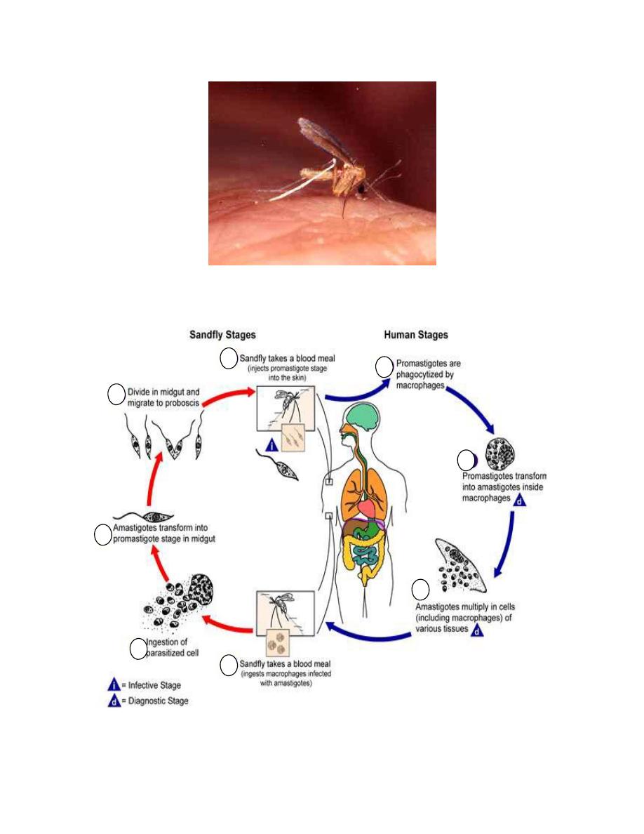
Life cycle
Involves an alternative existence in a vertebrate (man,…ect) and an insect
(sand fly).The flagellated promastigote enter the body (skin) of the final
host through infected sand fly bite
→ the parasites engulfed by
macrophage and endothelial cells of skin capillaries
→promasitgote
develops into amastigote (Leishman-Donovan (LD) bodies)
→amastigote
multiply inside macrophages by binary fission
→ cell burst → free
amastigote either infect other cells (macrophages) in skin as in cutaneous
leishmaniasis or other cells in skin and the adjacent cells in mucous
membrane as in mucocutaneousleishmaniasis or pass to different organ by
blood stream (spleen, liver, bone marrow and lymph nodes) as in visceral
leishmaniasis, then amastigotes engulfed by new reticuloendothelial cells
→a blood sucking sand fly (female) draws amastigotes (L.D bodies) with
its blood meal (by bites of proboscis)
→amastigotes develop in
promastigote forms in the mid gut of sand fly
→ multiply by longitudinal
binary fission
→ solid mass of promastigotes fill up the anterior end of the
mid gut and the esophagus , extending up to the pharynx
→ a heavy
pharyngeal infection of the sand fly is known as anterior station
development , which may block esophagus
→ at the time of sucking blood
, regurgitation of promastigotes from their buccal cavity in the skin
puncture by proboscis (biting organ)
→ infection of man.
Anterior station development is the anterior migration of the parasites
from the mid gut to foregut, pharynx and buccal cavity of insect vector (as
sand fly in Leishmania species).
6
Posterior station development is the posterior migration of the parasite
from the midgut to the hindgut and excreted with feces if insect vector (as
Ruduviid bug in Trypanosoma cruzi).
Barabasal body
blepharoplast
kinetoplast
Nucleus
axoneme
flagellum
Ant. end
post. end
Undulatig membrane
Ant. end
post..end
Ant. end
post.end
Developmental forms (stages) of Leishmania
7
Sand fly
-Life cycle of leishmania spp.