Lec.2
Dr. Inas Khalifa Al-Sharquie
2012-2013
1
Bacteriology
Cell Structure
Learning Outcomes:
• Review molecules and structures of the bacterial cell.
• Describe the roles played by each of the major components of bacterial cell.
• Discuss briefly the features and the structures of the cytoplasmic membrane
and cell well of bacterial cell.
3-Cytoplasmic membrane
The bacterial cell membrane is a typical "unit membrane" composed of
phospholipid bilayer, similar in microscope to that in eukaryotic cells, and
upward of 200 different kinds of proteins.
The prokaryotic and eukaryotic cell membranes are chemically similar, but
eukaryotic membranes contain sterols, whereas prokaryotes generally do
not. The only prokaryotes that have sterols in their membrane are members
of the genus Mycoplasma.
Functions of cytoplasmic membrane:
1. Selective permeability and transport of solutes
The cytoplasmic membrane forms a hydrophobic barrier impermeable to
most hydrophilic molecules. However, several mechanisms (transport
systems) exist that enable the cell to transport nutrients into and waste
products out of the cell. These transport systems work against a
concentration gradient to increase the concentration of nutrients inside the

Lec.2
Dr. Inas Khalifa Al-Sharquie
2012-2013
2
cell, a function that requires energy in some form. There are three general
transport mechanisms involved in membrane transport:
A. Passive transport:
This mechanism relies on diffusion, uses no
energy, and operates only when the solute is at higher concentration
outside than inside the cell. Simple diffusion accounts for the entry of
very few nutrients including dissolved oxygen, carbon dioxide, and
water itself. Simple diffusion provides neither speed nor selectivity.
Facilitated diffusion also uses no energy so the solute never achieves
an internal concentration greater than what exists outside the cell.
However, facilitated diffusion is selective. Channel proteins form
selective channels that facilitate the passage of specific molecules.
Facilitated diffusion is common in eukaryotic microorganisms (eg,
yeast), but is rare in prokaryotes.
B. Active transport: Many nutrients are concentrated more than a
thousand fold as a result of active transport.
C. Group translocation: Is a process in which an organic molecule such
as glucose or mannose is transported into the cell while being
chemically modified.
2. Electron transport & oxidative phosphorylation: The cytochromes
and other enzymes and components of the respiratory chain are located
in the cell membrane. The bacterial cell membrane is thus a functional
analog of the mitochondrial membrane in eukaryotic.
3. Excretion of hydrolytic exoenzymes & pathogenicity proteins:
All
organisms that rely on macromolecular organic polymers as a source of
nutrients (eg, proteins, polysaccharides, lipids) excrete hydrolytic
enzymes that degrade the polymers to subunits small enough to
penetrate the cell membrane.

Lec.2
Dr. Inas Khalifa Al-Sharquie
2012-2013
3
4. Biosynthetic functions: The cell membrane is the site of the carrier
lipids and enzymes used in cell wall synthesis.
5. Chemotactic systems: Attractants and repellents bind to specific
receptors in the bacterial membrane e.g., flagella.
4- Cell wall
Most prokaryotes have a rigid external cell wall that contains peptidoglycan,
a polymer of amino acids and sugars. Eukaryotes on the other hand, do not
contain peptidoglycan.
The cell wall is the outermost component common to all bacteria (except
Mycoplasma species, which bounded by a cell membrane, not a cell wall).
The cell wall is located external to the cytoplasmic membrane.
Cell wall functions:
1- Osmotic protection.
2- Plays an essential role in cell division as well as serving as a primer
for its own biosynthesis.
3- Various layers of the wall are the sites of major antigenic
determinants of the cell surface, and one component—the
lipopolysaccharide of gram-negative cell walls—is responsible for the
nonspecific endotoxin activity of gram-negative bacteria.
4- Not selectively permeable.
5- Bacteria are classified to G+ve or G-ve according to their response to
the differential stains method called Gram’s stains. The difference
reside on the cell wall differences.

Lec.2
Dr. Inas Khalifa Al-Sharquie
2012-2013
4
Peptidoglycan
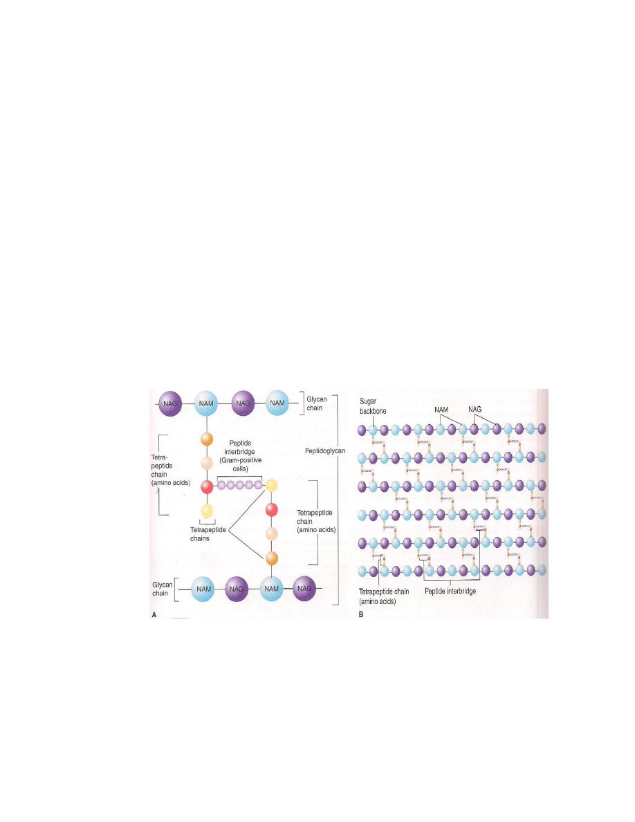
• It is a complex polymer consists of a backbone that composed of alternating
N-acetyl glucosamine (NAG) and N-acetyl muramic acid (NAM).
• A set of identical tetrapeptide side chains attached to N-acetylmuramic acid.
• A set of identical peptide cross-bridges (Figure 1).
• Because peptidoglycan is present in bacteria but not in human cells, it is a
good target for antibacterial drugs. Several of these drugs, such as
penicillins, cephalosporins, and vancomycin, inhibit the synthesis of
peptidoglycan by inhibiting the transpeptidase that makes the cross-links
between the two adjacent tetrapeptides.
• Scientists describe two basic types of bacterial cell walls: gram-positive and
gram-negative.
Figure 1: Peptidoglycan structure. A: Peptidoglycan is composed of a glycan
chain (NAM&NAG), a tetrapeptide chain, and a cross-link (peptide interbridge).
B: Three-dimensional structure. (Levinson W. Review of Medical Microbiology
and Immunology.12
th
ed. Copyright 2012, McGraw-Hill.)

Lec.2
Dr. Inas Khalifa Al-Sharquie
2012-2013
5
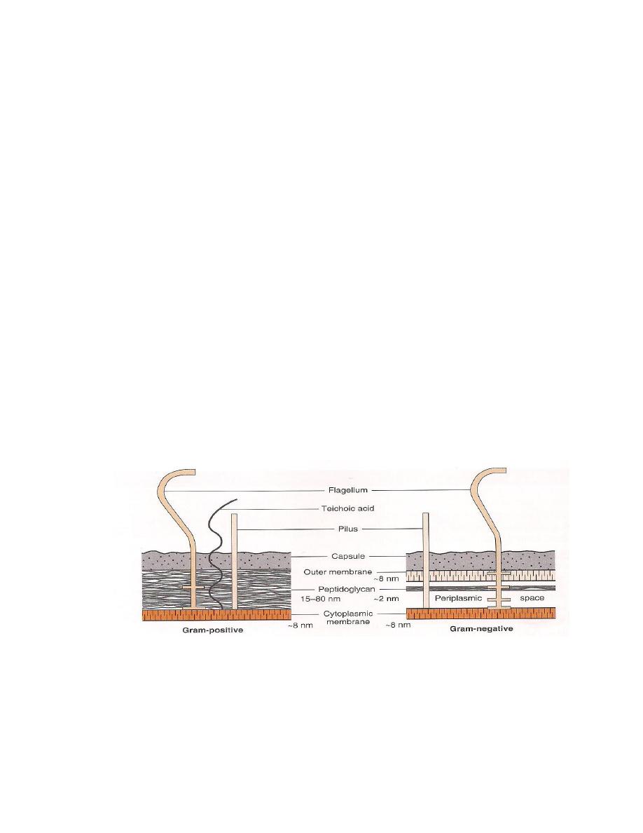
Cell walls of Gram-Positive and Gram-Negative Bacteria
The structure, chemical composition, and thickness of the cell wall differ in gram-
positive and gram-negative bacteria.
Gram-positive
The basal structure of cell wall is the peptidoglycan layer.
Relatively thick layer of peptidoglycan.
Contains fibers of teichoic acids.
There are 2 types of teichoic acid:
a. Wall teichoic acid that linked to peptidoglycans.
b. Membrane teichoic acid that linked to membrane glycolipid.
Because the latter are intimately associated with lipids, they have been
called lipoteichoic acids (LTA). (Figure 2)
The medical importance of teichoic acids lies in their ability to induce septic
shock when caused by certain gram-positive bacteria; that is, they activate
the same pathways as does endotoxin (LPS) in gram negative bacteria.
Teichoic acids also mediate the attachment of staph to mucosal cells. Gram
positive bacteria do not have teichoic acids.
Retains crystal violet dye in Gram staining procedure; appear purple.
Gram-negative
Have only a thin layer of peptidoglycan
Have a complex outer layer consisting of Lipopolysaccharide (LPS),
lipoprotein, and phospholipid.
Lipoprotein: that cross link the outer membrane and peptidoglycan layer and
stabilize them together.

Lec.2
Dr. Inas Khalifa Al-Sharquie
2012-2013
6
Outer membrane: it’s a bilayer structure: and inner leaflet resembles in
composition the cell membrane. While the phospholipids of the outer leaflet
are replaced by lipopolysaccharides molecules. Thus this bilayer differs
from the bilayer of cell membrane. The outer membrane protects the cell
from hydrophobic as well as hydrophilic molecules. The outer membrane
has special channels called porins that permit the passive diffusion of low
molecular weight hydrophilic compounds as sugars and amino acid also
large antibiotic molecules penetrate the outer membrane slowly that makes
G-ve bacteria much resistant to antibiotics.
Periplasmic Space: Located between outer membrane and cell membrane
Contains water, nutrients, and substances secreted by the cell, such as
digestive enzymes. The periplasmic proteins include binding proteins for
amino acids, vitamins, sugars hydrolytic enzymes and detoxifying enzymes
that inactivate certain antibiotics (Figure 2)
Following Gram staining procedure, cells appear pink
Figure 2: Cell wall of gram-positive and gram-negative bacteria (Levinson W.
Review of Medical Microbiology and Immunology.12
th
ed. Copyright 2012,
McGraw-Hill.)

Lec.2
Dr. Inas Khalifa Al-Sharquie
2012-2013
7
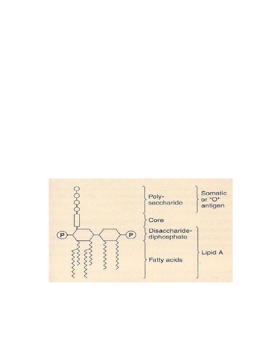
Lipopolysaccharide (LPS)
• The LPS of the outer membrane of the cell wall is endotoxin.
• It is responsible for many of the features of disease, such as fever and shock
caused by these organisms.
• Composed of three distinct units: (Figure 3)
1. A phospholipid called lipid A, which is responsible for the toxic effects.
2. A core polysaccharide of five sugars linked to lipid A.
3. An outer polysaccharide consisting of up to 25 repeating units of three to
five sugars. This outer polymer is the important somatic, or O, antigen of
several gram-negative bacteria that used to identify certain organisms in the
clinical laboratory.
Figure 3: Endotoxin (LPS) structure. The O-antigen polysaccharide is
exposed on the exterior of the cell, whereas the lipid A faces the interior.
(Levinson W. Review of Medical Microbiology and Immunology.12
th
ed.
Copyright 2012, McGraw-Hill.)

Lec.2
Dr. Inas Khalifa Al-Sharquie
2012-2013
8
Structures outside the Cell Wall
1- Capsule
• A gelatinous layer covering the entire bacteria.
• Composed of polysaccharide, except anthrax bacillus, which contain
polymerized D-glutamic acid.
• The capsule is important because:
1- It is a determinant of virulence of many bacteria since it limits the ability
of phagocytes to engulf the bacteria.
2- Capsular polysaccharide is used as the antigens in certain vaccines
because they are capable of eliciting protective antibodies.
3- Specific identification of an organism can be made by using antiserum
against the capsular polysaccharide.
4- The capsule may play a role in the adherence of bacteria to human
tissues, which is an important initial step in causing infection.
2- Flagella
• Thread-like appendages composed of protein.
• Three types of arrangement are know:
Monotrichous : single polar Flagellum
Lophotrichous : multiple polar Flagellum
Peritrichous : Flagella are distributed over the entire cell.
• Flagellum proteins called flagellin that aggregate to form a helical structure.
They are highly antigenic (H antigens) and some of immune responses to

Lec.2
Dr. Inas Khalifa Al-Sharquie
2012-2013
9
infection are directed against these proteins. Flagella are structure
composed of a hook and body.
• Flagella play a role in the pathogenesis.
• Same species of bacteria (e.g., Salmonella) are identified in the clinical Lab.
By the use of specific antibodies against flagellar protein.
• Some species of motile bacteria (e.g., E.coli and Proteus species) are
common causes of urinary tract infections. Flagella may play a role in
pathogenesis by propelling the bacteria up the urethra in to the bladder.
3- Pili (Fimbriae)
Hair like filaments that extend from the cell surface.
Shorter and straighter than flagella.
Composed of subunits of pilin, a protein arranged in helical strand.
Found mainly on gram-negative organism.
Ordinary pili mediate the attachment of bacteria to specific receptors on cell
surface, which is a necessary step in the initiation of infection for some
organisms. Mutant of Neisseria gonorrhoeae that do not form pili are
nonpathogenic.
Sex pili responsible for the attachment of donor and recipient cells in
bacteria conjugation.
Pilli are present in motile and non-motile strains of many species of
bacteria.

Lec.2
Dr. Inas Khalifa Al-Sharquie
2012-2013
10
4-
Glycocalyx
Polysaccharide (slime layer) secreted by certain bacteria.
It attached bacteria firmly to the surface of human cells and to the surface of
catheters, prosthetic joints, and heart valves.
The medical importance of the Glycocalyx is illustrated by the findings that
it is the Glycocalyx-producing strains of Pseudomonas aeruginosa, which
cause respiratory tract infections in cystic fibrosis patient.
Endospore
• Members of several bacteria genera are capable of forming endospores.
Commonly the G+ve rods.
• Under conditions of nutritional depletion each cell forms a single internal
spore
that is liberated when the mother cell under goes autolysis.
• Highly resistant to heat and chemicals when returned to favourable
nutritional conditions and activated, the spore germinates to produce a
single vegetative cell.
• Sporulation accurse in culture that have terminated growth as a result of
depletion of carbon and N sources.
The spore consists of:
1- The core: spore protoplasm.
2- Cell envelope of spore composed of :
3- Spore wall.
4- Cortex.
5- Coat: keratin like protein.

Lec.2
Dr. Inas Khalifa Al-Sharquie
2012-2013
11
Summary
• The cytoplasmic membrane of bacteria consist of a phospholipid bilayer
(without sterols) located just inside the peptidoglycan.
• It regulates the transport of nutrients into the cells and the secretion of toxins
out of the cells.
• Cell wall is peptidoglycan Polymer (amino acids + sugars). Sugars; NAG &
NAM.
• G+ vs. G- bacteria.
• G+ Thicker cell wall, Teichoic Acids
• G- Endotoxin – LPS
• Between the inner cell membrane and the outer membrane of G-ve bacteria
lies the periplasmic space
• The cell wall of mycobacterium has more lipid than either G+ or G- ve.
• Capsule is anti-phagocytic.
• Pili are filaments of protein that extended from the bacterial surface and
mediate attachment of bacteria to the surface of human cells.
• The Glycocalyx is a polysaccharide (slime layer) secreted by certain
bacteria.
• Spores are medically important because. They are highly heat resistant and
are not killed by many disinfectants.
Main References:
1. Jawetz, Melnick and Adelberg’s Medical Microbiology (Brooks,
Butel,Morse), 2010.
2. Levinson W. Review of Medical Microbiology and Immunology.12
th
ed.
Copyright 2012, McGraw-Hill.
