
Muscle System
12
th
& 13
th
lectures
February 11 & 18, 2016
Muscle tissue, one of the four major tissue types, plays the vital role of providing
movement and heat generation to the organs of the body.
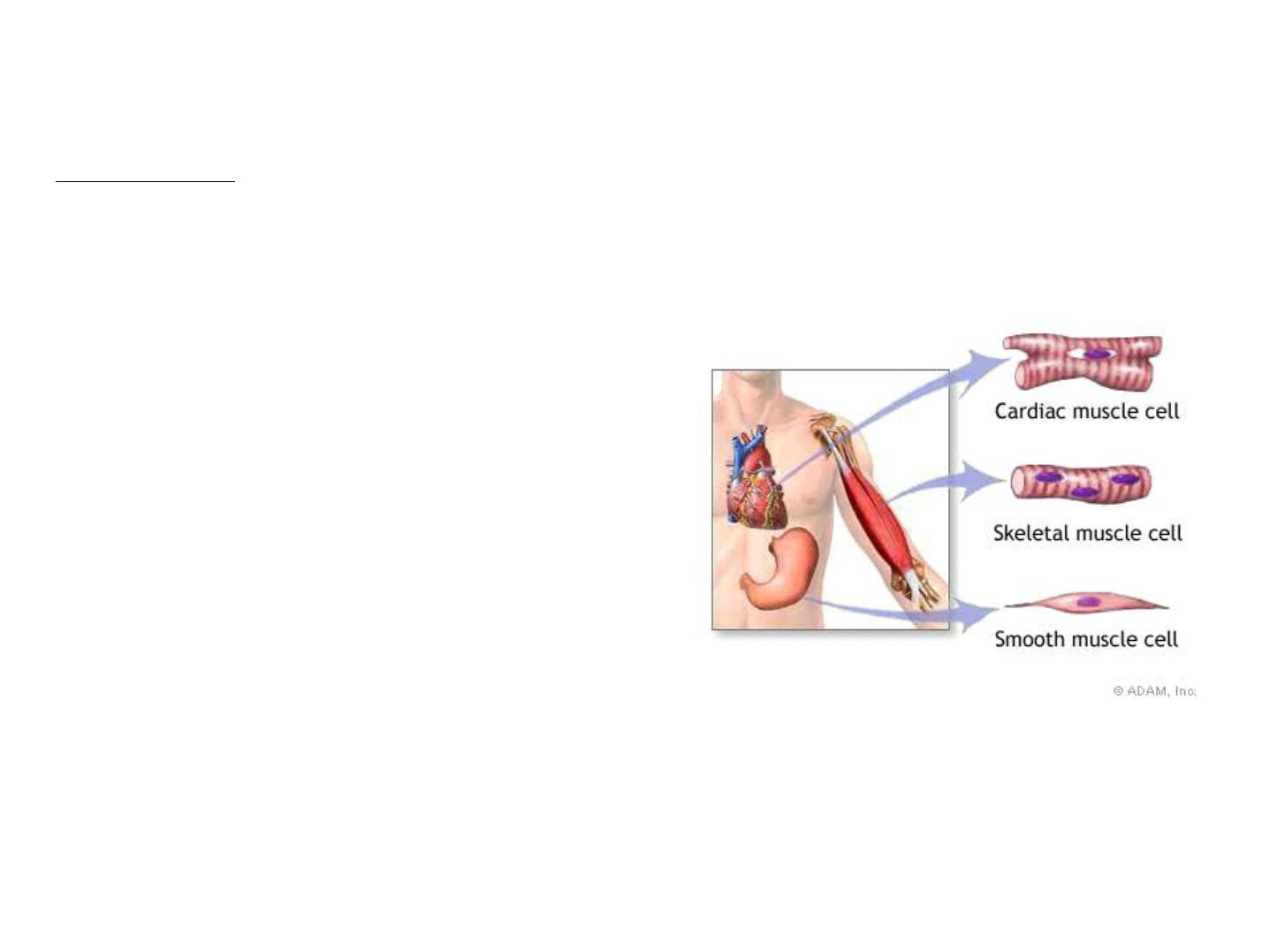
There are 3 types of muscle tissue
1. Skeletal muscle = voluntary striated muscle
2. Cardiac muscle = involuntary striated muscle
3. Smooth muscle = involuntary non-striated
muscle

Characteristics of all muscle tissues:
1. Specialized cells: elongated, high density of myofilaments = cytoplasmic
filaments of actin and myosin
2. Excitability/irritability: receive and respond to stimulus
3. Contractility: shorten and produce force upon stimulation
4. Extensibility: can be stretched
5. Elasticity: recoil after stretch
Skeletal Muscle Tissue -forms skeletal muscles (44% of body mass) A skeletal
muscle = an organ: composed of skeletal muscle cells (fibers), CT, nerves and blood
vessels.
Functions of skeletal muscles:
1. Produce skeletal movement
2. Maintain posture and upright position
3. Stabilize joints
4. Support soft tissues
5. Guard entrances and exits
6. Generate heat (maintain body temp)

Skeletal Muscle Fibers
-Huge cells: up to 100µm diameter, 30cm long
-Multinucleate
-Formed by fusion of 100 myoblasts-nuclei of
each myoblast retained to provide enough
mRNA for protein synthesis in large fiber
-Unused myoblasts in adult = satellite cells
-Satellite cells capable of division and fusion to
fiber for repair but cannot generate new fibers
-Cell membrane = sarcolemma

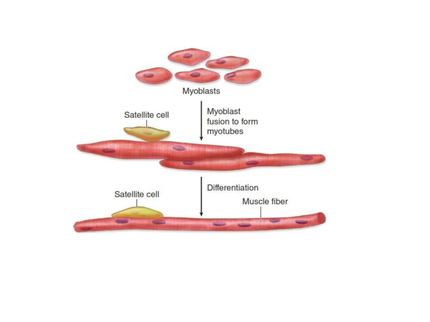
Development of skeletal muscle.
• Skeletal muscle begins to differentiate when mesenchymal
cells called myoblasts align and fuse together to make longer,
multinucleated tubes called myotubes.
• Myotubes synthesize the proteins to make up myofilaments
and gradually begin to show cross striations by light
microscopy.
• Myotubes continue differentiating to form functional
myofilaments and the nuclei are displaced against the
sarcolemma.
• Part of the myoblast population does not fuse and
differentiate, but remains as a group of mesenchymal cells
called muscle satellite cells located on the external surface of
muscle fibers inside the developing external lamina.
• Satellite cells proliferate and produce new muscle fibers
following muscle injury.
Development of skeletal muscle

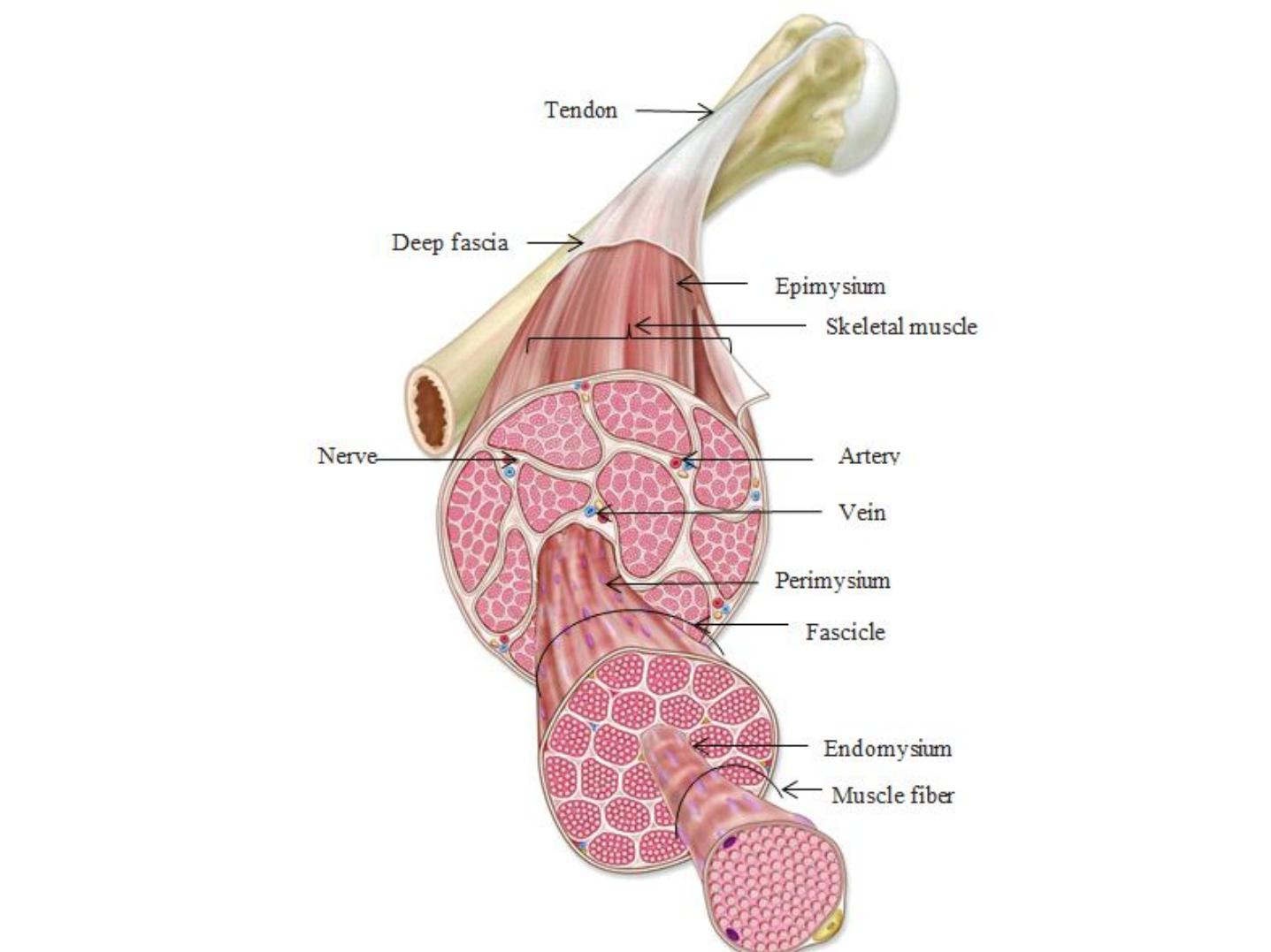
Structure or Parts of Skeletal Muscle
Endomysium –a delicate connective tissue sheath that encloses each
skeletal muscle fiber.
Perimysium – a coarse fibrous membrane that wraps the sheathed
muscle fibers.
Fascicle –bundle of fibers formed from group of sheathed muscle fibers
wrapped by perimysium.
Epimysium –a tough overcoat of connective tissue that bounds together
fascicles. This is the connective tissue that covers the entire muscle.
Tendons –these are cordlike structures that are formed from epimysia.
These are composed of mostly collagenic fibers that can cross rough bony
projections. Aside from anchoring muscles, tendons are very important in
providing durability.
Aponeuroses –these are sheet like structures that attaches muscles
indirectly to bones, cartilages or connective tissue coverings of each other.
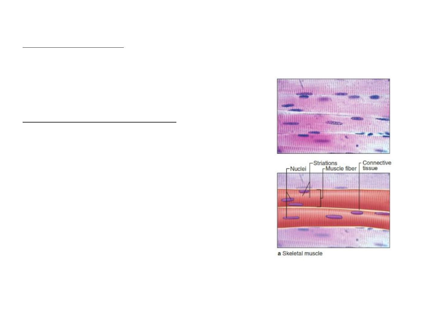
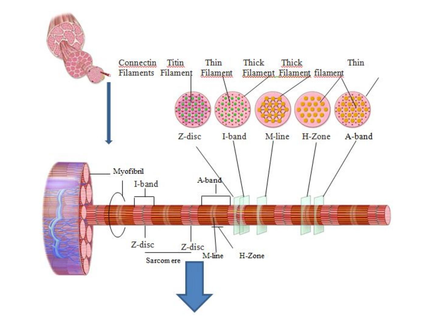
Organization of skeletal muscle
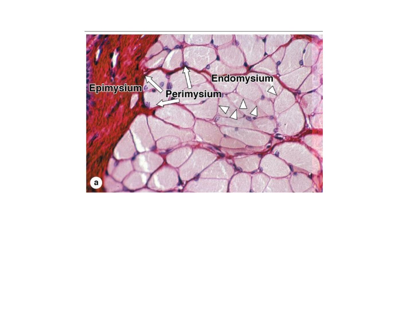
(a): Micrograph shows a cross section of striated muscle demonstrating connective
tissue and cell nuclei. The endomysium around individual muscle fibers is indicated
by arrowheads. At left is a portion of the epimysium. All three of these tissues
contain collagen types I and III (reticulin). X200. H&E.

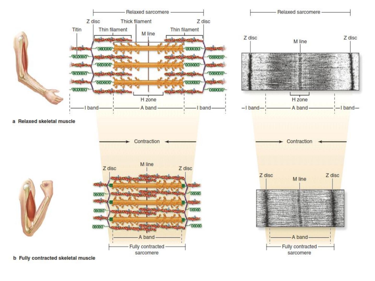
Mechanism of Contraction
• Resting sarcomeres consist of partially overlapping thick and
thin filaments.
• During contraction, neither the thick nor thin filaments changes
their length.
• Contraction is the result of an increase in the amount of overlap
between the filaments caused by the sliding of thin and thick
filaments past one another.
• Contraction is induced by an action potential produced at a
synapse, the neuromuscular junction, between the muscle fiber
and a terminus of a motor axon.
• Key molecular events in muscle contraction are summarized in
the next slide

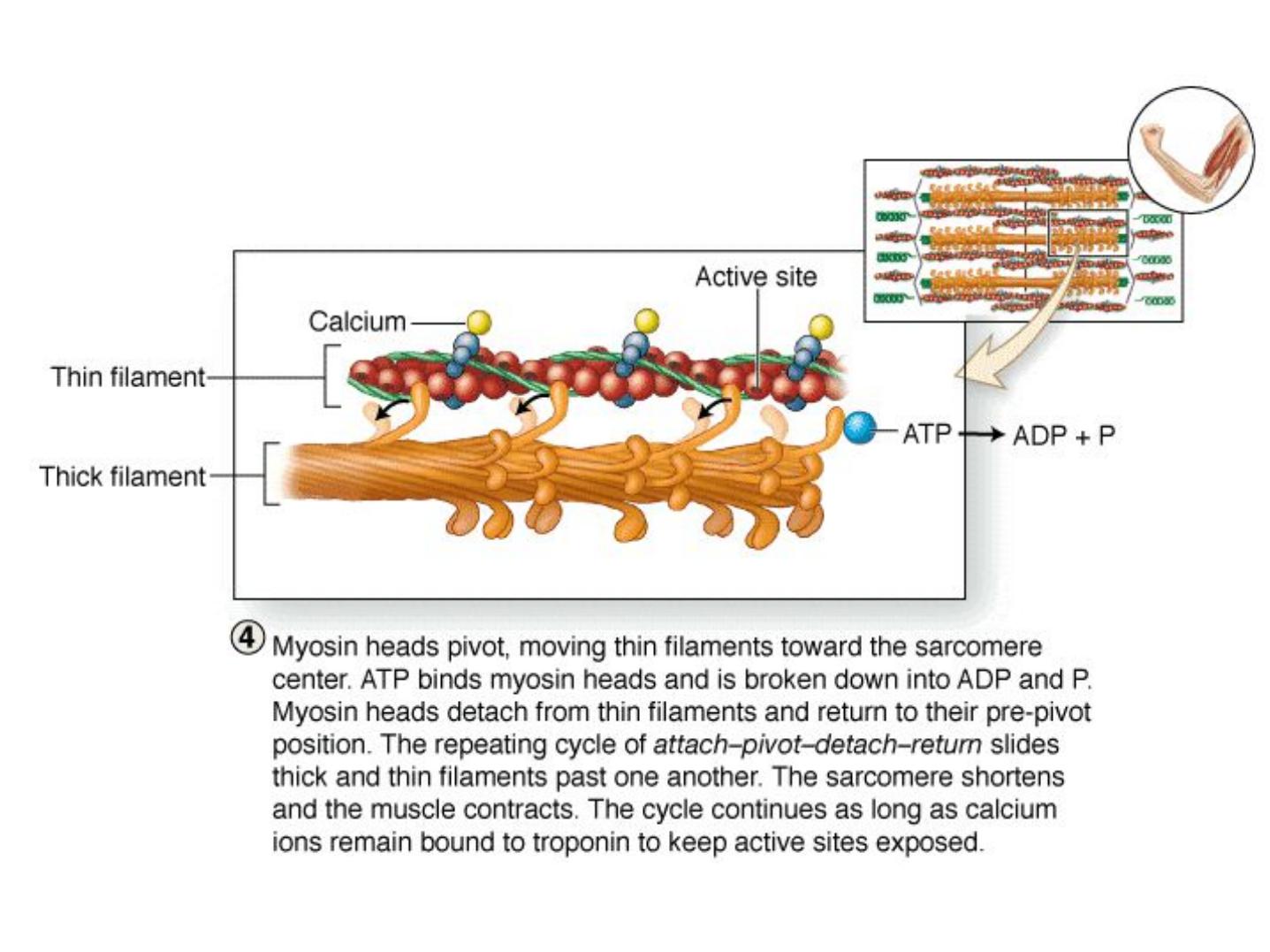
Sliding Filament Theory
-Contraction of skeletal muscle is due to thick filaments and
thin filament sliding past each other, not compression of the
filaments
-evidence:
1. H-zones and I-bands decrease width during contraction
2. Zones of overlap increase
3. Z-lines move closer together
4. A-band remains constant
-Sliding causes shortening of every sarcomere in every
myofibril in every fiber
-Overall result = shortening of whole skeletal muscle

Skeletal muscle fibers of humans are classified into three types based on their physiological,
biochemical, and histochemical characteristics
All three fiber types are normally found throughout most muscles.
§Type I or slow, red oxidative fibers contain many mitochondria and abundant myoglobin, a
protein with iron groups that bind O
2
and produce a dark red color. they are adapted for slow,
continuous contractions over prolonged periods, as required for example in the postural
muscles of the back.
§Type IIa or fast, intermediate oxidative-glycolytic fibers have many mitochondria and
much myoglobin, but also have considerable glycogen. They are adapted for rapid
contractions and short bursts of activity, such as those required for athletics.
§Type IIb or fast, white glycolytic fibers have fewer mitochondria and less myoglobin, but
abundant glycogen, making them very pale in color. They are typically small muscles with a
relatively large number of neuromuscular junctions, such as the muscles that move the eyes
and digits.
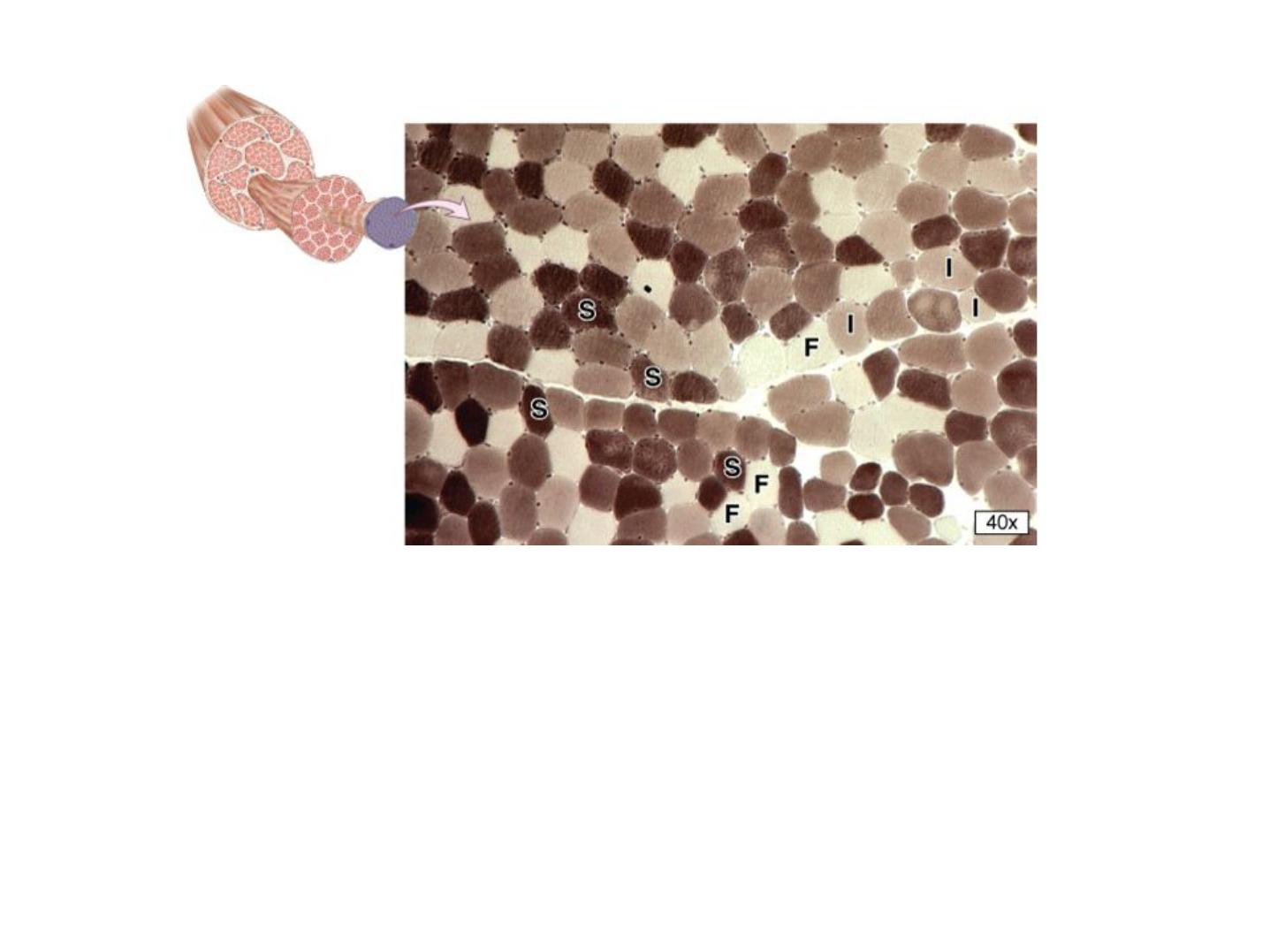
Cross-section of skeletal muscle stained histochemically to detect the density of myofibrillar
myosin-ATPase can be used to demonstrate the distribution of slow (S) type I fibers,
intermediate (I) type IIa fibers, and fast (F) type IIb fibers.
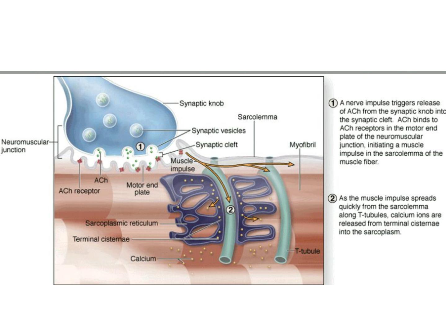
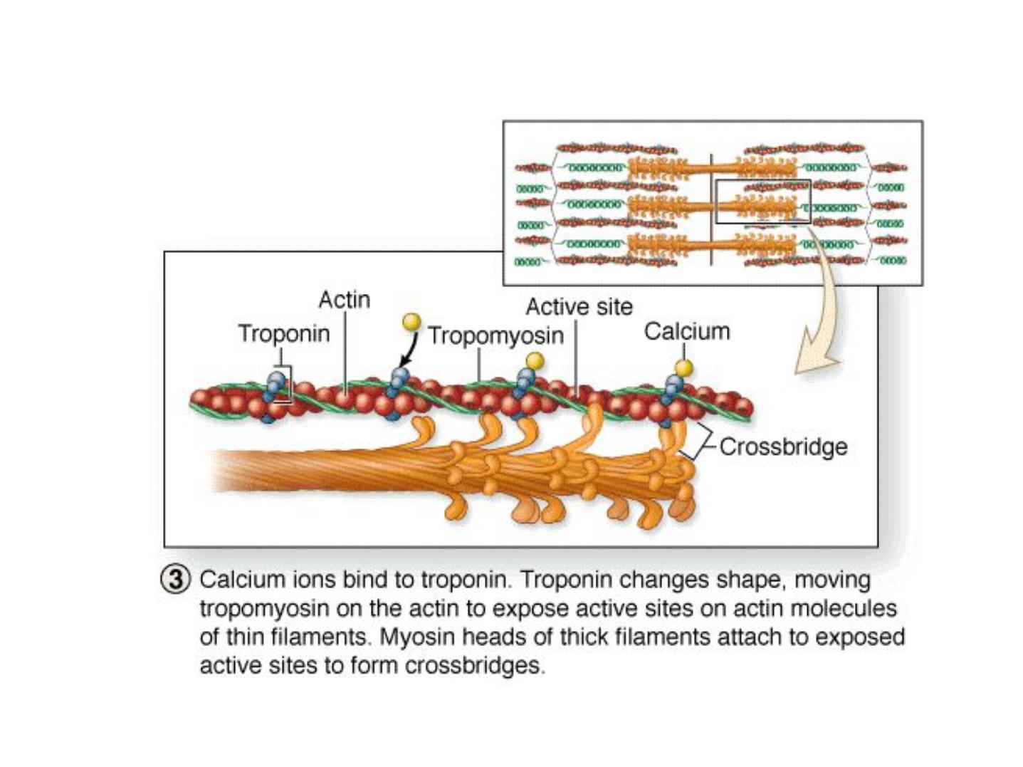
Events of muscle contraction
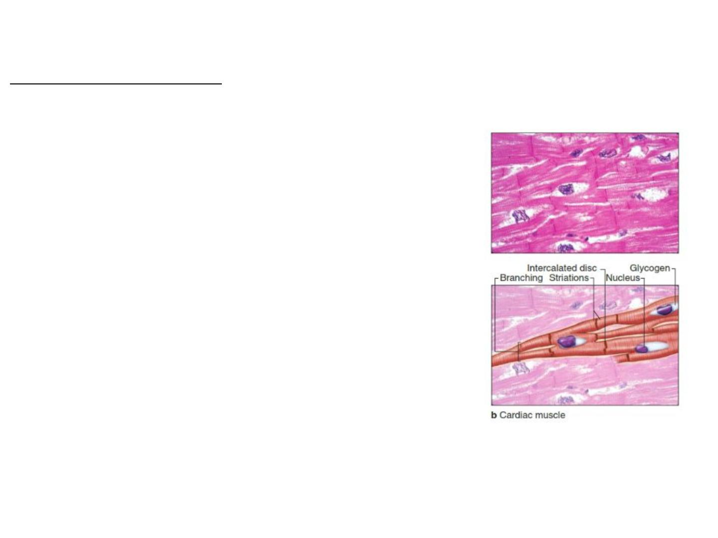
Cardiac Muscle Tissue
-forms the majority of heart tissue cells
=cardiocytes
-one or two nuclei
-no cell division
-long branched cells
-myofibrils organized into sarcomeres (striated)
-aerobic respiration only
-mitochondria and myoglobin rich
-glycogen and lipid energy reserves
-intercalated discs at cell junctions (gap junctions
and desmosomes) allow transmission of action
potentials & link myofibrils from one cell to next

Features of cardiac muscle:
1. Can contract without neural stimulation; automaticity due to
pacemaker cells that generate action potentials spontaneously
2. Pace and amount of tension can be adjusted by nervous
system
3. Contractions 10X longer than skeletal muscle
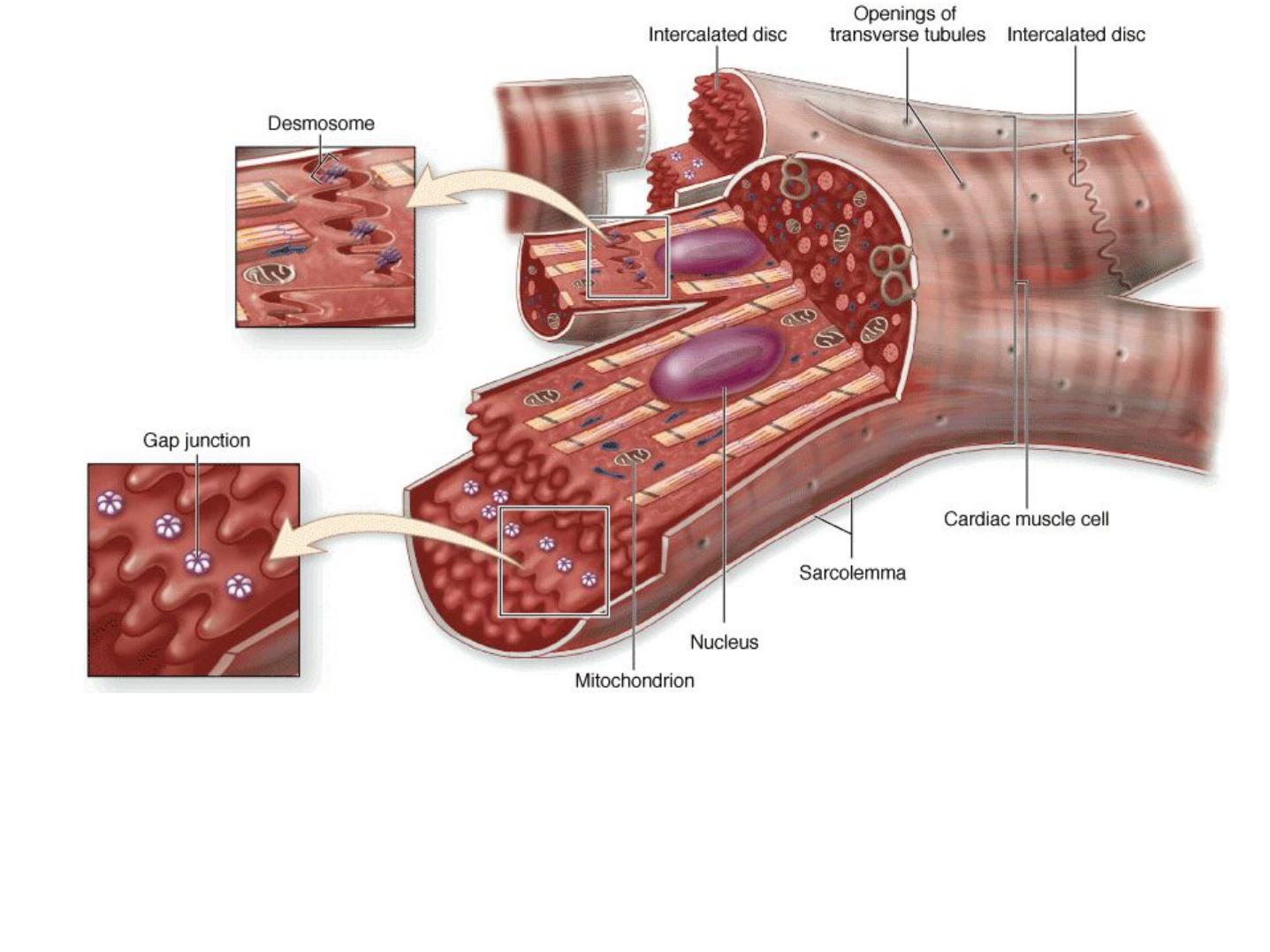
Diagram of cardiac muscle cells indicates characteristic features of this muscle type. The
fibers consist of separate cells with interdigitating processes where they are held together.
These regions of contact are called the intercalated discs, which cross an entire fiber between
two cells.
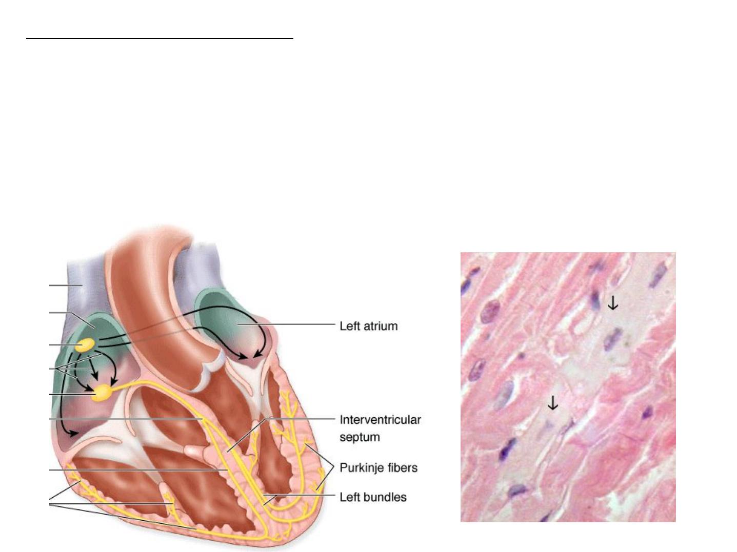
Other Heart Tissue Structures
1- Epicardium: CT layer that surrounds the heart
2- Purkinje Fibers: the purkinje fibers are found in the sub-endocardium.
They are larger than cardiac muscle cells, but have fewer myofibrils, lots of
glycogen and mitochondria. These cells are connected together by desmosomes and
gap junctions, but not by intercalated discs. They are specialized conducting fibers,
which is responsible for conduction of signal used for heart contraction; located at
bottom (base) of heart.
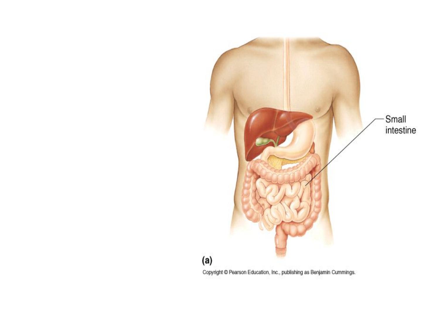
• IV. Smooth Muscle- A
tissue formed by
uninucleated spindle
shaped cells found in
six areas of the body:
blood vessel walls,
respiratory tract,
digestive tubes,
urinary organs,
reproductive organs,
and the eye.
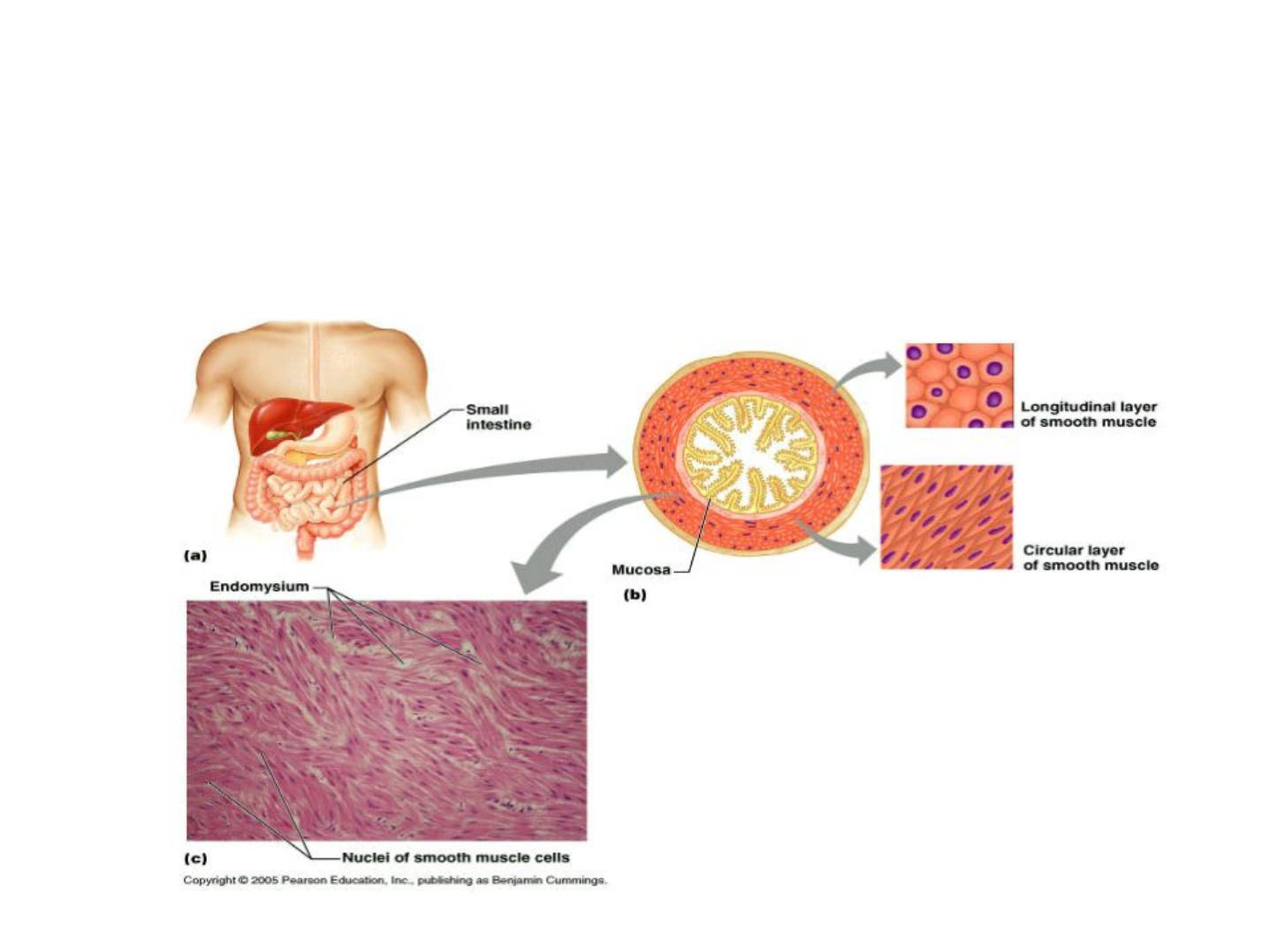
A. It exist as two layers with fibers running perpendicular to each other.
One layer, the longitudinal layer is parallel to the axis, the circular layer is
perpendicular. As they alternate contractions they shorten and constrict
the organ.
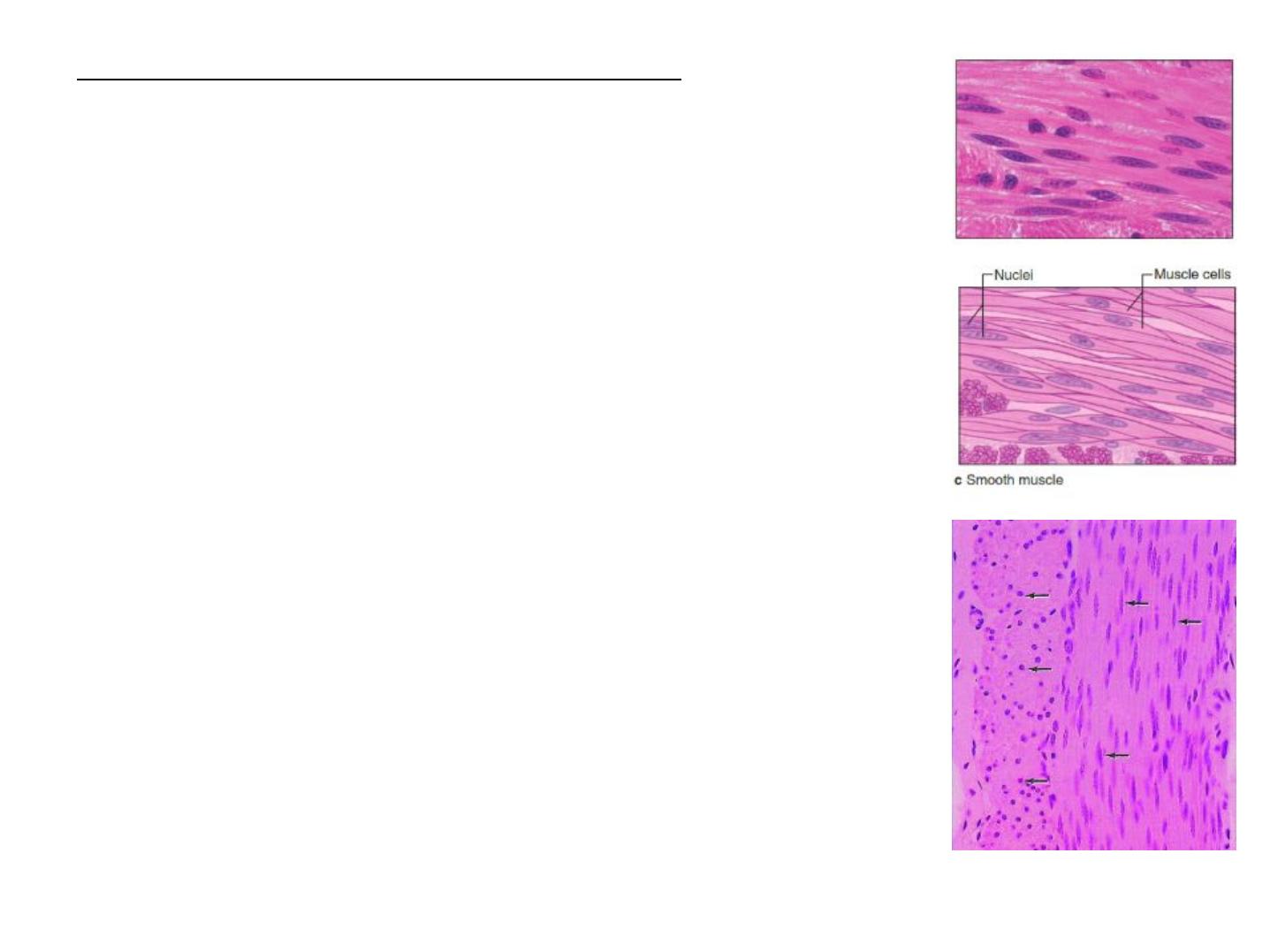
Main Charectristic of Smooth Muscle Tissue
-lines hollow organs: regulates blood flow and movement of
materials in organs
-forms arrectorpili muscles
-usually organized into two layers: Circular & longitudinal
-spindle shaped cells
-central nucleus
-cells capable of division
-no myofibrils, sarcomeres thick filaments scattered
-thin filaments attached to dense bodies on desmin cytoskeleton
(web)
-adjacent cells attach at dense bodies with gap junctions (firm
linkage and communication)
-no tendons
-contraction compresses whole cell
B. There are no striations and no sarcomeres. Calcium ions signal contraction
and the forces are not high. It can contract for a long time before fatiguing.
Typically cells are not individually innervated and contraction may be
signaled by stretching or hormones.

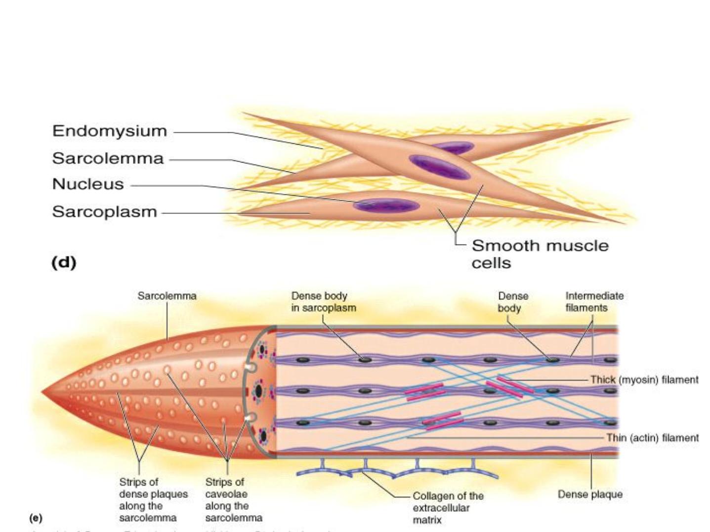
Most molecules that allow contraction are
similar in the three types of muscle, but
the filaments of smooth muscle are
arranged differently and appear less
organized.
The diagram shows that thin filaments
attach to dense bodies located at the cell
membrane and deep in the cytoplasm.
Dense bodies contain a-actinin for thin
filament attachment.
Dense bodies at the membrane are also
attachment
sites
for
intermediate
filaments and for adhesive junctions
between cells. This arrangement of both
the cytoskeleton and contractile apparatus
allows the multicellular tissue to contract
as a unit, providing better efficiency and
force.
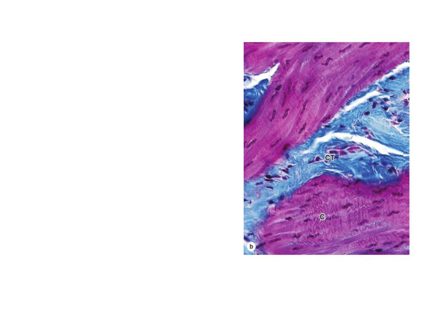
(b) Micrograph showing a contracted (C)
region of smooth muscle, with contraction
decreasing the cell length and deforming
the nuclei. The long nuclei of individual
fibers assume a cork-screw shape when
the fibers contract, reflecting the reduced
cell length at contraction. Connective
tissue (CT) of the perimysium outside the
muscle fascicle is stained blue. X240.
Mallory trichrome.

Copyright 2009, John Wiley & Sons, Inc.
Regeneration of Muscular Tissue
• Hyperplasia
- An increase in the number of fibers
• Skeletal muscle has limited regenerative abilities
- Growth of skeletal muscle after birth is due mainly to
hypertrophy
- Satellite cells divide slowly and fuse with existing fibers
• Assist in muscle growth
• Repair of damaged fibers
• Cardiac muscle can undergo hypertrophy in response
to increased workload
- Many athletes have enlarged hearts
• Smooth muscle in the uterus retain their capacity for
division

Copyright 2009, John Wiley & Sons, Inc.
Aging and Muscular Tissue
• Aging
- Brings a progressive loss of skeletal muscle mass
- A decrease in maximal strength
- A slowing of muscle reflexes
- A loss of flexibility
• With aging, the relative number of slow oxidative
fibers appears to increase
• Aerobic activities and strength training can slow
the decline in muscular performance

Muscle tissue throughout life
1. Mesoderm cells called myoblast fuse to form skeletal muscle tissues or
join at cellular ends to form cardiac and smooth muscle.
2. Cardiac muscle contracts by the week 3 and skeletal muscle by week 7
of development!
3. Mitosis= skeletal muscle stops dividing once formed but has limited
regenerated capacity in case of injury, cardiac muscle stops dividing by
age 9, and smooth muscle divides as needed and has great regenerative
abilitiy.
4. Muscle tissue is replaced with connective tissue as one ages. This is
called sarcopenia and is reversible with exercise