
Acute Charcot footEASILY MISSED?
BMJ 14 March 2012د. حسين محمد جمعه
اختصاصي الامراض الباطنة
البورد العربي
كلية طب الموصل
2012
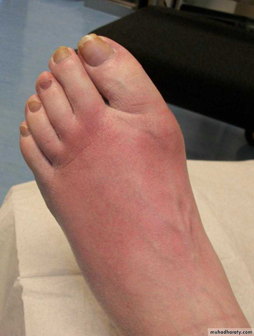
A 38 year old man was referred by his general practitioner to our diabetes foot clinic with a swollen red foot (fig 1⇓). He had had type 1 diabetes for 25 years, complicated with retinopathy,
peripheral neuropathy, and nephropathy, and was being worked up for dialysis following a failed pancreas-kidney transplant.
The absence of pain together with preserved pulses and intact skin raised a suspicion of acute Charcot foot.
A plain radiograph
of the foot showed fractures through the necks of the first three metatarsals (fig 2⇓). We offloaded the foot in a total contact cast and advised the patient to limit weight bearing.Magnetic resonance imaging (MRI) subsequently confirmed neuroarthropathic changes of acute Charcot (fig 3⇓).
What is acute Charcot foot?
Charcot’s neuroarthropathy is a destructive process of bone and joint, typically seen in a foot that has lost its protective sensory innervation. The classic description of this disabling condition by Jean-Martin Charcot in 1883 was in patients with tabes dorsalis, but nowadays most cases of Charcot foot occur as a
complication of diabetes mellitus. The chronic stage of the disease is easily recognisable, but the acute phase can present a diagnostic challenge.
In a recent series the diagnosis of acute Charcot foot was missed before specialist referral in 19 of 20
patients. In another report, referring clinicians failed to diagnose Charcot foot in 19 of 24 cases seen in a specialist diabetes foot clinic.
Why is acute Charcot foot missed?
The acute phase of a Charcot foot may not be considered or may be mistaken for more common causes of leg or foot swelling, such as cellulitis, gout, deep venous thrombosis, or sprains. The misdiagnosis of ankle sprain is particularly common if the patient recalls a history of trivial injury. Standard radiographs may show no abnormalities at this stage, contributing to delays in diagnosisWhy does this matter?
The delay in correct diagnosis is harmful because during the acute phase the foot bones are vulnerable to fragmentation and dislocation. If the patient continues to walk on an insensitive foot, this may lead—sometimes within weeks—to irreversible deformities, such as mid-foot dislocation or collapse and inversion of the plantar arch, the so called rocker-bottom foot.These deformities may, in turn, predispose to skin ulcer, an established risk factor for amputation.4 If the disease is diagnosed in the acute phase, bone and joint damage can largely be prevented by avoiding weight bearing.
Timely recognition may also identify patients with diabetes who are at increased risk of mortality owing to the severe neuropathy associated with
Charcot foot. In one series, patients with acute Charcot foot or neuropathic foot ulcers had a 5-year mortality rate of 40%. Mortality may relate to co-existent renal disease in some patients, but neuropathy is also believed to independently increase cardiovascular risk by promoting vascular calcification.
How is acute Charcot foot diagnosed?
Clinical featuresThe usual presentation is a red, swollen, warm foot in which pulses are preserved (fig 1). Owing to neuropathy, pain is not always present or is less than expected for the severity of the clinical findings. Longstanding diabetes, either type 1 or type
2, or a history of renal transplantation confer a particularly high risk. The patient may be thought at this stage to have gout, ankle sprain, or deep venous thrombosis, but the most common misdiagnosis is infection.
The presence of an ulcer favours the diagnosis of cellulitis or osteomyelitis, particularly if this can
be probed to bone. Absence of skin break, stable insulin requirements, and normal white blood cell counts or C-reactive protein levels are more suggestive of acute Charcot than infection. Neuro-arthropathy and infection are, however, not
mutually exclusive and if any doubt exists the patient should be treated for both conditions until the true diagnosis is established
Investigations
Standard radiographs are an important first line investigation.The finding of fractures or bony misalignment in the absence of obvious trauma is highly suggestive of Charcot foot (fig 2).
The initial radiograph may be normal, but this should not divert from the diagnosis if the clinical suspicion is high. A radioisotope bone scan or MRI can show bone disease even when radiographic changes are subtle (fig 3).
The choice between nuclear imaging and MRI is largely based on local availability and experience. Bone scan has less specificity than MRI (25-38% v 80-100%), but the diagnostic sensitivity of either test approaches 100%. The mid-foot region is the most common site of disease, although hind-foot involvement carries a particularly severe prognosis owing to the risk of ankle
instability. MRI is the investigation of choice in patients with ulcers and a high probability of deep infection. However, the differentiation of Charcot from osteomyelitis may occasionally be difficult even with MRI.
How is acute Charcot foot managed?
If acute Charcot foot is suspected, seek urgent referral to aspecialist foot clinic and advise patients to avoid weight bearing pending evaluation. Early offloading of the foot with total contact casting is the gold standard of treatment. Casting is usually needed for three to six months, and healing is indicatedby resolution of oedema and warmth.
Radiographic or MRI evidence of healing assists the clinical decision to discontinue casting and transfer the patient into a bespoke shoe. A lifelong
programme of patient education and routine foot care should form an essential component of treatment. Reconstructive surgery is currently reserved for patients in whom attempts at conservative care have failed to prevent major deformities.
Earlier surgical intervention could nonetheless be considered for patients with ankle disease owing to the often limited success of conservative measures in this form of disease.
Bisphosphonates have been proposed to counteract the excess bone turnover that characterises the acute Charcot foot, but the evidence for their benefit is still inconclusive.
Key points
• Suspect acute Charcot foot in a patient with diabetes and neuropathy who presents with a swollen warm foot
• If acute Charcot foot is suspected, arrange for offloading of the foot (to minimise further damage) and refer to a specialist foot clinic
immediately
• Plain radiographs may be normal in the early stages of the disease
• Magnetic resonance imaging should be considered when the suspicion of acute Charcot foot is high
How common is acute Charcot foot?
• The true incidence of acute Charcot foot is difficult to establish, because few population based studies have been published and nodiagnostic criteria have been universally agreed
• The reported annual incidence of Charcot arthropathy in patients with diabetes has varied from 0.3% in population studies11 to 12.0%
in referral centre studies7
• Studies in specialist units report that Charcot arthropathy was present in 9% of patients with diabetic neuropathy and foot ulcer12 and
in 12% of diabetic patients who had received pancreas-kidney transplants
Acute Charcot foot

Plain radiograph of the foot showing fractures through the necks of the first three metatarsals