Panorama 5In Medicine
د. حسين محمد جمعهاختصاصي الامراض الباطنة
البورد العربي
كلية طب الموصل
2012
Pulmonary embolism may cause a range of ECG abnormalities (or be associated with a normal ECG). Most commonly, sinus tachycardia is seen, but other ECG findings may include atrial fibrillation or flutter and supraventricular tachycardias. Right heart strain may be evident as right bundle branch block or, uncommonly, with an acute right heart strain pattern (the SI QIII TIII pattern). Pulmonary embolism is suggested by the finding of
hypoxia with clear lung fields, tachycardia, Cardiac troponin rises are not uncommon
Cigarette smoking is the most important modifiable risk factor for the development of thyroid eye disease. Thyroid eye disease is four times more common in smokers or ex-smokers than in patients who have never smoked. The outcome after surgery for thyroid eye disease is also worse in smokers.
Cigarette smoking also increases the risk of a worsening of ophthalmopathy after radioiodine therapy, so you should advise smokers to stop before they have this treatment. prednisolone may prevent worsening of ophthalmopathy after radioiodine therapy.
Table 2: Features of Graves' disease
• Diffuse goitre• Thyroid eye disease
• Pretibial myxoedema
Thyroid acropachy
Symptoms and signs of thyroid eye disease
Pain
A feeling of pressure behind the eye
A gritty sensation in the eye
Double vision
Photophobia
Common signs are:
Swelling and/or erythema of the conjunctiva (can be mistaken for conjunctivitis, and hence leads to delayed diagnosis)
Oedema and/or erythema of the eyelid
Retraction of the eyelid
Exophthalmos
Divergent or convergent squint due to involvement of the extraocular muscles.
Learning bite
Antithyroglobulin and antimicrosomal antibodies are typically raised in patients with Graves' disease (but can be negative in 5% to 10%).
Propylthiouracil can also cause neutropenia, but carbimazole is a more common cause of this side effect.
No authority recommends therapeutic intervention in all patients with subclinical thyroid disease based on laboratory testing alone.
Abnormal thyroid tests in critically ill patients have been shown to be predictive of mortality. When used alone, a free T4 level <3.1 µg/dl has a 75% sensitivity and 80% specificity for predicting death. When a free T4 level <3.1 µg/dl is combined with an 8 am serum cortisol level >30 µg/dl, sensitivity for predicting death rises to 100% and specificity to 100%. The predictive value for death from these two tests is better than the currently used APACHE II scoring system.
The recent clinical practice guideline on venous
thromboembolism prophylaxis from the American College of Physicians indicates that forevery 1000 patients treated the absolute benefit is a reduction of three pulmonary embolisms but with an absolute increase of nine haemorrhages,
four of them major.
There is thus no net benefit and no significant reduction in mortality.
Oral anticoagulation (OAC) is the recommended
treatment for patients at risk of thromboembolic events due toatrial fibrillation,
mechanical heart valves,
deep vein thrombosis, pulmonary embolism and left ventricular thrombi. The number of patients who have an indication for both Dual antiplatelet therapy DAT and OAC is increasing, since more patients who are already on OAC are scheduled for PCI and some patients who are on DAT will develop a medical condition which requires OAC.
Accordingly, the current guidelines for heart failure
do not support the general use of OAC in thosepatients with heart failure and no history of atrial
fibrillation or a prior thromboembolic event.
Since DES and BMS differ in terms of endothelialisation
and the recommended duration of DAT to prevent
subacute stent thrombosis, it has been proposed
that BMS should be the preferred stent type so that
the duration of triple therapy might be limited to
4 weeks. So far no study has primarily addressed
the outcome of patients with BMS compared to
DES and an indication for OAC.
You should normally stop drug treatment of peripheral lymph node tuberculosis after six months, regardless of the appearance of new nodes, residual nodes, or sinuses draining during treatment.
If there is a persisting discharging sinus then it may be sensible to reinvestigate with culture.
You should treat patients with active peripheral lymph node tuberculosis who have had an affected gland surgically removed with the standard recommended regimen. A six month, four drug initial regimen (six months of isoniazid and rifampicin supplemented in the first two months with pyrazinamide and ethambutol) should be used to treat active respiratory TB in: adults not known to be HIV positive, adults who are HIV positive, children.
Learning bite
groups of people with latent tuberculosis who are at increased risk of going on to develop active tuberculosis, include people who:Have had a HIV infection
Have had a solid organ transplantation
Are injecting drug users
Have a haematological malignancy
Have had a jejuno-ileal bypass
Have had a gastrectomy
Are receiving treatment with anti-tumour necrosis factor alpha. you should give six months of isoniazid
Directly observed therapy is not necessary for most patients with active tuberculosis.
DOT should be considered for patients who have adverse factors on their risk assessment, in particular:
• Street or shelter dwelling homeless people with active TB .
• Patients with likely poor adherence, in particular those who have a history of non-adherence .
• All prisoners with active or latent tuberculosis.
According to randomised controlled trials treatment for six months is sufficient for tuberculosis at any site except the central nervous system( twelve months).
Conversion of sputum from smear and culture positive to negative is the only reliable way of determining the effectiveness of treatment.
The new guidelines are clear: any person younger than 16 years with a positive tuberculin skin test but normal chest x ray should be given preventive therapy with three months of isoniazid and rifampicin or six months of isoniazid.
A number of studies have shown that smoking increases the risk of tuberculosis two- to fivefold. A study in India showed that tuberculosis is the most common cause of death in smokers.
This is probably due to suppression of alveolar macrophages by substances in the cigarette smoke.
In about 50% of patients with pulmonary tuberculosis the sputum smear test will be negative. 30% of patients treated for pulmonary tuberculosis are culture negative.
This is because over 10 000 bacteria per ml of sputum are needed to give a positive smear. Increasingly in the white population of the UK a positive smear results from an environmental mycobacterium such as M kansasii or M avium, not M tuberculosis. So a sputum smear test cannot definitively confirm the diagnosis of tuberculosis.
HIV infection is by far the strongest risk factor for tuberculosis: it increases risk by about 100-fold.
The next highest risk is jejunal bypass surgery, which increases risk by about 20-fold for reasons we don't understand.
Diabetes and smoking both increase risk by between two- and fivefold (by depressing host immunity).
Being tall and thin increases risk by about
1.3- to 1.5-fold.
Having chronic renal failure, silicosis increases the risk of developing active tuberculosis.
The BCG vaccination usually renders the tuberculin skin test weakly positive .
The new gamma interferon blood tests will help to distinguish between a skin test rendered positive by BCG vaccination alone and one rendered positive by genuine infection with M tuberculosis because the
blood tests use antigens specific to the tuberculosis bacteria and not to the BCG vaccine.
BCG is a live attenuated vaccine. Patients with HIV infection should not receive it.
Predicted six month mortality
Risk of future adverse cardiovascular events1.5% or below
Lowest
>1.5 to 3.0%
Low
>3.0 to 6.0%
Intermediate
>6.0 to 9.0%
High
over 9.0%
Highest
• Unstable angina and non-ST segment elevation myocardial infarction (NSTEMI) - in association with NICE
Mortality of upper GI haemorrhage depends on
• The rate of haemorrhage• The duration of haemorrhage
• The site of haemorrhage: Varices (MR 30%), PU (MR 10%) and MW syndrome (MR < 1%)
• The patient's co-morbidities (often reflected in the age of the patient) 2% MR up to age 60, and Steep rise in MR above 60
• Death is rarely due to exsanguination and instead occurs due to decompensation of co-morbid conditions
Re-bleeding is associated with 10 times increased MR
Re-bleeding is associated with:
Atheroma – vessels unable to contract effectively
Posterior wall DU
High lesser curve GU
Vessel visible in ulcer base.
You should be aware that a postural drop in systolic blood pressure of more than 20 mm Hg on standing is an early indicator of loss of blood volume, and will occur before the patient becomes hypotensive.
You should give additional fresh frozen plasma to patients receiving a transfusion of more than four units of blood, as the transfused blood does not contain clotting factors.
Patients with ulcers with adherent clots should be treated by clot removal, followed by endoscopic treatment of the lesion that lies beneath the clot. This has been shown to decrease the rate of recurrent bleeding.14
Patients with Mallory-Weiss tears tend to stop bleeding spontaneously.
What time window do you have to give thrombolysis?
The time frame for thrombolysis for pulmonary embolism is different to that for myocardial infarction. Trials have delivered thrombolysis up to seven days after the embolic event and demonstrated improvements in pulmonary perfusion. However delivering therapy as early as possible makes good sense - and is recommended by the British Thoracic Society guidelines. Indeed 75% of those who die from pulmonary embolism do so within one hour of the event.Oestrogens and menopause
Oestrogens have potential protective cardiovasculareffects through high density lipoprotein (HDL)
and LDL cholesterol modulation, inhibition of
smooth muscle proliferation, enhanced nitric
oxide, prostacyclin and vascular endothelial
growth factor synthesis, and progenitor cell stimulation
; however, they also have potential detrimental
effects (increasing triglycerides and
inflammatory and prothrombotic markers).
Whether the lower prevalence of IHD among premenopausal women compared to age matched
men can be attributed specifically to a protective
role of endogenous oestrogens is still not clear.
Randomised trials testing exogenous oestrogens for
the prevention of IHD showed no benefit or evenharm in terms of cardiovascular events.
To explain these findings, a‘‘timing hypothesis’’ has
been proposed,
whereby oestrogens may be cardioprotective only before the development of advanced atherosclerotic lesions.
Autonomic balance
Women, unlike men, have aprevailing parasympathetic autonomic cardiac tone.This is consistent with a higher female rate of syncope, hypotension,and bradycardia after MI, and, conversely, with more malignant post-MI tachyarrhythmias and ahigher incidence of sudden cardiac death among men.
Women experience longer door-to-balloon
delays after diagnosis.Primary PCI seems to offer better myocardial salvage in women, suggesting greater myocardial tolerance to hypoxia than in men.
Cardiologists must pay particular attention to women hospitalised for IHD because, on average, they are older, with multiple risk factors and comorbidities, and therefore at high risk. Nonetheless, the lower overall
prevalence of coronary disease in women and its
occurrence at a more advanced age suggest a
protective effect of female gender on the development
of IHD.
In perspective, Elizabeth I (1533–1603) might well have cherished her heart of aqueen when she humbly said, ‘‘I have the body of aweak and feeble woman, but the heart of aking’’
Lung dual blood supply:
• Bronchial vessels (systemic circulation; supply the supporting structures of the lung)
• Pulmonary vessel (low pressure; supply alveoli)
The risk of death from
Unstable angina is around 5-8% in the following six months.Myocardial infarction without ST elevation
One in eight patients will die within six monthsFor patients who are admitted to hospital with an acute myocardial infarction, the subsequent risk of death is 12-15% in the following six months.
Learning bite: the right ventricle
There are a number of anatomical and physiological properties of the right ventricle that differentiate it from the left ventricle:The right ventricle has only 15% of the muscle mass of the left ventricle, but has the same cardiac output. This reflects its role in perfusion of the low pressure pulmonary circulation
Coronary perfusion of the right ventricle occurs biphasically in both systole and diastole. Coronary perfusion of the left ventricle only occurs in diastole
The right ventricle has a lower coronary resistance than the left ventricle, with a tendency for left to right collateral vessel formation.
These differences allow the right ventricle to have a lower oxygen demand and better oxygen delivery, so it is more resilient to ischaemia than the left ventricle.
The presence of atrial fibrillation increases morbidity or mortality by around twofold - this usually results
from stroke and heart failure.
The risk of embolic stroke increases fivefold in the presence of atrial fibrillation.
In patients where urgent reversal to sinus rhythm is necessary but where the duration of arrhythmia is either unclear or more than 48 hours you need to arrange a transoesophageal echocardiography to prove the absence of thrombus in the left atrium before electrical cardioversion
Electrical cardioversion
Direct current cardioversion involves delivery of synchronised electrical energy across the chest wall. Success rates vary between 65% and 90%. The success of direct current cardioversion appears to be greater with anteroposterior paddle positioning (sternum and left subscapular) than with anteroapical (ventricular apex and right infraclavicular).Brief arrhythmias can arise immediately following direct current cardioversion. These are mainly ventricular and supraventricular premature beats, bradycardia, and short periods of sinus arrest. Ventricular tachycardia or fibrillation can be precipitated in patients with hypokalaemia and digitalis intoxication.
Patients with underlying conduction defects are at risk of developing profound bradycardia, complete heart block, or asystolic periods following cardioversion. These patients are identified by having a slow ventricular response to atrial fibrillation in the absence of rate reducing medications. External or temporary pacemaker facilities must be at hand before attempting cardioversion.
In clinical practice, amiodarone is a reasonable alternative to class Ic agents and is the drug of choice in patients with ventricular dysfunction and ischaemic heart disease. Amiodarone also has an added advantage of providing prompt rate control in addition to its antiarrhythmic effect. This rate controlling effect (beta blockade and calcium channel blockade properties) is observed early following intravenous loading; the class III antiarrhythmic properties taking effect between eight and 24 hours.
Which patients with atrial fibrillation should be offered rate control?
It is necessary to provide long term rate control when atrial fibrillation is regarded as permanent.Rate control is also a preferred strategy in patients with paroxysmal atrial fibrillation who are stable at presentation; a significant proportion of these patients can spontaneously revert to sinus rhythm within 24-48 hours. The aim of rate control is to improve symptoms and prevent worsening of ventricular dysfunction. Recent evidence from randomised trials (AFFIRM, PIAF, RACE, and STAF) has shown that rate control is at least as effective as rhythm control in improving symptoms and functional capacity, particularly in people older than 65 years
Recent advances: what if rate control fails?
It may at times be difficult to control the ventricular rate and related symptoms in certain patients, particularly with permanent atrial fibrillation. There is then an option to completely block the AV conduction by AV node radiofrequency or cryoablation followed by the implantation of a permanent ventricular pacemaker.Young patients with structurally normal hearts and no other identifiable risk factors (lone atrial fibrillation) are not at substantial risk and may be treated with aspirin.
Patients with paroxysmal atrial fibrillation should receive long term anticoagulation if they have risk factors for thromboembolism (Old age, hypertension, diabetes mellitus, previous cerebrovascular accidents, left ventricular dysfunction, co-existent ischaemic or valvular heart disease, hyperthyroidism, and presence of prosthetic valves are considered as high risk factors) and not on the basis of frequency or severity of paroxysms.
The circuit involved in typical atrial flutter lies within the right atrium and can be mapped accurately. The flutter circuit usually involves a narrow bridge of tissue between the tricuspid valve and the inferior vena cava (cavotricuspid isthmus).
Ablation across this area effectively blocks atrial flutter, achieving almost 100% long term success.
If there is no response to your voice, he may still respond to pain. The following techniques cause pain without causing serious harm:
• Pressing a knuckle into the sternum
• Pinching an earlobe
• Squeezing a nail bed.
AVPU assessment scale
A Alert
V Responds to voice
P Responds to pain
U Unconscious
In the AVPU system, an A score is equivalent to a score of 15/15 on the Glasgow coma scale, and a U score is equivalent to 3/15.
• Basic life support
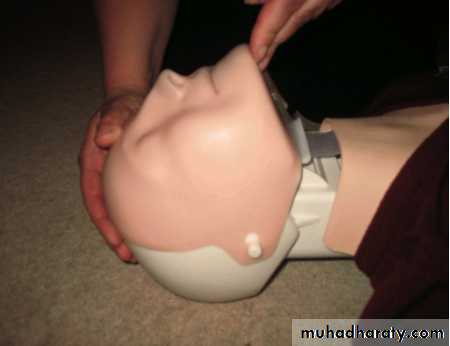
Position of your hands when performing head tilt/chin lift manoeuvre
As a lay provider, even if you suspect a cervical spine injury, you should still use the head tilt/chin lift technique, but perform it gently and only as far as necessary to provide a patent airway. Healthcare professionals should use a jaw thrust.Jargon buster
First degree relatives: mother, father, daughter, son, sister, brother.Second degree relatives: grandparent, grandchild, aunt, uncle, niece, nephew, half sister, and half brother.
Third degree relatives: first cousins.
The Renal Association recommends you offer primary prevention (statins and aspirin) if the 10 year cardiovascular disease risk reaches 20%.
Measure serum calcium, phosphate, and PTH concentrations in people
with stage 4 or 5 chronic kidney disease (GFR less than < 30 ml/min/1.73 m2).The routine measurement of calcium, phosphate, parathyroid hormone (PTH), and vitamin D levels in people with stage 1, 2, 3A, or 3B chronic kidney disease is not recommended.
When vitamin D supplementation is indicated in people with chronic kidney disease offer:
Cholecalciferol or ergocalciferol to people with stage 1, 2, 3A, or 3B chronic kidney disease
1-alpha-hydroxycholecalciferol (alfacalcidol) or 1,25-dihydroxycholecalciferol (calcitriol) to people with stage 4 or 5.
The diagnosis is confirmed by spirometry.
The presence of a postbronchodilator FEV1< 80% of the predicted value in combination with an FEV1/FVC < 70% confirms the presence of airflow limitation that is not fully reversible.COPD
There are three cardinal symptoms of COPD
exacerbation• Increased shortness of breath
• Increased cough
• Increased sputum volume or purulence
You should treat exacerbations with antibiotics, bronchodilators, and corticosteroids (ABC)
Respiratory failure is frequently present during exacerbations and you may need to start non-invasive ventilation.
Fifty to seventy per cent of exacerbations are due to respiratory infections
About 10% are due to pollution
In about 30% the cause cannot be identified.
Viral infections are thought to cause 20-40% of exacerbations. The most commonly isolated organisms are:
Rhinovirus (23%)Respiratory syncytial virus (11%)Influenzae, parainfluenzae, adenovirus, and coronavirus (6%).
Bacterial infections are thought to cause about 30% of infective exacerbations. Haemophilus influenzae (11%) Streptococcus pneumoniae (10%)Moraxella catarrhalis (10%)Haemophilus parainfluenzae (10%)Pseudomonas aeruginosa (10%).
medical conditions that can aggravate symptoms or mimic an exacerbation of chronic obstructive pulmonary disease. These include:
Pneumonia
Pneumothorax
Pleural effusion
Lung cancer
Upper airway obstruction
Rib fracture
Bronchiectasis
Pulmonary embolism
Congestive heart failure
Cardiac arrhythmia
You should give oxygen through a venturi facemask, to maintain the saturation of arterial blood at more than 90%.
The port size of the venturi valve ensures that the correct proportions of oxygen and entrained air are mixed to obtain a fixed oxygen concentration.
Low flow devices, such as nasal cannulae, are not able to deliver a fixed oxygen concentration as they are dependent on a patient's respiratory rate and tidal volume. They deliver a variable inspired oxygen concentration, which can result in suppression of respiratory drive, carbon dioxide narcosis, and respiratory arrest. In hypercapnic respiratory failure, you should start at an oxygen concentration of 24%.
You should aim to increase the PaO2 sufficiently to maintain optimal levels above 8.0 kPa without risking detrimental carbon dioxide retention and acidosis. After giving oxygen for 30 to 60 minutes, you should recheck arterial blood gases
Both Spiriva (Tiotropium) and Ipratropium (Atrovent) are anticholinergic agents that are used for maintenance treatment of bronchospasm associated with COPD .Spiriva has an additional indication for reducing COPD exacerbations based on the outcomes of the large UPLIFT clinical trial. Both medications are not indicated for acute treatment of a bronchospasm and should never be used as rescue medications. The major difference between the two drugs is that Spiriva is a long acting medication and Ipratropium is a short acting drug. Spiriva is used once daily, while Ipratropium may be used every 4 to 6 hours.
Key points
The choice of investigation and treatment of Crohn's disease is guided by the site, activity, and behaviour of disease70% of patients will have surgery at some point in their illness
Smokers are more likely to require surgery for their disease and have a higher risk of relapse after it
Smoking: Crohn's disease is more prevalent among smokers (in contrast to ulcerative colitis) and smokers have more surgery for their disease. Smoking cessation is associated with a 65% reduction in risk of relapse.
Smokers have a 2.5-fold increased risk of relapse after resection compared to non-smokers
Anaemia in Crohn's disease
• Malabsorption (B12 and folate deficiency)• Bleeding (iron deficiency)
• Anaemia of chronic disease
• Medication (eg azathioprine)
Faecal calprotectin
Faecal calprotectin (a neutrophil derived protein) is becoming more widely used as a tool for monitoring disease activity and treatment response in Crohn's disease. It appears to be a more sensitive marker for disease activity than CRP.
High levels of serum natriuretic peptides can have causes other than heart failure (left ventricular hypertrophy, ischaemia, tachycardia, right ventricular overload, hypoxaemia (including pulmonary embolism), renal dysfunction (GFR <60 ml/minute), sepsis, COPD, diabetes, age >70 years, and cirrhosis of the liver).
A serum BNP level less than 100 pg/ml (29 pmol/l) or an NTproBNP level less than 400 pg/ml (47 pmol/l) in an untreated patient makes a diagnosis of heart failure unlikely .
The level of serum natriuretic peptide does not differentiate between heart failure due to left ventricular systolic dysfunction and heart failure with preserved left ventricular ejection fraction.
ACE inhibitor therapy should generally be instituted before beta blocker therapy in heart failure.i…....d.
Aspirin (75-150 mg once daily) should be prescribed for patients with the combination of heart failure and atherosclerotic arterial disease (including coronary heart disease).
However, even in patients with heart failure in sinus rhythm, anticoagulation should be considered for those with
• a history of thromboembolism,
• left ventricular aneurysm, or
• intracardiac thrombus.
Indications for beta blockers
HypertensionAngina
Mitral valve prolapse
Cardiac arrhythmia
Afibrillation
Heart failure
Myocardial infarction
Glaucoma
Migraine prophylaxis
Symptomatic control (tachycardia, tremor) in anxiety and hyperthyroidism
Essential tremor
Phaeochromocytoma, in conjunction with α-blocker
Beta blockers have also been used in the following conditions:
HOCM
Acute dissecting aortic aneurysm
Marfan(treatment with propranolol slows progression of aortic dilation and its complications)
Prevention of variceal bleeding in portal hypertension
Possible mitigation of hyperhidrosis
Social anxiety disorder and other anxiety disorders
Prognosis
Within one year of having a first myocardial infarction, it has been reported that 25% of men and 38% of women will die.
Within six years of having a first myocardial infarction, 18% of men and 35% of women will have another myocardial infarction, 22% of men and 46% of women will have heart failure, and 7% of men and 6% of women will have sudden death.
The term dementia covers several syndromes of largely irreversible neurodegenerative disorders that result in severe cognitive decline, personality and behavioural changes, disability, and premature death. The main causes of dementia of late onset are:
• Alzheimer's disease
• Vascular dementia
• Dementia with Lewy bodies.
Many have mixed Alzheimer's and vascular dementia. Dementia affects 5% of people aged 65 and 20% of people aged 80. in a general hospital with 500 beds 100 patients will have dementia.
Biochemically, Alzheimer's disease is associated with a deficit in the neurotransmitter acetylcholine.
Parkinson's disease is associated with a deficit in the neurotransmitter dopamine. Dementia with Lewy bodies is associated with deficits in both acetylcholine and dopamine.
Every 30 seconds someone somewhere has their leg amputated due to diabetes. Most of these started as diabetic foot ulcers.
According to the UK's Department of Health (2001), diabetes is the leading cause of blindness, end stage renal failure, and amputations that are not accident related.
Diabetes mellitus, both type 1 and type 2, accounts for 50% of amputations in the UK
The mortality within three years of an amputation is 50%.
25% of diabetes have not been diagnosed.
25% of people with diabetes are likely to develop diabetic foot ulcers at some time in their life.
Table 1: Classification of the diabetic foot into neuropathic foot or neuroischaemic foot7
Indicator
Neuropathic foot
Neuroischaemic foot
Temperature
Warm
Cool
Pedal pulse
Bounding
Absent
Sensation
Numb
May be painful
Site
Plantar
Marginated
Callus
Present around ulcer
Very little callus
Colour/appearance
Distended dorsal veins
Foot is pink
Skin condition
Dry skin
Atrophic skin
LADA Latent autoimmune diabetes in adults
has also been referred to as late onset autoimmune diabetes of adulthood, slow onset type 1 diabetes, and type 1.5 (one and ahalf) diabetes. It is defined by three features:
adult age at diagnosis, presence of diabetes associated autoantibodies, and delay between diagnosis and the need for insulin to manage hyperglycaemia. Patients who develop diabetes in adulthood (with and without autoantibodies) vary in age of onset and phenotype and are therefore difficult to distinguish from each other.
The main causes of death in the adult Diabetic ketoacidosis
population include severe hypokalaemia; adult respiratory distress syndrome; and comorbid states such as pneumonia, acute myocardial infarction, and sepsis. Pulmonary oedema has been reported only rarely in DKA. Elderly patients and thosewith impaired cardiac function are at particular risk, and monitoring of central venous pressure should be considered.
Cerebral oedema is the most common cause of death in young children and adolescents, but it is rare in adults during treatment of DKA. Cerebral oedema is associated with a mortality rate of 20-40%.
Table. Biochemical criteria for diabetic ketoacidosis and hyperosmolar non-ketotic state
Diabetic ketoacidosisHyperosmolar non-ketotic state
Serum glucose
Elevation is variable
At least 30 mmol/l
Serum bicarbonate
15 mmol/l or less
Above 15 mmol/l
Urine ketones
(Test sticks measure acetoacetate, which is converted from 3-hydroxybutyrate)
At least ++
+ or less
Osmolality (calculated from 2 x ((Na+) +(K+)) +(urea) + (glucose))
Variable
At least 340 mmol/l
In patients with inferior myocardial infarction the following triad has a high specificity, but a low sensitivity, for the diagnosis of right ventricular infarction:
• Raised jugular venous pressure
• Clear lungs
• Hypotension
These signs have a low sensitivity for the diagnosis of right ventricular infarction because conditions such as cardiac tamponade, constrictive pericarditis, and pulmonary embolus share similar clinical features.
You must assess the patient’s hydration status as volume depletion may mask these signs. The key physical signs may only become apparent after volume repletion
Kussmaul’s sign (distension of the jugular vein on inspiration) is an important predictor of right ventricular infarction. It is both highly specific and sensitive.
Learning bite: right sided ECG
You should include a right sided ECG as part of the routine evaluation of all patients with an inferior myocardial infarction, as this will confirm any clinical suspicion of right ventricular ischaemiaST elevation in the right sided leads V3R to V6R has a high sensitivity, specificity, and predictive value for right ventricular infarction.
From the results of numerous studies, a 1 mm ST segment elevation in V4R has been shown to have the highest sensitivity and specificity for right ventricular infarction .
Learning bite: echocardiography
extremely useful non-invasive tool to aid you in the diagnosis of right ventricular infarction. The key abnormal echocardiographic features are:• Right ventricular dilation
• Asynchrony of the right ventricular free wall
• Paradoxical movement of the interventricular septum.
• The short axis views have the highest sensitivity (82%) and specificity, ranging from 62% to 93% for haemodynamically significant right ventricular impairment.
• Echocardiography can also rule out pericardial conditions and cardiac tamponade, which are the main differential diagnoses of right ventricular infarction.
Drugs that block the atrioventricular node, such as adenosine, verapamil, digoxin, and beta blockers, are contraindicated in patients with Wolff-Parkinson-White syndrome. These drugs will accelerate conduction of the atrial impulses down the accessory pathway, resulting in a fast ventricular response. This can degenerate into ventricular fibrillation.
It is preferable to use drugs that reduce conduction through the accessory pathway in patients in atrial fibrillation associated with Wolff-Parkinson-White syndrome. These include:
• Intravenous flecainide. You should not use this drug if the patient has structural or ischaemic heart disease as it is negatively inotropic and can cause ventricular arrhythmias in this group of patients
• Intravenous procainamide
• Intravenous amiodarone (given slowly).
Learning bite
You should treat patients with a single antiepileptic drug whenever possible.You should consider drug therapy after a first unprovoked seizure if:
• The patient has a neurological deficit
• The EEG shows unequivocal epileptic activity
• The patient or their family or carers consider the risk of having a further seizure unacceptable
• Brain imaging shows a structural abnormality.
• There is status epilepticus at onset
Patients should generally start drug therapy after a second epileptic seizure.
Intravenous diazepam is potentially dangerous. It is best reserved for specialist centres with intensive care facilities.
Intravenous clonazepam
Intravenous clonazepam is similarly dangerous.
The best traditional anti-epileptic drugs for partial seizures include:
CarbamazepineLamotrigine.
The best standard anti-epileptic drugs for generalised seizures include3:
Valproate
Lamotrigine
Levitiracetam.
After one seizure, the risk of recurrence is only about 35% at five years. However, this can be much higher if risk factors are present, in which case treatment can be considered even after one seizure.
Once a second seizure occurs, 75% of untreated people will have a third event.
What is the prognosis?
For most the prognosis is good.About 70% go into remission, defined as being free of seizures for five years on or off treatment.
This leaves 20-30% who develop chronic epilepsy, which is often treated with many anti-epileptic drugs.
A small minority of these patients may require surgical treatment for their epilepsy.
The combination of raised T4 and undetectable thyroid stimulating hormone, although consistent with a diagnosis of hyperthyroidism, is also found in patients with non-thyroidal illness (for example, in patients with acute exacerbation of chronic obstructive pulmonary disease, rheumatoid arthritis, and heart failure).
But serum T3 is always elevated in hyperthyroidism, whereas it is normal or slightly low( reduced conversion of T4 to T3 )in people without thyroid disease.
Intolerance to carbimazole is an indication for using propylthiouracil. The dose of propylthiouracil is 400 mg daily, usually given as 200 mg twice a day.
It is best to give propylthiouracil rather than carbimazole during breastfeeding because propylthiouracil is excreted in the milk to a lesser extent. Even after an 18 month course of antithyroid drugs, more than 50% of patients will relapse. Most of these will relapse in the first one or two years.
Iodine-131
Iodine-131 is the first choice of treatment for patients older than 40. But it is increasingly prescribed for younger patients in some centres. Euthyroidism is restored in80-90% of patients after three months of a standard dose of 400 MBq (approximately 11 mCi) iodine-131. After more than 50 years of use there is no evidence of a significant risk of leukaemia or cancer of the bladder or gastrointestinal tract.
Iodine-131 therapy is contraindicated in pregnancy because of the risk of fetal hypothyroidism.
There is some evidence that of all the treatments for Graves' disease, iodine-131 therapy is most likely to be associated with deterioration of ophthalmopathy.
This risk can be diminished by giving prednisolone 30-40 mg daily for six weeks following iodine-131 therapy. These patients need to be reviewed closely by a specialist.
Surgery
Subtotal thyroidectomy is the treatmentof choice for:
• Young patients with Graves' disease with large vascular goitres and moderate to severe hyperthyroidism
• People who have relapsed after a course of antithyroid drugs
• People who are poorly compliant .
• toxic nodular goitre if this is causing mediastinal compression.
There is an increasing list of drugs which interfere with the absorption of thyroxine (oral iron preparations, calcium carbonate, proton pump inhibitors) or increase its metabolic breakdown (antidepressants). It is sensible, therefore, in this case to advise that the thyroxine is taken last thing at night and that the final dose of ferrous sulphate is taken around teatime.
Previously, patients with subclinical hyperthyroidism were just reviewed annually until they developed overt hyperthyroidism. But we now know that subclinical hyperthyroidism may be a risk factor for osteoporosis and atrial fibrillation. These patients should therefore probably be treated in the same way as those with more overt disease.
The diagnosis of hypothyroidism must be confirmed by measuring serum thyroid stimulating hormone and serum free thyroxine .
Amiodarone contains a large amount of iodine (75 mg in each 200 mg tablet) and can induce thyroid dysfunction. Clinical hypothyroidism can occur in up to 20% of patients on long term amiodarone.
Lithium is associated with clinical hypothyroidism in up to 15% of patients.
In a population-based study of over 30 000 subjects with no previous history of thyroid dysfunction, the prevalence of overt and subclinical hypothyroidism among women who were current smokers was significantly lower than for never-smokers.
Drug interactions with thyroxine
Patients on warfarin may need careful monitoring and adjustment of their warfarin dose when their thyroxine dose is altered. Thyroxine can enhance the anticoagulant effect of warfarinThyroxine may increase the metabolism of propranolol
Anticonvulsants and rifampicin increase hepatic metabolism of thyroxine and may increase thyroxine replacement requirements
Side effects of thyroxine in subclinical hypothyroidism
Several randomised controlled trials have evaluated thyroxine treatment in people with subclinical hypothyroidism.
The evidence of benefit is less clear than for clinical hypothyroidism.
Two randomised controlled trials reported side effects with thyroxine.
One trial found worse anxiety scores in patients treated with thyroxine, compared with patients treated with placebo
The second trial reported that 11% of people on thyroxine withdrew because of complications (worsening angina or atrial fibrillation)
Subclinical hypothyroidism in pregnancy
Women with subclinical hypothyroidism who are trying to get pregnant should be treated with thyroxine. Untreated hypothyroidism in pregnancy is associated with an increased risk of fetal loss, stillbirth, and premature labour
Learning bite
Non-fasting results can produce higher triglycerides and lower high density lipoprotein cholesterol concentration.Non-fasting measurements slightly overestimate CHD risk but are regarded as sufficiently accurate to use in screening and are more convenient for patients. Routine use of a tourniquet has no significant effect on lipid measurements.
Testing lipids
How often should patients' lipids be tested after starting lipid lowering treatment?A review of the international literature suggests intervals of:
8 (±4) weeks after starting drug treatment
8 (±4) weekly after adjustments to treatment until within the target range.
How often should cholesterol or lipids be tested once a patient has reached target or optimal cholesterol?
Annually (unless there is a specific reason for more frequent reviews).
How often should liver enzymes be routinely measured in patients taking statins?
Measure alanine aminotransferase:
Before treatment with a statin
Eight weeks after starting a statin or after any dose increase.
What if liver enzymes become raised in a person taking a statin?
If ≤3 x upper limit of normal:Continue statin
Recheck liver enzymes in 4-6 weeks
No extra monitoring required unless values rising.
If ≥3 x upper limit of normal (depending on magnitude of rise):
Stop statin or reduce dose, recheck liver enzymes within 4-6 weeks
Cautious reintroduction of statin may be considered (for example at a lower dose).
How often should creatine kinase be measured in patients taking statins?
Pre-treatmentBefore starting treatment with a statin
If the baseline creatine kinase level is >5 times the upper limit of normal, do not start statin. Routine monitoring of creatine kinase is not necessary .Check creatine kinase if patient develops myalgia.
What if creatine kinase becomes raised in a person taking a statin?
If ≥5 x upper limit of normal:Stop treatment, check renal function, and monitor creatine kinase fortnightly
Consider secondary causes of myopathy if creatine kinase remains elevated.
If ≤5 x upper limit of normal:
If no muscle symptoms, continue statin (patients should be alerted to report symptoms; consider further checks of creatine kinase)
If muscle symptoms, monitor symptoms and creatine kinase regularly if creatine kinase continues to rise.
AST/ALT ratio in these patients is typically 2:1 or more.
increased GGT levels with normal ALP levels and macrocytosis make alcohol induced liver damage more likely. Urea levels are often low in alcohol related cirrhosis due to reduced hepatic metabolism of protein and reduced protein intake.
Alcoholic liver disease
Hazardous drinking is defined as a quantity or pattern of alcohol consumption that places patients at risk for adverse health events, whil
harmful drinking is defined as alcohol consumption that results in adverse events (eg, physical or psychological harm).
Heavy drinking is defined as a quantity of alcohol consumption that exceeds an established threshold value.
hazardous drinking was defined as an average consumption of 21 drinks or more per week for men (or 7drinks per occasion at least 3 times a week), and 14 drinks or more per week for women (or 5 drinks per occasion at least 3 times a week).
Because hazardous and heavy drinking are similarly defined (ie, a quantity or pattern of alcohol consumption that exceeds a specific threshold and may increase risk for adverse health events), we will use 1 term, hazardous drinking, to define this type of drinking disorder.