Diabetic foot
د. حسين محمد جمعةاختصاصي الامراض الباطنة
البورد العربي
كلية طب الموصل
2010
Key points
leave the foot at high risk of amputationIt is important to find the cause of the problem and differentiate between neuroischaemic ulcers, which occur on the margins of the foot, and neuropathic ulcers which occur on the plantar surface
Debridement of dead tissue is a crucial part of wound management in patients with diabetes
Multidisciplinary management is essential when managing people with diabetes.
Clinical tips
Patients with neuroischaemia should never apply direct heat to the feet but wear warm socks without a constricting ankle band.You must refer patients with neuropathy or neuroischaemia to podiatry for advice about appropriate footwearDebridement is a key method of microbiological control of the diabetic foot.It carries a high level of clinical risk and should therefore only be carried out by trained practitioners when managing patients with diabetes.
Pain can occur in neuroischaemia when the neuropathy is mild, and the patient then experiences pain from an ulcer
Opioids may be necessary for pain control. You should use these with caution in patients with autonomic neuropathy who are more susceptible to respiratory depression
You should check the temperature gradient using the back of your hand moving from the pre-tibial region down towards the foot. In this way it is possible to detect hot spots, cold areas, and any areas of asymmetry in temperature. An icy foot indicates acute ischaemia, whereas a warm foot may be neuroischaemic if co-existent autonomic neuropathy is present.
Introduction
Every 30 seconds someone somewhere has their leg amputated due to diabetes. Most of these started as diabetic foot ulcers.According to the UK's Department of Health (2001), diabetes is the leading cause of blindness, end stage renal failure, and amputations that are not accident related.
For a patient with a diabetic foot ulcer to be managed effectively, a multidisciplinary team, rather than a healthcare professional working in isolation, is necessary to ensure these complex wounds are treated appropriately.
Recognising early signs of ulcer development, together with ensuring that patients control their diabetes well, prevent potentially catastrophic patient outcomes.
Statistics
Diabetes mellitus, both type 1 and type 2, accounts for 50% of amputations in the UKThe mortality rate within three years of an amputation is 50%.
In 2005, 4.7% of the UK's population (2.35m people) were estimated to have either type 1 or type 2 diabetes.
25% of diabetes have not been diagnosed.
25% of people with diabetes are likely to develop diabetic foot ulcers at some time in their life.
Socio-economic data
Type 2 diabetes is:Six times more common in people of South Asian origin
Three times more common in people of African and Afro-Caribbean descent.
Complications
Complications are 3.5 times more common in areas of social deprivation in the UK. People with poorly controlled diabetes are at greater risk of developing complications. The main complications that occur in a patient with a diabetic foot ulcer are:
• Infection
• Gangrene
• Amputation
• Death.
Risk factors for development of a diabetic foot ulcer
The presence of the following factors increases the risk of a person with diabetes developing a foot ulcer:• Neuropathy
• Arterial disease
• Abnormal pressure loads (areas of high pressure on the soles of the feet).
Other significant risk factors are the presence of:
• Callus• Deformity
• Swelling.
Classifying and staging the foot
Classifying and staging the foot will allow you to manage the foot correctly. Using the Edmonds and Foster Simple Staging System, you first classify the foot as neuropathic or neuroischaemic.We recommend this classification as a quick, simple, and practical system.
Table 1: Classification of the diabetic foot into neuropathic foot or neuroischaemic foot7
Indicator
Neuropathic footNeuroischaemic foot
Temperature
Warm
Cool
Pedal pulse
Bounding
Absent
Sensation
Numb
May be painful
Site
Plantar
Marginated
Callus
Present around ulcer
Very little callus
Colour/appearance
Distended dorsal veins
Foot is pink
Skin condition
Dry skin
Atrophic skin
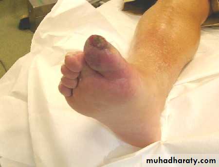
• Figure 1: A neuroischaemic ulcer. Note the absence of callus and the marginated ulcer on tip of toe
•
• Figure 2: A neuropathic foot
• Note the distended veins due to arteriovenous shunting that occurs with autonomic neuropathy and the presence of callus on the lateral plantar surface. There is also some staining due to venous hypertension.•
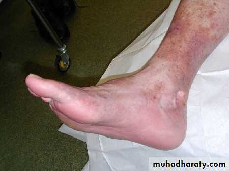
Table 2: Staging system for the diabetic foot7 This staging system, one of several which have been devised over the past two decades, is widely used because of its simplicity and practicality.
Stage
DescriptionStage 1
Low risk foot with no neuropathy and no ischaemia
Stage 2
High risk foot
Stage 3
An ulcerated foot
Stage 4
An infected foot
Stage 5
A necrotic foot
Stage 6
An unsalvageable foot
Once you have identified the stage, your management should aim to prevent a foot from progressing to the next stage.
Assessment
All people with diabetes should have an annual foot inspection as part of the regular checking and monitoring regimen.
You should examine each foot in turn, observing the dorsum, sole, medial and lateral borders, back of the heel, malleoli, and interdigital areas.
On examination, you should specifically look for the following:
Skin changes
Look for breaks in the integrity of the skin. No damage should be dismissed as minor. You need to stage and manage all damage.Dry fissured skin is common in neuropathic ulcers. You should not confuse these with eczema or psoriasis although these conditions can co-exist in people with diabetes.
Distended veins indicate neuropathy.
Skin atrophy can indicate ischaemia.
Other skin lesions are
necrobiosis lipoidica diabeticorum with its clearly demarcated liver-coloured lesions.
Webspace maceration is a sign of tinea pedis.
It is best to keep foot lesions covered and dry; this prevents the spread of infection which can have disastrous results, such as gangrene and amputation. For this reason, you should advise patients to not let a foot ulcer get wet, for example, by using a bath or shower. Dressings which keep wounds moist are contraindicated.Callus
Indicates areas of high pressure and friction. They should not be allowed to become too thick as it can be a precursor to ulceration. Callus forms a plaque, while corns are smaller discrete areas.You should refer all patients with callus and corns to a podiatrist for removal to prevent later problems with ulceration. You should discourage patients from removing their own corns or calluses as doing so carries a high risk of injury.
Nails
subungual bleeding or haematoma presents as a purple discoloration and should be further investigated. Malignant lesions can also occur under nails. In acute ischaemia, nail beds are pale. Also observe if nails are thickened which could cause pressure on the nail bed from a shoe. Look for discharge from nails which may indicate an underlying ulcer. Fungal nail infections are common. Note the crumbling appearance of these nails, due to fungal infection and ischaemia.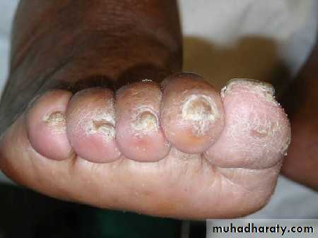
Swelling
Any swelling of a diabetic foot causes a high risk of ulceration due to poorly fitting, too tight shoes. There are many causes of swelling and it is important to establish whether this is bilateral or unilateral. Swelling also hampers the healing of existing foot ulcers. You should refer diabetic patients with a swollen foot to a podiatrist.Deformity
like hammer toe and claw toes should be noted as they are likely to cause pressure on footwear and subsequent ulceration. The Charcot foot is a specific deformity resulting in bone and joint destruction. Patients who develop Charcot's osteoarthropathy may have symptoms of peripheral or autonomic neuropathy. In its acute stage it often follows recent trauma, presenting with a unilateral erythema and swelling, with the affected foot being at least 2° hotter than the unaffected foot.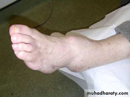
The following picture shows a typical late stage Charcot deformity.
ColourRedness of the foot may indicate cellulitis, critical ischaemia, Charcot foot.Redness of the toes may indicate cellulitis, osteomyelitis, ischaemia, chilblain.Blue discoloration of the foot may indicate cardiac failure, chronic obstructive airways disease, venous insufficiency.Blue discoloration of the toe may indicate severe infection or ischaemia.A white foot, especially on elevation, may indicate ischaemia.Black areas may indicate ischaemia, emboli, tumour, application of henna .
Pulses
If it is difficult to find pedal pulses manually, you should use a hand held Doppler. This measures the ankle-brachial pressure index by comparing the resting systolic pressures of the pedal pulses and dividing these by the resting brachial systolic pressure. The highest measure of each leg is divided by the highest brachial reading.The normal range is 0.92-1.25 and readings within this range indicate absence of arterial disease.
less than 0.92 indicates the presence of ischaemia.
An ankle-brachial pressure index of
less than 0.80 indicates moderate disease, and a reading of
0.60 or less indicates severe disease.
A normal or elevated ankle-brachial pressure index does not necessarily exclude ischaemia. This is because patients with diabetes may have calcified arteries which do not compress, leading to a falsely elevated ankle-brachial pressure index in such patients. The ankle-brachial pressure index readings are also inaccurate if the examination is carried out in a cold room.
Temperature
Use the back of your hand to compare the skin temperature of both feet and legs. Assess temperature gradient by moving the back of your hand from the shin down towards the dorsum of the foot. In this way, you can detect hot spots which may indicate infection.A unilateral temperature increase on the foot may indicate Charcot's osteoarthropathy. Gradient asymmetry may indicate unilateral ischaemia on the colder limb.
An icy cold foot indicates acute ischaemia .
while autonomic neuropathy
in a neuroischaemic foot may keep the foot warm.
Sensation
Testing for protective pain with a 10 g monofilament is an important part of assessment. Patients who lack protective pain sensation will be unaware if they injure their feet or overload areas. This can lead to callus formation and ulceration. Patients will continue to walk on an ulcerated foot without complaining of pain.It is essential to detect the patient who lacks protective pain sensation by testing both surfaces of the feet, along with the malleoli and toes. Avoid testing over an area of callus. You need to place the monofilament at 90° to the skin and then angle it until the filament buckles. At this point 10 g of pressure is exerted on the skin.
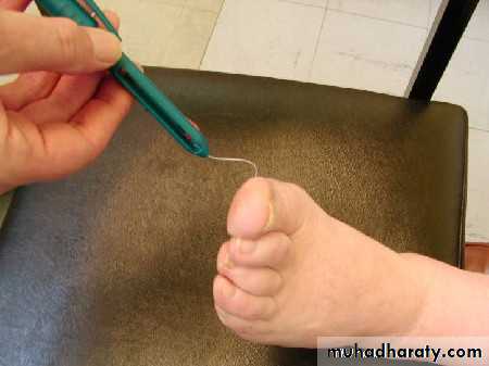
Figure 3: Using a 10 g monofilament to test for protective pain sensation
Footwear assessmentlook at the patient's shoes to see if they fit properly. Poorly fitting shoes or socks may cause pressure on toes. Look for anything that might cause injury such as a tight sock band or a ridge in the sock.
As a general rule, slip on shoes do not provide sufficient support. They compress the toes and lead to areas of high pressure on the soles and borders of the foot, which can lead to ulceration. If the patient is at Stage 2 or higher and is not already seeing a podiatrist, you need to refer them to a podiatrist for specialist advice.
Management of the diabetic foot
There are six areas of control that shape the management of the diabetic foot. These are:Mechanical: offloading the foot, and prevention of ulceration
Microbiological: managing and preventing infection
Metabolic: control of blood glucose, blood pressure, cholesterol, and smoking cessation
Vascular: assessment of the ankle-brachial pressure index and referral for reconstructive surgery and/or debridement
Wound care: keeping the wound dry and debridement of dead tissue to prevent infection spread
Educational: advice and health promotion activities with the patient to prevent recurrence.
The following case histories summarise the management of a diabetic foot.
Management of the diabetic foot is the responsibility of a multidisciplinary specialist team to deliver optimum care as required by both the UK's National Service Framework for Diabetes and the joint report from the Department of Health and Diabetes UK Care Planning Working Group (2006).Clinical tips
When you suspect a fungal infection you should send nail clippings for microscopy, culture, and sensitivity.
Eradicating the fungus is difficult. Left untreated these infections may cause chronic pain and secondary infection.
Thinning or debulking of the nail by a podiatrist is sufficient treatment of most nail infections
Specks of blood elsewhere on a diabetic foot, particularly under a callus, are danger signals. These calluses should be removed. Often an ulcer is present underneath a discoloured callus.
Learning bite
Gangrene occurring in a patient with diabetes with a neuroischaemic foot is associated with very high morbidity and mortality.Management should include vascular intervention (angioplasty or distal bypass) or conservative care with the aim of achieving auto-amputation.
Clinical tips
Ischaemic feet worsen in cold weatherDirect heat should never be applied to the feet
Patients with neuropathy may burn their feet
Patients with neuroischaemia will deteriorate because of the increased metabolic rate of warmer tissues, which cannot be met when perfusion is poor.
Atrophic nails can be an indication of ischaemic disease since the nail bed is poorly perfused in patients with ischaemic feet.
Missing digits are a key sign that a patient has neuroischaemia.
Learning bite
Patients with diabetes have an increased risk of developing Charcot's osteoarthropathy, osteomyelitis, and septic arthritisDistinguishing between Charcot's osteoarthropathy and infection can be challenging. Half of all patients with diabetes with severe infections are apyrexial and normoglycaemic.
You should also treat patients for infection until Charcot's osteoarthropathy is excluded.
You should suspect Charcot's osteoarthropathy in patients presenting with small fibre neuropathy, with autonomic neuropathy, and a warm, swollen foot and treat them for Charcot's osteoarthropathy until the diagnosis is confirmed or refuted.
Treatment consists of application of a total contact cast until the temperature of the two feet is within 2°C and x ray shows consolidation, with fracture healing, remodelling, and bone sclerosis, which is followed by careful rehabilitation in suitable boots.
Clinical tips
Thirty per cent of episodes of acute Charcot's osteoarthropathy are painful.Patients with type 1 diabetes and Charcot's osteoarthropathy may have pre-existing osteoporosis and may present with fractures.Patients with type 2 diabetes and Charcot's osteoarthropathy do not tend to have pre-existing osteoporosis and present with varying degrees of dislocation. The reason for this is unclear
You should ask about recent trauma in the history: this is a common finding in patients with Charcot's osteoarthropathy.
The most common site of neuropathic ulceration is on the toes. A traumatic lesion, as in this patient, frequently leads to ulceration. Plantar lesions associated with neglected callus are also common.
Although patients with neuropathy often do not experience pain they may be aware of a throbbing sensation if the foot is infected.
Patients with neuropathy may complain of a stabbing pain and their feet may feel cold although they are well perfused. Fever and elevated white cell counts are not present in half the patients with a deep infection of the foot.
Clinical tips
You should identify infection early and treat it effectively. Absence of overt signs of clinical infection in a patient with diabetes at an early stage does not exclude the possibility of underlying infection.Neuropathy and ischaemia may mask signs and symptoms of infection. So if you suspect infection in a diabetic foot, it is always useful to take a swab.
Offloading pressure is essential for patients with diabetes with cellulitis or foot infection. This can be achieved through rest or by casts that provide access for foot inspections.
Any foot with ulceration or infection should be offloaded. A patient with cellulitis needs bed rest.
Podiatrists should remove heavy callus to prevent the risk of ulceration underneath.
A visit to the podiatrist and advice on cutting nails is necessary for ingrowing toenail.
Antifungal preparations are necessary for nail infections. It is often a few months before improvement is seen.
Unilateral erythema, oedema, and warmth all point to a diagnosis of Charcot's osteoarthropathy. The difference in temperature between both feet is at least 2º hotter in the affected foot in all patients with active Charcot's osteoarthropathy.
Deformity is not an early feature of Charcot's osteoarthropathy; pain is present in this condition but the patient may still be able to weight bear, so this is not a definitive diagnostic feature for Charcot's osteoarthropathy. However, patients with this condition should not be weight bearing since this increases risk of further bony damage.
Pallor, coolness to touch, and pain are common in ischaemia but pain is the only one of these three signs that is consistent with Charcot's osteoarthropathy. Heat is a characteristic sign, typically 2º-5º hotter than the unaffected foot. Erythema, rather than pallor, is seen in the Charcot foot.
It is necessary to remove dead tissue to prevent a focus for infection, and ensure that the wound bed can be adequately assessed. Managing pain is an important aspect of wound care.
Diabetic foot ulcers should not be moistened: this may lead to spread of infection and development of wet gangrene which may have disastrous consequences.
Compression is the mainstay of management of venous leg ulcers; many diabetic wounds have an arterial or mixed arterial and venous component which means that compression may be contraindicated.
Dressings containing iodine (like Inadine) or silver (like Actisorb Silver) do not hydrate the wound and are useful for reducing the risk of infection. Diabetic foot ulcers should be kept as dry as possible. Systemic antibiotics are needed if the wound is infected. Debridement of dead tissue is very important to prevent the tissue from becoming a focal point of infection.
The tissue in a neuroischaemic wound is hypoxic and topical antibiotics are unlikely to be helpful.
Patients with painful neuropathy only have symptoms and do not have any clinical signs. It is known to severely affect the patient's quality of life. The pain may interfere with their sleeping and eating. The pain will not improve quickly unless the hyperglycaemia is controlled. After recovery, there may be residual numbness.
Learning bite
Neuropathic pain almost always improves within two years whether or not blood glucose is controlled, but tight control always achieves better outcomes, with a more rapid and greater reduction of pain. Pain may be superceded by numbness.
Clinical tips
There is also a large array of treatments including capsaicin cream, spinal stimulation, topical Opsite film, a bed cradle to keep the bed clothes from contact with the skin, and cooling the feet in a bowl of cold water that can be used to manage neuropathic pain.Gabapentin and tricyclic antidepressants are useful in managing neuropathic pain; intravenous lignocaine should be given under specialist supervision only.
Opioids are reserved for those occasions where other treatments fail.
Autonomic neuropathy may present in the diabetic foot as distended veins, due to arteriovenous shunting.