Urinary
incontinence
D. Hasanain Farhan

Definition
is the involuntary loss of
urine that is objectively
demonstrated with social
and hygienic problem

Classification
Anatomic or genuine urinary
stress incontinence
Urge incontinence
Neuropathic incontinence
Congenital incontinence
False (overflow) incontinence
Iatrogenic incontinence
Fistulous incontinence

Stress incontinence
is an involuntary loss of urine that
occurs during physical activity, such as
coughing, sneezing, laughing, , sudden
changes of position or exercise.
bet. 15-30% of women over age 65 yr
have urinary incontinence
&stress
incontinence is the most common type
30%to 50% of women with stress
incontinence also complain of urinary
frequency, urgency, and/or urge
incontinence

Types
Classic or genuine stress incontinence
is caused by pelvic prolapse, urethral
hyper mobility or displacement of the
urethra and bladder neck from their
normal anatomic alignment(also called
anatomic stress incontinence)
Stress incontinence can also occur as
a result of intrinsic sphincter
deficiency, in which the sphincter is
weak because of neurologic insult ,
previous surgery, estrogen deficiency ,
radiation damage or trauma.

Anatomy:
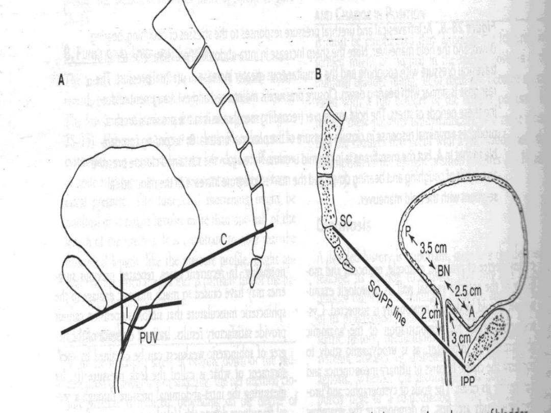
The anatomic feature is that of
hypermobility or a lowering of the
position of the VU segment
Various relations between the
urethra, bladder, and bony landmarks
have been studied
Posterior vesicourethral angle
Axis of inclination (urethral line vs.
vertical plane)
UV junction and the SCIPP


Risk Factors
1) Gender
:urinary incontinence is much more
common in women than men.
2) Genetics
:several studies suggested
genetic predisposition for stress
incontinence.
3) Race, culture, and environment
—stress
incontinence was reported to be more
common in whites than blacks.
4) Overweight
:
causes more pressure on
pelvic floor.

5) Pregnancy&Childbirth
:
increasing weight of baby puts extra
stress on pelvic floor , the hormone
relaxin softens the muscles of the
pelvic floor ready for the birth, In
vaginal delivery the nerves around
pelvic floor become stretched and
bruised ,women who'd had a tear or
episiotomy had a three-fold risk of
developing urinary incontinence.

6) Smoking:
a chronic cough puts pressure
on the pelvic floor and makes SUI worse.
7) Age:
stress incontinence is not a normal
part of aging ; physical changes
associated with aging as the weakening
of the muscles make elderly more
susceptible to stress incontinence
8) Medications:
can affect the pelvic floor.
Examples are alpha-blockers used to
treat high blood pressure, some
antidepressants and sedatives, and
some muscle-relaxant drugs.

DIAGNOSTIC EVALUATION
causes of transient incontinence should be
ruled out
1) Drug side effects
2) Delirium or hypoxia
3) Impaired mobility
4) Urinary tract infection
5) Atrophic vaginitis
6) psychological problems
7) Excessive fluid intake

8) Recent prostatectomy
9) Stool impaction.
EVALUATION include:
History
Physical examination
Urinalysis
Measurement of postvoid residual (PVR)
urine volume
Micturition Diary
Pad Test
Urodynamic Evaluation

History
is important in assessing the characteristics
and severity of incontinence as well as its
impact on quality of life.
It is also important in identifying risk
factors and/or transient causes of
incontinence
patient history alone is not an accurate tool
in the diagnosis of sphincteric incontinence
and should not be used as the sole
determinant of diagnosis or treatment

Physical Examination
Neurourologic examination
begins by
observing the patient's gait
The lumbosacral nerve roots should be
assessed by checking deep tendon reflexes,
lower extremity strength, sensation , anal
sphincter tone & genital sensation.
The abdomen and flanks
should be
examined for masses, ascites &
organomegaly which can influence intra-
abdominal pressure.

Rectal examination
will disclose the
size and consistency of the
prostate&anal sphincter tone
Cough test
: the bladder full in the
lithotomy position, the patient is asked
to cough in an attempt to reproduce the
incontinence
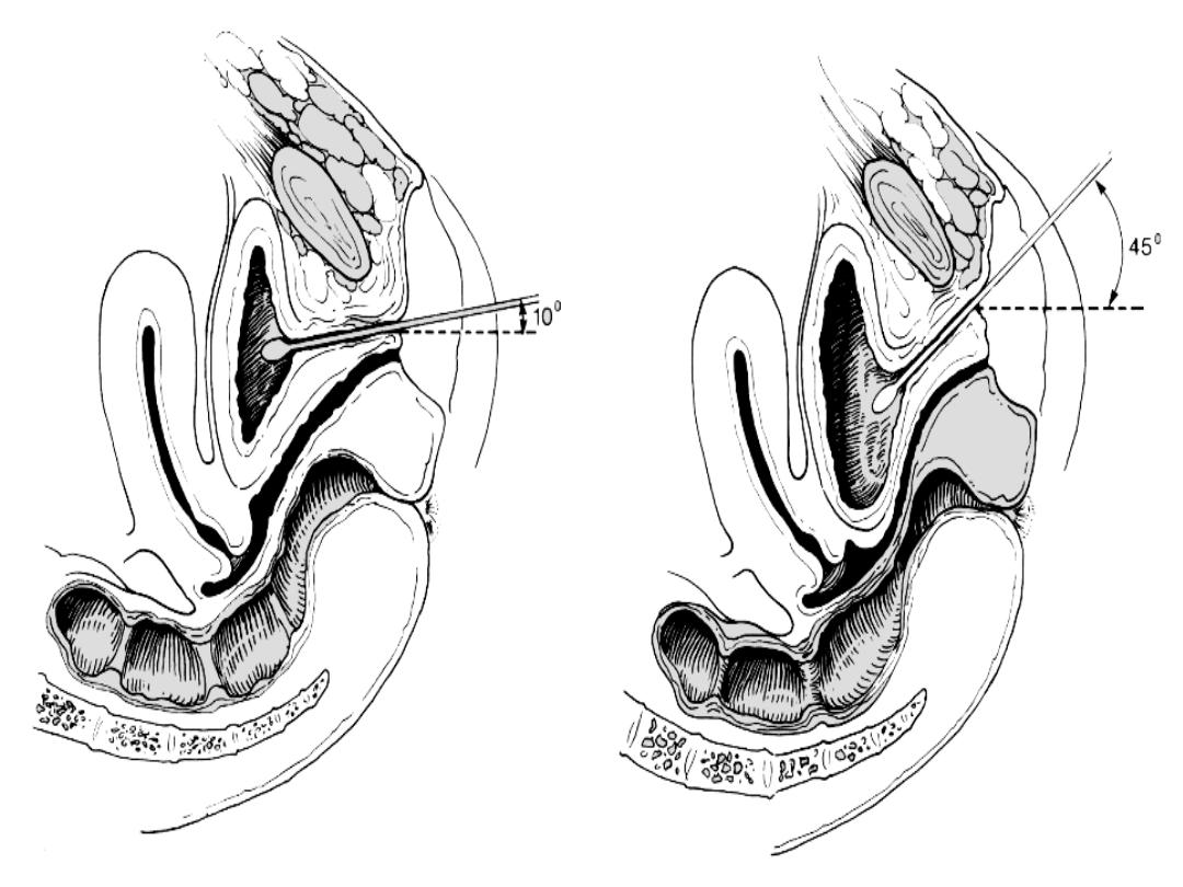
the Q-Tip test
: assess the degree of
urethral hypermobility by inserting a
lubricated sterile cotton-tipped
applicator gently through the urethra
into the bladder,the patient is then
asked to strain and the degree of
rotation is assessed. Hypermobility is
defined as a resting or straining angle
of greater than 30 degrees from the
horizontal.

Vaginal examination:
anterior vaginal wall is examined to assess
cystocele
posterior vaginal wall and vault are
examined for the presence of a rectocele or
enterocele.
Pelvic floor strength is assessed
Because the urethra and trigone are
estrogen-dependent tissues. The most
common signs of inadequate estrogen
levels are thinning and paleness of the
vaginal epithelium, loss of rugae,
disappearance of the labia minora and
presence of a urethral carbuncle.

Urinalysis
Urinalysis can identify acute urinary tract
infection ,the condition reversible with
treatment.
Residual Urine Measurement
It is usually measured by catheterization or
ultrasonography. A postvoid residual of less
than 50 ml is considered normal( in Stress
incontinence) , and a postvoid residual of
more than 200 ml is considered abnormal.
Values between 50 and 200 ml require
clinical correlation In interpreting the results

Micturition
Diary
• Micturition diaries and pad tests make it
possible to document voiding patterns in
the patient's own environment and during
various daily activities.
• the following measurements to be included
in a micturition diary: time of micturition,
time and type of incontinence, and voided
volume .
• 24-hour studies are adequate for the
evaluation of lower urinary tract symptoms.

Pad Test
a
semiobjective measurement of urine loss
over a given period of time.
A weight gain a sanitary towel of up to 8 g
over a 24-hour pad test is considered
normal.

Urethral Pressure Profilometry
The classical
pressure changes in stress
incontinence:
1) Low urethral closure pressure.
2) Short urethral functional length
3) Weak response to stress.
Cystometry
leak with cough
Flowmetry

TREATMENT
Nonsurgical Treatment
1) Behavior Modification
2) Pelvic Floor Exercises
3) Biofeedback
4) Electrical Stimulation
have all been reported to cause
improvement in 30% to 75% of patients.
5)
α-adrenergic agonists ,SRI
6) Estrogens

Surgical Treatment
if hypermobility ,treatment is:
Suspension of the bladder neck &
proximal urethra which is either
1) Retropubic Suspensions Marshall-
Marchetti-Krantz (MMK) and Burch
colposuspension or
2)Transvaginal suspensions

if (ISD) exists
suspension alone is not adequate &
treatment is:
1)Pubovaginal sling (Autologous Tissues
as Rectus Fascia or Nonautologous Tissues
as pericardium or Synthetic Materials as
Monofilament Polypropylene Tape the tension-
free vaginal tape (TVT) procedure or TOT
2)Periurethral injections
3)Sphincter prostheses

Urge Incontinence
• The basic feature is detrusor instability
and loss of urine while attempting to
inhibit micturition
• The bladder is described to be
overactive with clinical symptoms of
urgency,frequency, and nocturia
• The bladder overactivity can be
idiopathic or result from bladder
inflammation,tumour,obstruction,
neurological and trauma

Urodynamic Features
•
Flowmetry
High flow rate
•
Cystometry
Detrusor hyperirritability
with increase intravesical pressure
,decrease capacity and uninhibited
contraction
•
Urethral closure pressure
Normal or
high, normal response to stress and
normal urethral fuctional length

Treatment
• Behavior Modification.
• Anticholenergic drugs.
• Intravesical botulinum toxin injection.
• Surgery
SNS,augmentation cystoplasty, and
diversion.

THE END
THANK YOU
